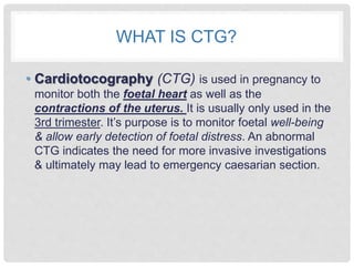
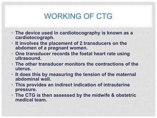
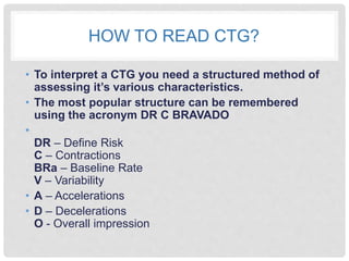
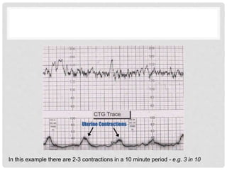
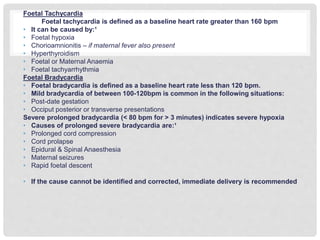
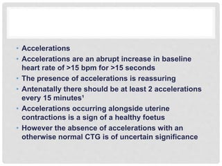
CARDIOTOCOGRAPHY
WHAT IS CTG?
• Cardiotocography (CTG) is used in pregnancy to
monitor both the foetal heart as well as the
contractions of the uterus. It is usually only used in the
3rd trimester. It’s purpose is to monitor foetal well-being
& allow early detection of foetal distress. An abnormal
CTG indicates the need for more invasive investigations
& ultimately may lead to emergency caesarian section.
WORKING OF CTG
• The device used in cardiotocography is known as a
cardiotocograph.
• It involves the placement of 2 transducers on the
abdomen of a pregnant women.
• One transducer records the foetal heart rate using
ultrasound.
• The other transducer monitors the contractions of the
uterus.
• It does this by measuring the tension of the maternal
abdominal wall.
• This provides an indirect indication of intrauterine
pressure.
• The CTG is then assessed by the midwife & obstetric
medical team.
HOW TO READ CTG?
• To interpret a CTG you need a structured method of
assessing it’s various characteristics.
• The most popular structure can be remembered
using the acronym DR C BRAVADO
•
DR – Define Risk
C – Contractions
BRa – Baseline Rate
V – Variability
• A – Accelerations
• D – Decelerations
O - Overall impression
DEFINE RISK
• You first need to assess if this pregnancy is high or low risk
• This is important as it gives more context to the CTG reading
• e.g. If the pregnancy is high risk, your threshold for intervening may be lowered
• Reasons a pregnancy may be considered high risk are shown below¹
Maternal medical illness
• Gestational diabetes
Hypertension
Asthma
Obstetric complications
• Multiple gestation
Post-date gestation
Previous cesarean section
Intrauterine growth restriction
Premature rupture of the membranes
Congenital malformations
Oxytocin induction/augmentation of labor
Pre-eclampsia
Other risk factors
• No prenatal care
Smoking
• Drug abuse
CONTRACTIONS
• Record the number of contractions present in a 10
minute period - e.g. 3 in 10
• Each big square is equal to 1 minute, so you look
how many contractions occurred in 10 squares
• Individual contractions are seen as peaks on the part
of the CTG monitoring uterine activity
• You should assess contractions for the following:
• Duration – how long do the contractions last?
• Intensity – how strong are the contractions? (assessed
using palpation)
In this example there are 2-3 contractions in a 10 minute period - e.g. 3 in 10
BASELINE RATE OF FOETAL HEART
• The baseline rate is the average heart rate of the
foetus in a 10 minute window
• Look at the CTG & assess what the average heart
rate has been over the last 10 minutes
• Ignore any Accelerations or Decelerations
• A normal foetal heart rate is between 110-150 bpm¹
Foetal Tachycardia
Foetal tachycardia is defined as a baseline heart rate greater than 160 bpm
• It can be caused by:¹
• Foetal hypoxia
• Chorioamnionitis – if maternal fever also present
• Hyperthyroidism
• Foetal or Maternal Anaemia
• Foetal tachyarrhythmia
Foetal Bradycardia
• Foetal bradycardia is defined as a baseline heart rate less than 120 bpm.
• Mild bradycardia of between 100-120bpm is common in the following situations:
• Post-date gestation
• Occiput posterior or transverse presentations
Severe prolonged bradycardia (< 80 bpm for > 3 minutes) indicates severe hypoxia
• Causes of prolonged severe bradycardia are:¹
• Prolonged cord compression
• Cord prolapse
• Epidural & Spinal Anaesthesia
• Maternal seizures
• Rapid foetal descent
• If the cause cannot be identified and corrected, immediate delivery is recommended
VARIABILITY
• Baseline variability refers to the variation of foetal heart
rate from one beat to the next
• Variability occurs as a result of the interaction between
the nervous system, chemoreceptors, barorecptors &
cardiac responsiveness.
• Therefore it is a good indicator of how healthy the foetus
is at that moment in time.
• This is because a healthy foetus will constantly be
adapting it’s heart rate to respond to changes in it’s
environment.
• Normal variability is between 10-25 bpm³
• To calculate variability you look at how much the peaks &
troughs of the heart rate deviate from the baseline rate (in
bpm)
• Variability can be categorised as: 4
• Reassuring – ≥ 5 bpm
• Non-reassuring – < 5bpm for between 40-90 minutes
• Abnormal – < 5bpm for >90 minutes
• Reduced variability can be caused by: ³
• Foetus sleeping - this should last no longer than 40 minutes – most
common cause
• Foetal acidosis (due to hypoxia) – more likely if late decelerations
also present
• Foetal tachycardia
• Drugs – opiates, benzodiazipine’s, methyldopa, magnesium
sulphate
• Prematurity – variability is reduced at earlier gestation (<28 weeks)
• Congenital heart abnormalities
• Accelerations
• Accelerations are an abrupt increase in baseline
heart rate of >15 bpm for >15 seconds
• The presence of accelerations is reassuring
• Antenatally there should be at least 2 accelerations
every 15 minutes¹
• Accelerations occurring alongside uterine
contractions is a sign of a healthy foetus
• However the absence of accelerations with an
otherwise normal CTG is of uncertain significance
• Decelerations are an abrupt decrease in baseline
heart rate of >15 bpm for >15 seconds
• There are a number of different types of
decelerations, each with varying significance

Cardiotocography

  • 1.
  • 2.
    WHAT IS CTG? •Cardiotocography (CTG) is used in pregnancy to monitor both the foetal heart as well as the contractions of the uterus. It is usually only used in the 3rd trimester. It’s purpose is to monitor foetal well-being & allow early detection of foetal distress. An abnormal CTG indicates the need for more invasive investigations & ultimately may lead to emergency caesarian section.
  • 3.
    WORKING OF CTG •The device used in cardiotocography is known as a cardiotocograph. • It involves the placement of 2 transducers on the abdomen of a pregnant women. • One transducer records the foetal heart rate using ultrasound. • The other transducer monitors the contractions of the uterus. • It does this by measuring the tension of the maternal abdominal wall. • This provides an indirect indication of intrauterine pressure. • The CTG is then assessed by the midwife & obstetric medical team.
  • 4.
    HOW TO READCTG? • To interpret a CTG you need a structured method of assessing it’s various characteristics. • The most popular structure can be remembered using the acronym DR C BRAVADO • DR – Define Risk C – Contractions BRa – Baseline Rate V – Variability • A – Accelerations • D – Decelerations O - Overall impression
  • 5.
    DEFINE RISK • Youfirst need to assess if this pregnancy is high or low risk • This is important as it gives more context to the CTG reading • e.g. If the pregnancy is high risk, your threshold for intervening may be lowered • Reasons a pregnancy may be considered high risk are shown below¹ Maternal medical illness • Gestational diabetes Hypertension Asthma Obstetric complications • Multiple gestation Post-date gestation Previous cesarean section Intrauterine growth restriction Premature rupture of the membranes Congenital malformations Oxytocin induction/augmentation of labor Pre-eclampsia Other risk factors • No prenatal care Smoking • Drug abuse
  • 6.
    CONTRACTIONS • Record thenumber of contractions present in a 10 minute period - e.g. 3 in 10 • Each big square is equal to 1 minute, so you look how many contractions occurred in 10 squares • Individual contractions are seen as peaks on the part of the CTG monitoring uterine activity • You should assess contractions for the following: • Duration – how long do the contractions last? • Intensity – how strong are the contractions? (assessed using palpation)
  • 7.
    In this examplethere are 2-3 contractions in a 10 minute period - e.g. 3 in 10
  • 8.
    BASELINE RATE OFFOETAL HEART • The baseline rate is the average heart rate of the foetus in a 10 minute window • Look at the CTG & assess what the average heart rate has been over the last 10 minutes • Ignore any Accelerations or Decelerations • A normal foetal heart rate is between 110-150 bpm¹
  • 10.
    Foetal Tachycardia Foetal tachycardiais defined as a baseline heart rate greater than 160 bpm • It can be caused by:¹ • Foetal hypoxia • Chorioamnionitis – if maternal fever also present • Hyperthyroidism • Foetal or Maternal Anaemia • Foetal tachyarrhythmia Foetal Bradycardia • Foetal bradycardia is defined as a baseline heart rate less than 120 bpm. • Mild bradycardia of between 100-120bpm is common in the following situations: • Post-date gestation • Occiput posterior or transverse presentations Severe prolonged bradycardia (< 80 bpm for > 3 minutes) indicates severe hypoxia • Causes of prolonged severe bradycardia are:¹ • Prolonged cord compression • Cord prolapse • Epidural & Spinal Anaesthesia • Maternal seizures • Rapid foetal descent • If the cause cannot be identified and corrected, immediate delivery is recommended
  • 11.
    VARIABILITY • Baseline variabilityrefers to the variation of foetal heart rate from one beat to the next • Variability occurs as a result of the interaction between the nervous system, chemoreceptors, barorecptors & cardiac responsiveness. • Therefore it is a good indicator of how healthy the foetus is at that moment in time. • This is because a healthy foetus will constantly be adapting it’s heart rate to respond to changes in it’s environment. • Normal variability is between 10-25 bpm³ • To calculate variability you look at how much the peaks & troughs of the heart rate deviate from the baseline rate (in bpm)
  • 13.
    • Variability canbe categorised as: 4 • Reassuring – ≥ 5 bpm • Non-reassuring – < 5bpm for between 40-90 minutes • Abnormal – < 5bpm for >90 minutes
  • 14.
    • Reduced variabilitycan be caused by: ³ • Foetus sleeping - this should last no longer than 40 minutes – most common cause • Foetal acidosis (due to hypoxia) – more likely if late decelerations also present • Foetal tachycardia • Drugs – opiates, benzodiazipine’s, methyldopa, magnesium sulphate • Prematurity – variability is reduced at earlier gestation (<28 weeks) • Congenital heart abnormalities
  • 16.
    • Accelerations • Accelerationsare an abrupt increase in baseline heart rate of >15 bpm for >15 seconds • The presence of accelerations is reassuring • Antenatally there should be at least 2 accelerations every 15 minutes¹ • Accelerations occurring alongside uterine contractions is a sign of a healthy foetus • However the absence of accelerations with an otherwise normal CTG is of uncertain significance
  • 18.
    • Decelerations arean abrupt decrease in baseline heart rate of >15 bpm for >15 seconds • There are a number of different types of decelerations, each with varying significance