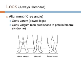
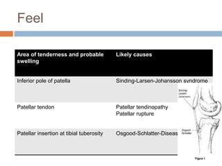
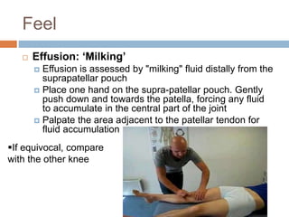
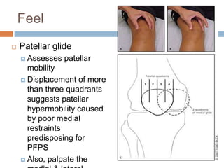
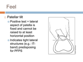
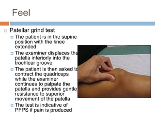
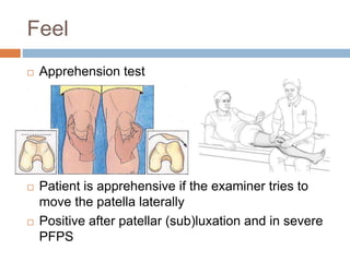
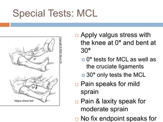
KNEE EXAMINATION
By:
Mohammed Ali Mousa, M.D, PhD.
14.09.2022
Objective
 Learn how to do a quick and thorough knee
exam
 Be able to identify
 Collateral ligament sprains/tears (LCL, MCL)
 Cruciate ligament sprains/tears (ACL, PCL)
 Meniscus injuries
 Patellofemoral syndrome
 Osteoarthritis
 Osgood Schlatter Disease
 Patellar tendinitis
 Pes anserine bursitis
 Illiotibial (IT) Band syndrome
Clinical Orthopedic Examination
 Look
 Feel
 Move
 Special tests
 Patella
 Menisci
 Collateral Ligs
 Cruciate ligs
 Don’t Forget NV, Joint Above and below
Look (Always Compare)
While Patient is standing from front, side, back
and other side then sitting and finally supine
 Skin
 erythema, ecchymosis
 Ms wasting
 Alignment
 Deformity
 Swelling/effusion
Look (Always Compare)
 Alignment (Knee angle)
 Genu varum (bowed legs)
 Genu valgum (can predispose to patellofemoral
syndrome)
Feel
 Palpate the patient in the sitting position
 Start with the joint line
Feel
 Joint line tenderness:
 Meniscal injury
 Osteoarthritis
 Fracture
Feel
 Next, find the inferior pole of the patella and
palpate the patella tendon down to its insertion
at the tibial tuberosity
Feel
Area of tenderness and probable
swelling
Likely causes
Inferior pole of patella Sinding-Larsen-Johansson syndrome
Patellar tendon Patellar tendinopathy
Patellar rupture
Patellar insertion at tibial tuberosity Osgood-Schlatter-Disease
Feel
 When you are
at the tibial
tuberosity
palpate with
your finger 1-2
cm medially
 If the patient is
tender there,
consider Pes
anserine
bursitis
Feel
 Have the patient lay supine
 Start with palpating:
 Medial and lateral femoral and tibial (epi)condyles
& the fibular head:
 Medial collateral ligament (MCL) sprain
 Lateral collateral ligament (LCL) sprain
 Iliotibial (IT) band syndrome
 Tibial plateau fracture
 Fibular head fracture
Feel
 IT-band syndrome
Feel
 Effusion: ‘Milking’
 Effusion is assessed by "milking" fluid distally from the
suprapatellar pouch
 Place one hand on the supra-patellar pouch. Gently
push down and towards the patella, forcing any fluid
to accumulate in the central part of the joint
 Palpate the area adjacent to the patellar tendon for
fluid accumulation
If equivocal, compare
with the other knee
Feel
 Effusion: Ballottement
 A ballotable patella may be
palpated after similar effusion
milking
 Place one hand on the supra-
patellar pouch. Gently push
down and towards the patella,
forcing any fluid to accumulate
in the central part of the joint
 Gently push down on the
patella with your thumb
 If there is a sizable effusion, the
patella will feel as if it is floating
and ‘bounce’ back up when
pushed down
Feel
 After a trauma there is effusion (hemarthrosis) only if
structures in the joint are involved:
 Anterior cruciate or posterior cruciate ligament (ACL, PCL)
injury
 Meniscus injury
 Patellar dislocation
 Intra-articular fracture (e.g. tibial plateau fracture)
 Remember: Sole collateral ligament (MCL, LCL) sprains
will not cause an effusion
 Causes of non-traumatic effusion:
 Osteoarthritis, rheumatoid arthritis, gout, pseudo gout,
Reiter’s syndrome
 Infections e.g. gonorrhea
 Tumors
Feel
 Patellofemoral
syndrome (PFPS)
 The “J” sign
 The patient supine
or seated and the
knee extended from
a flexed position.
Lateral deviation of
the patella can be
observed during the
terminal phase of
extension
Feel
 Next, clinical tests for patellar mobility and position,
and provocative tests for pain should be performed:
 patellar glide
 patellar tilt and
 patellar grind tests
 Positive results on these tests are consistent with the
diagnosis of PFPS
 The patellar apprehension test is used to assess for
lateral instability and is positive when pain or
discomfort occurs with lateral translation of the patella
Feel
 Patellar glide
 Assesses patellar
mobility
 Displacement of more
than three quadrants
suggests patellar
hypermobility caused
by poor medial
restraints
predisposing for
PFPS
 Also, palpate the
Feel
 Patellar tilt
 Positive test = lateral
aspect of patella is
fixed and cannot be
raised to at least
horizontal position
 Indicates tight lateral
structures (e.g.: IT-
band) predisposing
for PFPS
Feel
 Patellar grind test
 The patient is in the supine
position with the knee
extended
 The examiner displaces the
patella inferiorly into the
trochlear groove
 The patient is then asked to
contract the quadriceps
while the examiner
continues to palpate the
patella and provides gentle
resistance to superior
movement of the patella
 The test is indicative of
PFPS if pain is produced
Feel
 Apprehension test
 Patient is apprehensive if the examiner tries to
move the patella laterally
 Positive after patellar (sub)luxation and in severe
PFPS
Move
 Test for active & passive ROM while the
patient is supine
 Flexion 140°
 Extension 0° /-10°
 Internal rotation 10°
 External rotation 10°
 Always compare to the other, “healthy” knee!
Special Tests: MCL
 Apply valgus stress with
the knee at 0° and bent at
30°
 0° tests for MCL as well as
the cruciate ligaments
 30° only tests the MCL
 Pain speaks for mild
sprain
 Pain & laxity speak for
moderate sprain
 No fix endpoint speaks for
Special Tests: LCL
 Apply varus stress with the
knee at 0° and bent at 30°
 0° tests for LCL as well as
the cruciate ligaments
 30° only tests the LCL
 Pain speaks for mild sprain
 Pain & laxity speak for
moderate sprain
 No fix endpoint speaks for
complete LCL rupture
Special Tests: ACL
 Lachman test
 Patient supine with knee flexed
30°
 Pull on the tibia towards you
 Anterior drawer test
 Patient supine with knee flexed
90°
 Stabilize patient’s foot by
sitting on it
 Cup your hands around
patient’s knee & draw the tibia
towards you
Special Tests: ACL
 Lachman & anterior drawer test are positive if
 There is increased amount of anterior tibial
translation compared with the opposite leg, and/or
 There is no firm end-point
Special Tests: ACL
Sensitivity Specificity
Lachman test 85%[1] 94%[1]
Anterior Drawer test 68%[2] 79%[2]
Pivot Shift test 24%[1] 98%[1]
[1] Benjaminse A, Gokeler A, van der Schans CP. Clinical diagnosis of an anterior cruciate ligament rupture: a meta-analysis.
J Orthop Sports Phys Ther. 2006 May;36(5):267-88.
[2] Kim SJ, Kim HK. Reliability of the anterior drawer test, the pivot shift test, and the Lachman test. Clin Orthop Relat Res. 1995 Aug;(317):2
Special Tests: PCL
 Posterior drawer test
 Stay in the same position as
for the anterior drawer test
 Push the tibia posteriorly
 If it moves backwards the PCL
is probably damaged
Special Tests: PCL
 Another way of testing the PCL is the
sag sign
Special Tests: PCL
 Sag sign
Special Tests: Meniscus
 Mc Murray
 Hold the knee with one hand, place your fingers
along the joint line and flex it to 90°
 Hold the foot by the sole with the other hand
Special Tests: Meniscus
 Mc Murray (cont)
 Provide a valgus stress
 Rotate the leg externally
 Extend the knee
 If pain or a ‘click’ is felt = ‘positive McMurray’ for a
medial meniscus tear
Special Tests: Meniscus
 Mc Murray (cont)
 Provide a varus stress
 Rotate the leg internally
 Extend the knee
 If pain or a ‘click’ is felt = ‘positive McMurray’ for a
lateral meniscus tear
Special Tests: Meniscus
 Apley grind test
 The patient lays prone and flexes his/her knee 90°
 Place your hand on the patient’s heel and push down
(to compress the menisci between femur & tibia) while
rotating internally & externally
 Depending on the area of pain medial or lateral
meniscus tear
Special Tests: IT-band
Syndrome
 Ober’s Test
 The patient lies with the unaffected side down
and the unaffected hip and knee at a 90-degree
angle. If the IT-band is tight, the patient will have
difficulty adducting the leg beyond the midline and
may experience pain at the lateral knee
Referred pain
 ‘Knee pain’ can be referred from the lumbar
spine (e.g. herniated disk) or from a hip or
ankle pathology, so don’t forget to
 Do a gross hip and ankle exam
 Test the motor strength, sensibility and deep
tendon reflexes of the lower extremities
 And always examine both knees!

Knee Examination.pptx

  • 1.
    KNEE EXAMINATION By: Mohammed AliMousa, M.D, PhD. 14.09.2022
  • 2.
    Objective  Learn howto do a quick and thorough knee exam  Be able to identify  Collateral ligament sprains/tears (LCL, MCL)  Cruciate ligament sprains/tears (ACL, PCL)  Meniscus injuries  Patellofemoral syndrome  Osteoarthritis  Osgood Schlatter Disease  Patellar tendinitis  Pes anserine bursitis  Illiotibial (IT) Band syndrome
  • 3.
    Clinical Orthopedic Examination Look  Feel  Move  Special tests  Patella  Menisci  Collateral Ligs  Cruciate ligs  Don’t Forget NV, Joint Above and below
  • 4.
    Look (Always Compare) WhilePatient is standing from front, side, back and other side then sitting and finally supine  Skin  erythema, ecchymosis  Ms wasting  Alignment  Deformity  Swelling/effusion
  • 5.
    Look (Always Compare) Alignment (Knee angle)  Genu varum (bowed legs)  Genu valgum (can predispose to patellofemoral syndrome)
  • 6.
    Feel  Palpate thepatient in the sitting position  Start with the joint line
  • 7.
    Feel  Joint linetenderness:  Meniscal injury  Osteoarthritis  Fracture
  • 8.
    Feel  Next, findthe inferior pole of the patella and palpate the patella tendon down to its insertion at the tibial tuberosity
  • 9.
    Feel Area of tendernessand probable swelling Likely causes Inferior pole of patella Sinding-Larsen-Johansson syndrome Patellar tendon Patellar tendinopathy Patellar rupture Patellar insertion at tibial tuberosity Osgood-Schlatter-Disease
  • 10.
    Feel  When youare at the tibial tuberosity palpate with your finger 1-2 cm medially  If the patient is tender there, consider Pes anserine bursitis
  • 11.
    Feel  Have thepatient lay supine  Start with palpating:  Medial and lateral femoral and tibial (epi)condyles & the fibular head:  Medial collateral ligament (MCL) sprain  Lateral collateral ligament (LCL) sprain  Iliotibial (IT) band syndrome  Tibial plateau fracture  Fibular head fracture
  • 12.
  • 13.
    Feel  Effusion: ‘Milking’ Effusion is assessed by "milking" fluid distally from the suprapatellar pouch  Place one hand on the supra-patellar pouch. Gently push down and towards the patella, forcing any fluid to accumulate in the central part of the joint  Palpate the area adjacent to the patellar tendon for fluid accumulation If equivocal, compare with the other knee
  • 14.
    Feel  Effusion: Ballottement A ballotable patella may be palpated after similar effusion milking  Place one hand on the supra- patellar pouch. Gently push down and towards the patella, forcing any fluid to accumulate in the central part of the joint  Gently push down on the patella with your thumb  If there is a sizable effusion, the patella will feel as if it is floating and ‘bounce’ back up when pushed down
  • 15.
    Feel  After atrauma there is effusion (hemarthrosis) only if structures in the joint are involved:  Anterior cruciate or posterior cruciate ligament (ACL, PCL) injury  Meniscus injury  Patellar dislocation  Intra-articular fracture (e.g. tibial plateau fracture)  Remember: Sole collateral ligament (MCL, LCL) sprains will not cause an effusion  Causes of non-traumatic effusion:  Osteoarthritis, rheumatoid arthritis, gout, pseudo gout, Reiter’s syndrome  Infections e.g. gonorrhea  Tumors
  • 16.
    Feel  Patellofemoral syndrome (PFPS) The “J” sign  The patient supine or seated and the knee extended from a flexed position. Lateral deviation of the patella can be observed during the terminal phase of extension
  • 17.
    Feel  Next, clinicaltests for patellar mobility and position, and provocative tests for pain should be performed:  patellar glide  patellar tilt and  patellar grind tests  Positive results on these tests are consistent with the diagnosis of PFPS  The patellar apprehension test is used to assess for lateral instability and is positive when pain or discomfort occurs with lateral translation of the patella
  • 18.
    Feel  Patellar glide Assesses patellar mobility  Displacement of more than three quadrants suggests patellar hypermobility caused by poor medial restraints predisposing for PFPS  Also, palpate the
  • 19.
    Feel  Patellar tilt Positive test = lateral aspect of patella is fixed and cannot be raised to at least horizontal position  Indicates tight lateral structures (e.g.: IT- band) predisposing for PFPS
  • 20.
    Feel  Patellar grindtest  The patient is in the supine position with the knee extended  The examiner displaces the patella inferiorly into the trochlear groove  The patient is then asked to contract the quadriceps while the examiner continues to palpate the patella and provides gentle resistance to superior movement of the patella  The test is indicative of PFPS if pain is produced
  • 21.
    Feel  Apprehension test Patient is apprehensive if the examiner tries to move the patella laterally  Positive after patellar (sub)luxation and in severe PFPS
  • 22.
    Move  Test foractive & passive ROM while the patient is supine  Flexion 140°  Extension 0° /-10°  Internal rotation 10°  External rotation 10°  Always compare to the other, “healthy” knee!
  • 23.
    Special Tests: MCL Apply valgus stress with the knee at 0° and bent at 30°  0° tests for MCL as well as the cruciate ligaments  30° only tests the MCL  Pain speaks for mild sprain  Pain & laxity speak for moderate sprain  No fix endpoint speaks for
  • 24.
    Special Tests: LCL Apply varus stress with the knee at 0° and bent at 30°  0° tests for LCL as well as the cruciate ligaments  30° only tests the LCL  Pain speaks for mild sprain  Pain & laxity speak for moderate sprain  No fix endpoint speaks for complete LCL rupture
  • 25.
    Special Tests: ACL Lachman test  Patient supine with knee flexed 30°  Pull on the tibia towards you  Anterior drawer test  Patient supine with knee flexed 90°  Stabilize patient’s foot by sitting on it  Cup your hands around patient’s knee & draw the tibia towards you
  • 26.
    Special Tests: ACL Lachman & anterior drawer test are positive if  There is increased amount of anterior tibial translation compared with the opposite leg, and/or  There is no firm end-point
  • 27.
    Special Tests: ACL SensitivitySpecificity Lachman test 85%[1] 94%[1] Anterior Drawer test 68%[2] 79%[2] Pivot Shift test 24%[1] 98%[1] [1] Benjaminse A, Gokeler A, van der Schans CP. Clinical diagnosis of an anterior cruciate ligament rupture: a meta-analysis. J Orthop Sports Phys Ther. 2006 May;36(5):267-88. [2] Kim SJ, Kim HK. Reliability of the anterior drawer test, the pivot shift test, and the Lachman test. Clin Orthop Relat Res. 1995 Aug;(317):2
  • 28.
    Special Tests: PCL Posterior drawer test  Stay in the same position as for the anterior drawer test  Push the tibia posteriorly  If it moves backwards the PCL is probably damaged
  • 29.
    Special Tests: PCL Another way of testing the PCL is the sag sign
  • 30.
  • 31.
    Special Tests: Meniscus Mc Murray  Hold the knee with one hand, place your fingers along the joint line and flex it to 90°  Hold the foot by the sole with the other hand
  • 32.
    Special Tests: Meniscus Mc Murray (cont)  Provide a valgus stress  Rotate the leg externally  Extend the knee  If pain or a ‘click’ is felt = ‘positive McMurray’ for a medial meniscus tear
  • 33.
    Special Tests: Meniscus Mc Murray (cont)  Provide a varus stress  Rotate the leg internally  Extend the knee  If pain or a ‘click’ is felt = ‘positive McMurray’ for a lateral meniscus tear
  • 34.
    Special Tests: Meniscus Apley grind test  The patient lays prone and flexes his/her knee 90°  Place your hand on the patient’s heel and push down (to compress the menisci between femur & tibia) while rotating internally & externally  Depending on the area of pain medial or lateral meniscus tear
  • 35.
    Special Tests: IT-band Syndrome Ober’s Test  The patient lies with the unaffected side down and the unaffected hip and knee at a 90-degree angle. If the IT-band is tight, the patient will have difficulty adducting the leg beyond the midline and may experience pain at the lateral knee
  • 36.
    Referred pain  ‘Kneepain’ can be referred from the lumbar spine (e.g. herniated disk) or from a hip or ankle pathology, so don’t forget to  Do a gross hip and ankle exam  Test the motor strength, sensibility and deep tendon reflexes of the lower extremities  And always examine both knees!