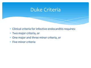
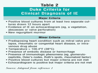
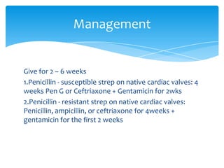
Infective Endocarditis
   http://crisbertcualteros.page.tl
occurs when microorganisms (bacteria or fungi)
  colonize the endocardial surface of the heart
  Classified into four groups:
1. Native Valve IE
2. Prosthetic Valve IE
3. Intravenous drug abuse (IVDA) IE
4. Nosocomial IE
Epidemiology


  The valves involved in IE:
1. Mitral 28-45%
2. Aortic 5-36%
3. Both 0-35%
4. Tricuspid 0-6%
5. Pulmonary <1%
Males > females
May occur at any age and increasingly common in
elderly
Mortality 20-30%
Predisposing Factors


IV drug use
Central line Prosthetic valve
Previous IE
Dental procedure
Rheumatic heart disease
High Risk


1.   Prosthetic cardiac valve
2.   Prior episodes of endocarditis
3.   Complex congenital cardiac defect
4.   Surgical systemic-pulmonary shunts
5.   Intravenous drug abuse
6.   Intravascular catheters
Moderate Risk


1.   PDA, VSD, primum ASD
2.   Co-Aorta
3.   Bicuspid aortic valve
4.   Hypertrophic cardiomyopathy
5.   Acquired valvular dysfunction
6.   MVP with mitral regurgitation
Low Risk


1. Isolated secundum atrial septal defect
2. ASD, VSD, or PDA > 6 months past repair
3. “Innocent” heart murmur by auscultation in the
   pediatric population
Pathophysiology


a. Turbulent blood flow disrupts the endocardium
   making it “sticky”
b. microorganisms infect the endocardial surface of
   the heart.
c. Adherence of the organisms to the endocardial
   surface
d. Eventual invasion of the valvular leaflets
Etiology


Common bacteria in children
 Strep. viridans–50%
 Staph aureus–40%
 Strep fecalis, Grp D Streptococcus(Enterococci)
Clinical Presentations


 High grade fever w/ chills
 SOB
 Arthralgias/myalgias
 Abdominal pain
 Pleuritic chestpain
 Backpain
 Anorexia
 Weight loss
 Fatigue
Signs

Fever
Heart murmur
petechiae, subungal or“splinter”hemorrhages
Clubbing
Splenomegaly
Neurologic changes
Janeway lesions
Roth Spots
Osler’s Nodes                   Janeway Lesions

Painful                          Erythematous
Erythematousnodules              Blanchingmacules
Located on pulp of fingers and   Nonpainful
toes
                                 Located in the palms and soles
Diagnostics


Blood Cultures: Minimum of 3 takings
3 separate venipuncture sites: 5 - 10mL
Detects >95% of cases
CBC          Immunologic tests:
ESR            Increase in
CRP            gammaglobulins
Urinalysis     Presence of cryoglobulin
               Low Complement
               levels(C3,C4)
               RF-positive
Imaging


 CXR: multiple focal infiltrates and calcification of
  heart valves
 ECG: Look for evidence of ischemia, conduction delay
  and arrhythmias
 Echocardiography: diagnostic tool for culture
  negative cases
Duke Criteria


Clinical criteria for infective endocarditis requires:
Two major criteria, or
One major and three minor criteria, or
Five minor criteria
Management


Give for 2 – 6 weeks
1.Penicillin - susceptible strep on native cardiac valves: 4
weeks Pen G or Ceftriaxone + Gentamicin for 2wks
2.Penicillin - resistant strep on native cardiac valves:
Penicillin, ampicillin, or ceftriaxone for 4weeks +
gentamicin for the first 2 weeks
5. Enterococcal infection on native valves - penicillin or
ampicillin + gentamicin for 4-6 weeks
6. (MSSA) on native valves : Nafcillin or oxacillin for at least 6
weeks + gentamicin for 3-5 days is optional
7. (MRSA) on native valves: Vancomycin for at least 6 weeks
with or without 3-5 days of gentamicin
8. MSSA infection on prosthetic valve : Nafcillin or Oxacillin +
Rifampin for at least 6 weeks, in combination with Gentamicin
for 2 weeks.
9. MRSA infection on prosthetic valve: Vancomycin + Rifampin
for at least 6 weeks, in combination with Gentamicin for 2
weeks
10. Gram Negative endocarditis due to HACEK: Ceftriaxone or
Ampicillin plus Gentamicin for 4 weeks

Infective Endocarditis

  • 1.
    Infective Endocarditis http://crisbertcualteros.page.tl
  • 2.
    occurs when microorganisms(bacteria or fungi) colonize the endocardial surface of the heart Classified into four groups: 1. Native Valve IE 2. Prosthetic Valve IE 3. Intravenous drug abuse (IVDA) IE 4. Nosocomial IE
  • 3.
    Epidemiology Thevalves involved in IE: 1. Mitral 28-45% 2. Aortic 5-36% 3. Both 0-35% 4. Tricuspid 0-6% 5. Pulmonary <1%
  • 4.
    Males > females Mayoccur at any age and increasingly common in elderly Mortality 20-30%
  • 5.
    Predisposing Factors IV druguse Central line Prosthetic valve Previous IE Dental procedure Rheumatic heart disease
  • 6.
    High Risk 1. Prosthetic cardiac valve 2. Prior episodes of endocarditis 3. Complex congenital cardiac defect 4. Surgical systemic-pulmonary shunts 5. Intravenous drug abuse 6. Intravascular catheters
  • 7.
    Moderate Risk 1. PDA, VSD, primum ASD 2. Co-Aorta 3. Bicuspid aortic valve 4. Hypertrophic cardiomyopathy 5. Acquired valvular dysfunction 6. MVP with mitral regurgitation
  • 8.
    Low Risk 1. Isolatedsecundum atrial septal defect 2. ASD, VSD, or PDA > 6 months past repair 3. “Innocent” heart murmur by auscultation in the pediatric population
  • 9.
    Pathophysiology a. Turbulent bloodflow disrupts the endocardium making it “sticky” b. microorganisms infect the endocardial surface of the heart. c. Adherence of the organisms to the endocardial surface d. Eventual invasion of the valvular leaflets
  • 10.
    Etiology Common bacteria inchildren  Strep. viridans–50%  Staph aureus–40%  Strep fecalis, Grp D Streptococcus(Enterococci)
  • 11.
    Clinical Presentations  Highgrade fever w/ chills  SOB  Arthralgias/myalgias  Abdominal pain  Pleuritic chestpain  Backpain  Anorexia  Weight loss  Fatigue
  • 12.
    Signs Fever Heart murmur petechiae, subungalor“splinter”hemorrhages Clubbing Splenomegaly Neurologic changes Janeway lesions Roth Spots
  • 13.
    Osler’s Nodes Janeway Lesions Painful Erythematous Erythematousnodules Blanchingmacules Located on pulp of fingers and Nonpainful toes Located in the palms and soles
  • 14.
    Diagnostics Blood Cultures: Minimumof 3 takings 3 separate venipuncture sites: 5 - 10mL Detects >95% of cases
  • 15.
    CBC Immunologic tests: ESR Increase in CRP gammaglobulins Urinalysis Presence of cryoglobulin Low Complement levels(C3,C4) RF-positive
  • 16.
    Imaging  CXR: multiplefocal infiltrates and calcification of heart valves  ECG: Look for evidence of ischemia, conduction delay and arrhythmias  Echocardiography: diagnostic tool for culture negative cases
  • 17.
    Duke Criteria Clinical criteriafor infective endocarditis requires: Two major criteria, or One major and three minor criteria, or Five minor criteria
  • 19.
    Management Give for 2– 6 weeks 1.Penicillin - susceptible strep on native cardiac valves: 4 weeks Pen G or Ceftriaxone + Gentamicin for 2wks 2.Penicillin - resistant strep on native cardiac valves: Penicillin, ampicillin, or ceftriaxone for 4weeks + gentamicin for the first 2 weeks
  • 20.
    5. Enterococcal infectionon native valves - penicillin or ampicillin + gentamicin for 4-6 weeks 6. (MSSA) on native valves : Nafcillin or oxacillin for at least 6 weeks + gentamicin for 3-5 days is optional 7. (MRSA) on native valves: Vancomycin for at least 6 weeks with or without 3-5 days of gentamicin
  • 21.
    8. MSSA infectionon prosthetic valve : Nafcillin or Oxacillin + Rifampin for at least 6 weeks, in combination with Gentamicin for 2 weeks. 9. MRSA infection on prosthetic valve: Vancomycin + Rifampin for at least 6 weeks, in combination with Gentamicin for 2 weeks 10. Gram Negative endocarditis due to HACEK: Ceftriaxone or Ampicillin plus Gentamicin for 4 weeks