1) Abdominal trauma is commonly encountered and can be life-threatening. Identification of serious intra-abdominal injuries can be challenging.
2) Motor vehicle accidents account for 60% of abdominal trauma, with blunt trauma more common than penetrating injuries.
3) Injuries to solid organs like the spleen, liver, and kidneys require careful assessment and may be graded based on severity. Conservative management is often attempted initially if the patient is stable.
4) Diagnostic tools like FAST ultrasound, CT scans, and laparoscopy can help identify injuries, but repeated examinations are often necessary. Management may involve surgery or conservative approaches depending on injury severity and patient stability.
Introduction to abdominal trauma, its significance in emergency care, and the challenges in assessment.
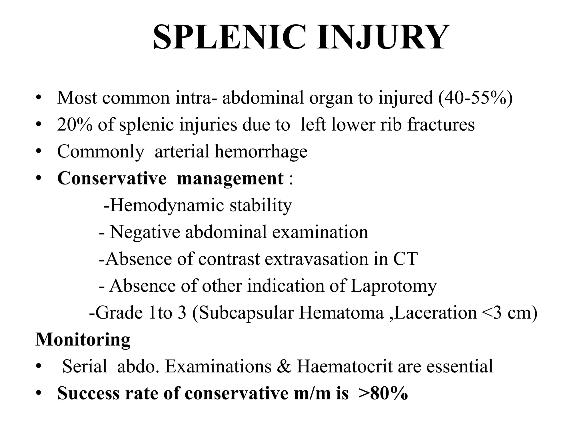
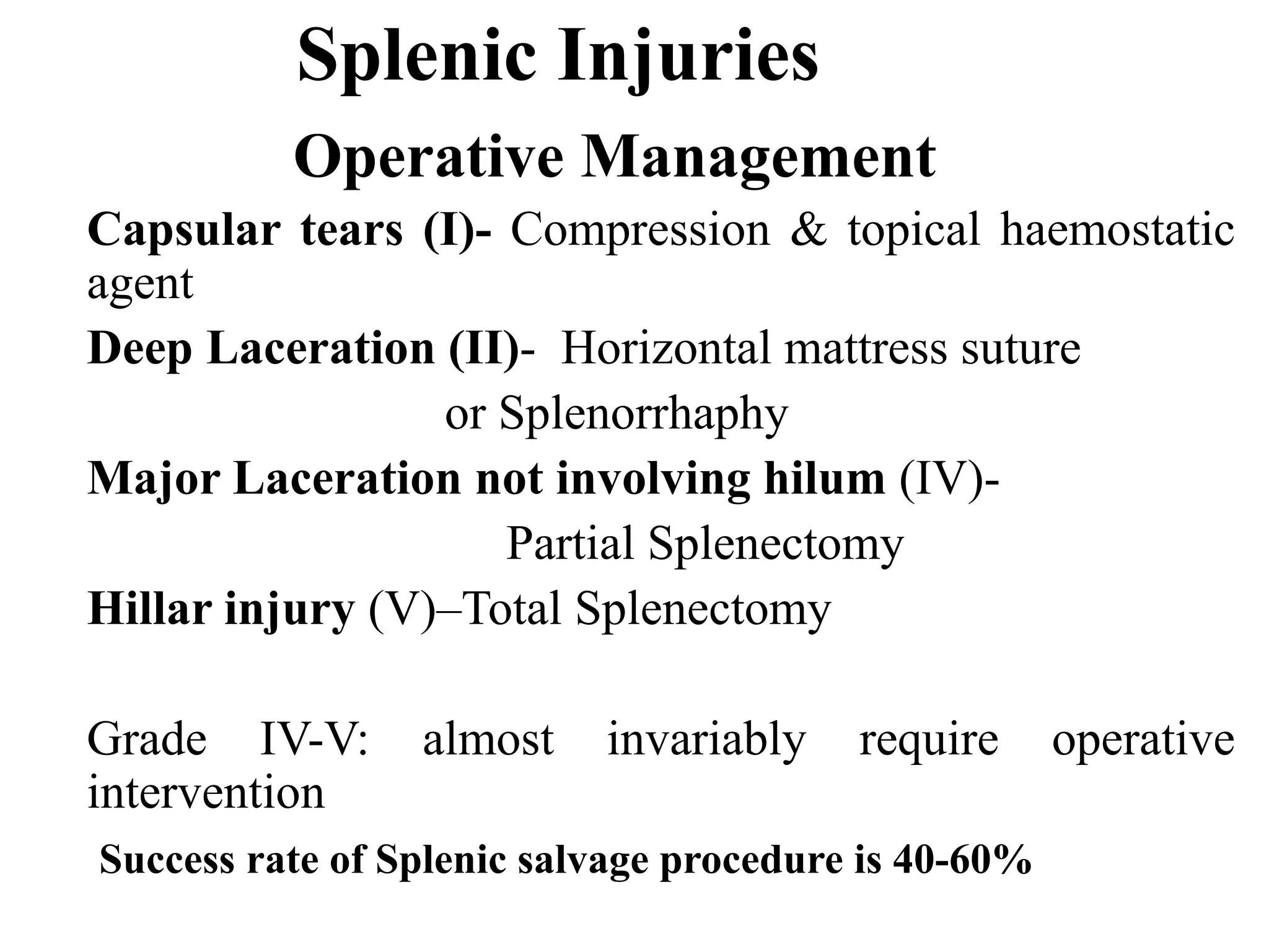
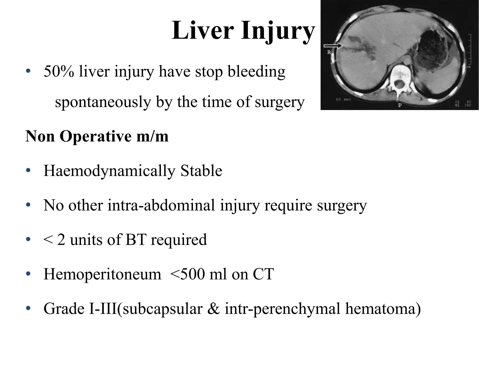
Statistics on the occurrence of abdominal trauma, peak incidence age, and global death rates due to injuries.
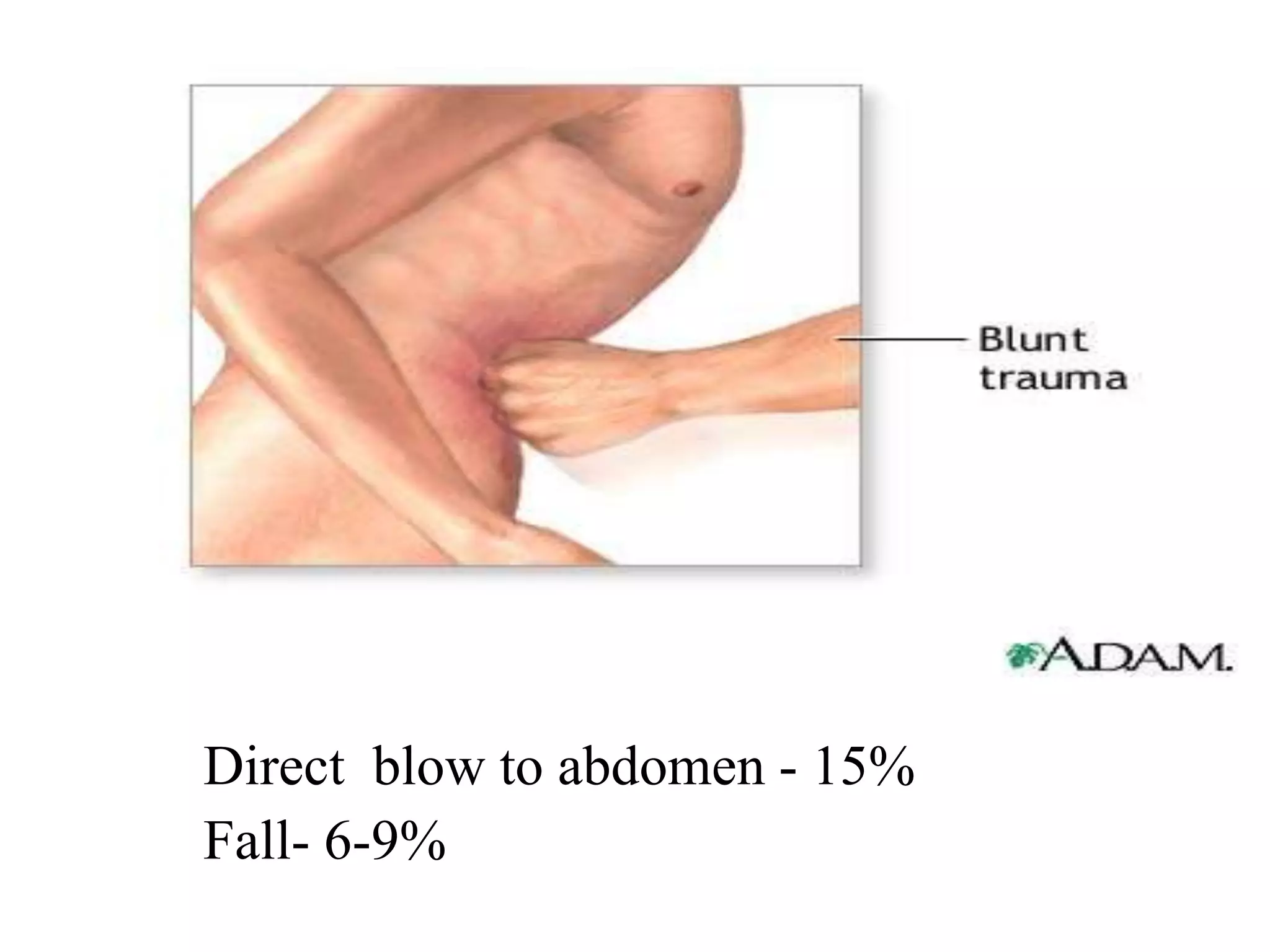
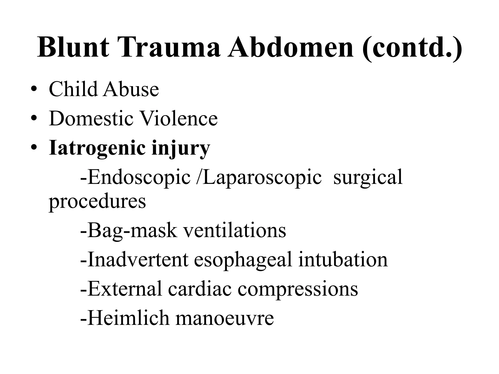
Different types of abdominal trauma, including blunt and penetrating injuries, and their causes such as accidents and domestic violence.
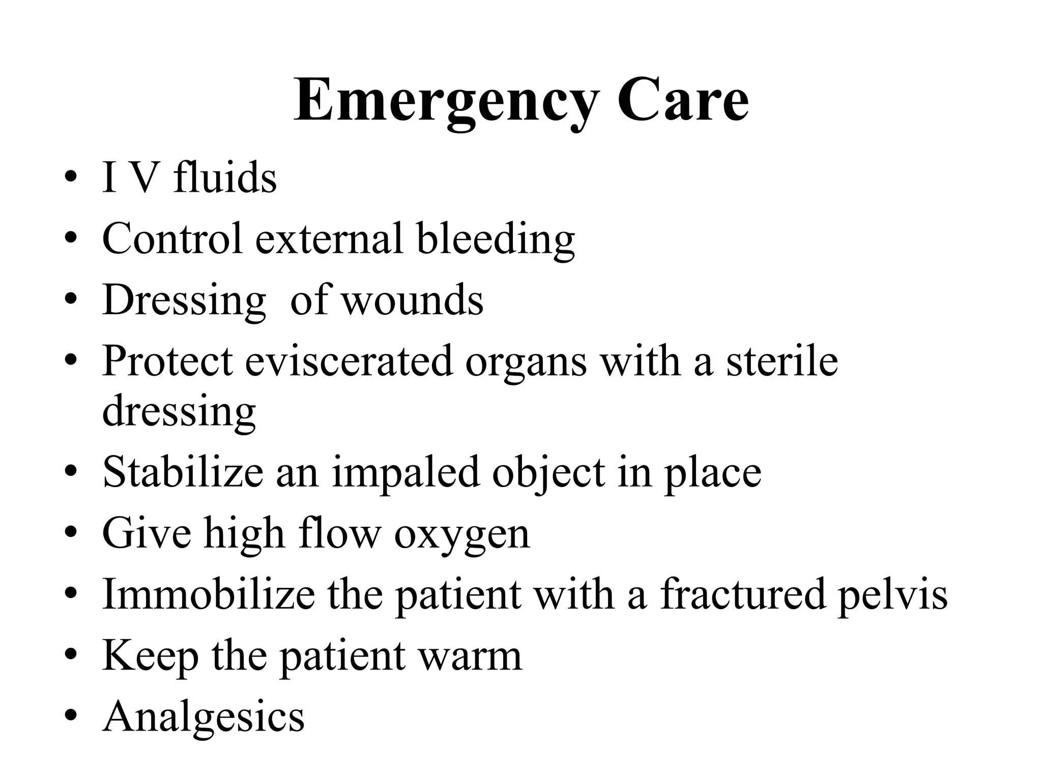
Focus on rapid transport to trauma centers, airway management, and essential initial care.
Primary survey process, identification of life-threatening conditions, and immediate care practices.
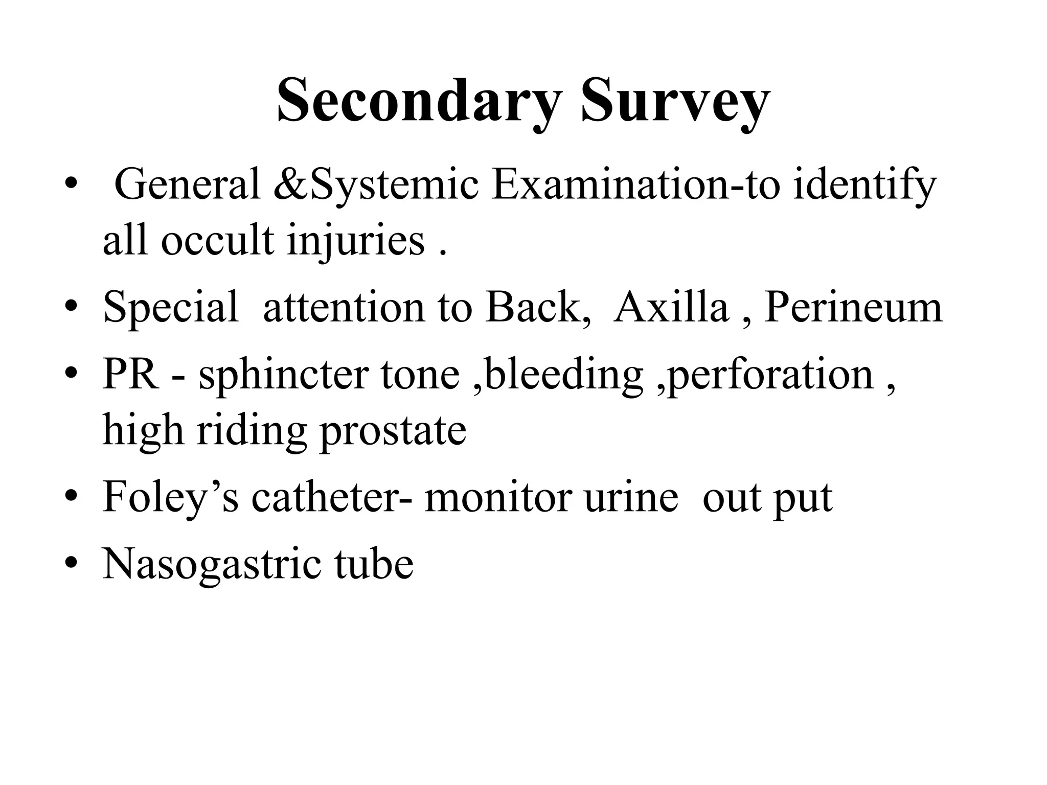
Detailed examination procedures, including systemic surveys and history taking to identify hidden injuries.
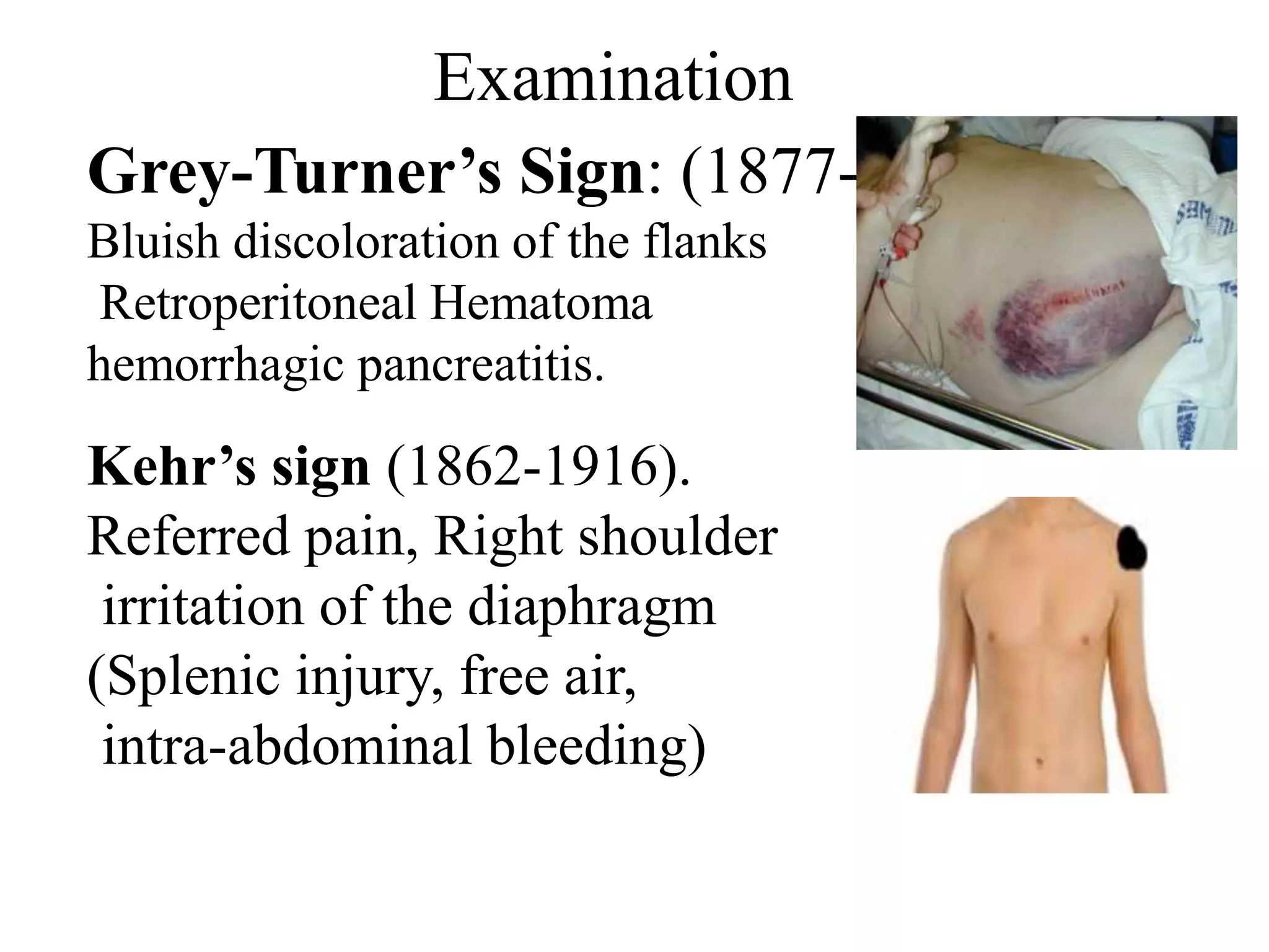
Clinical signs such as Cullen’s sign and Grey-Turner’s sign associated with abdominal trauma.
Methods for diagnosing abdominal injuries including FAST, X-Ray, and CT scan, highlighting their effectiveness.
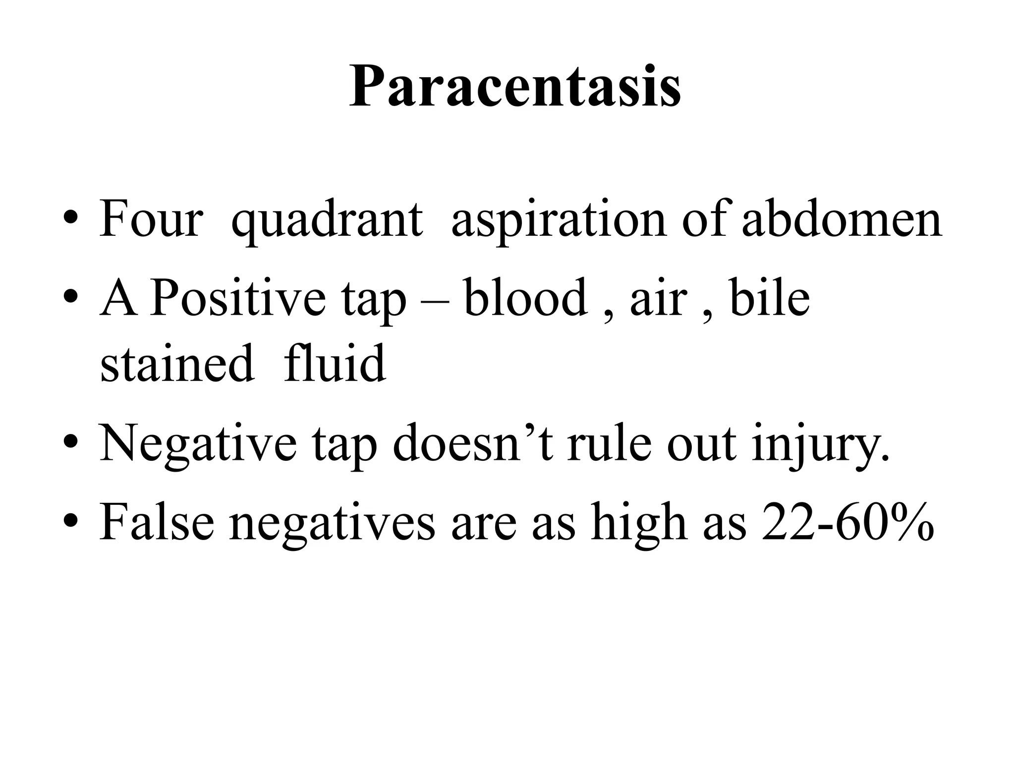
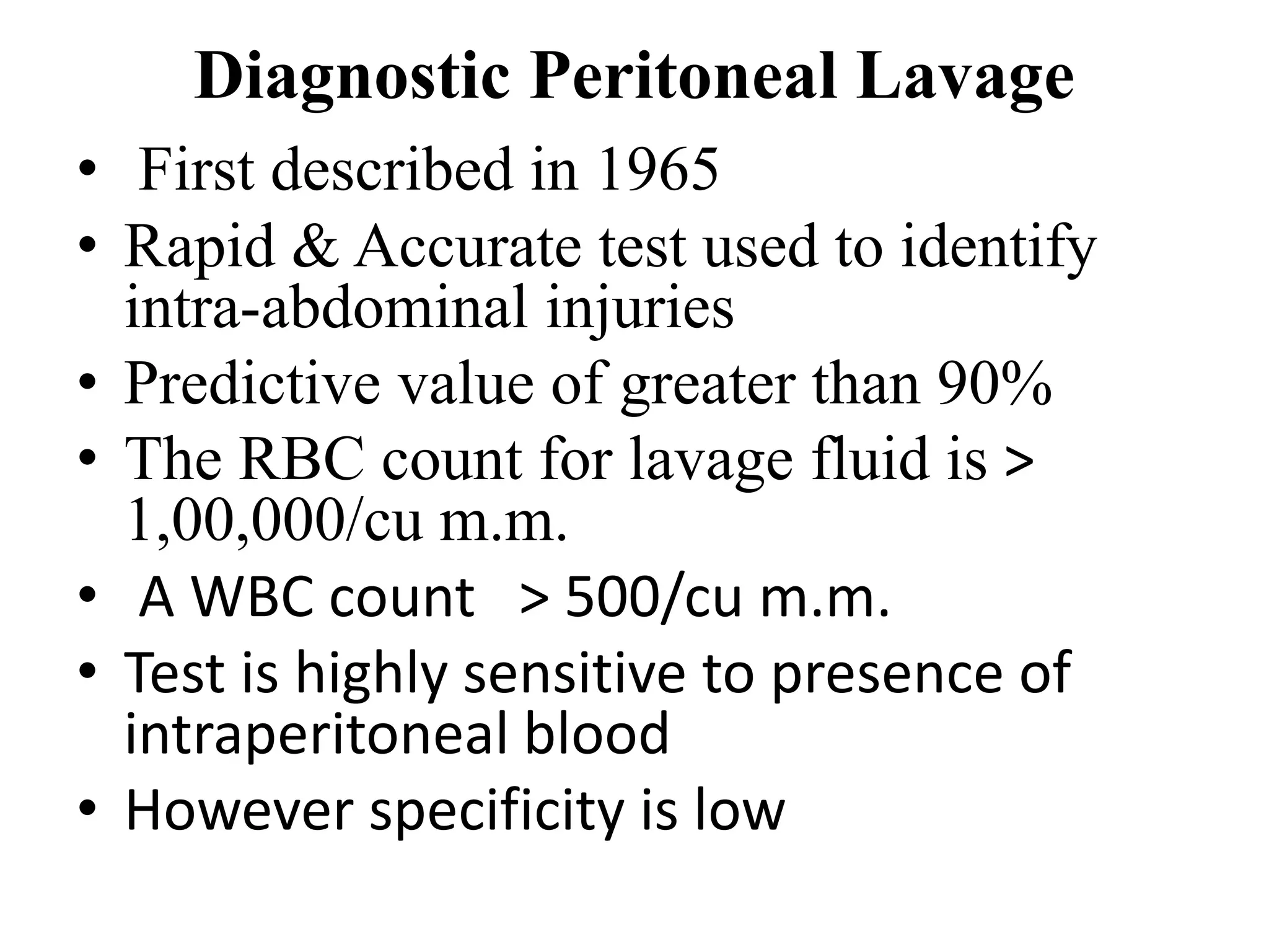
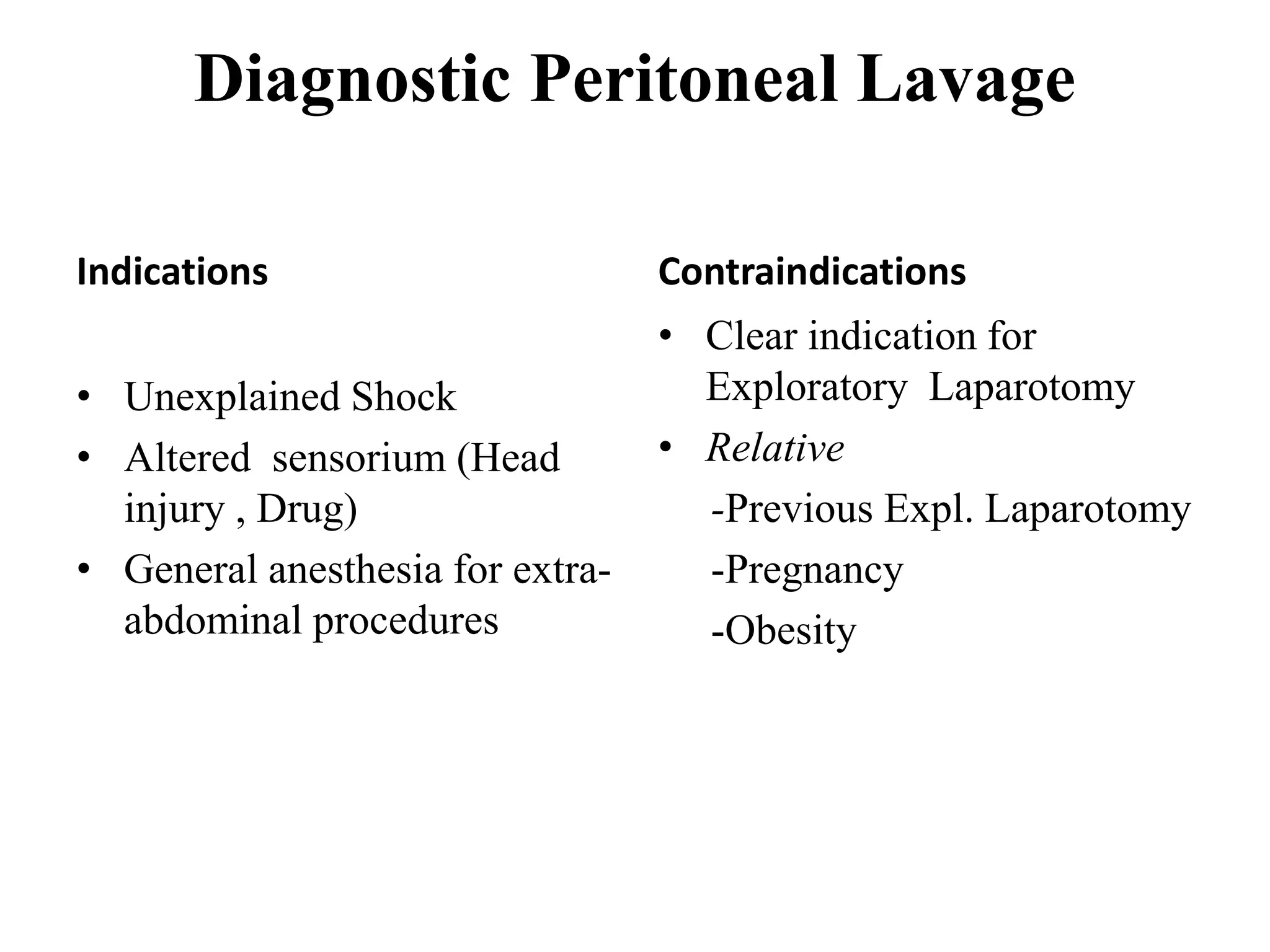
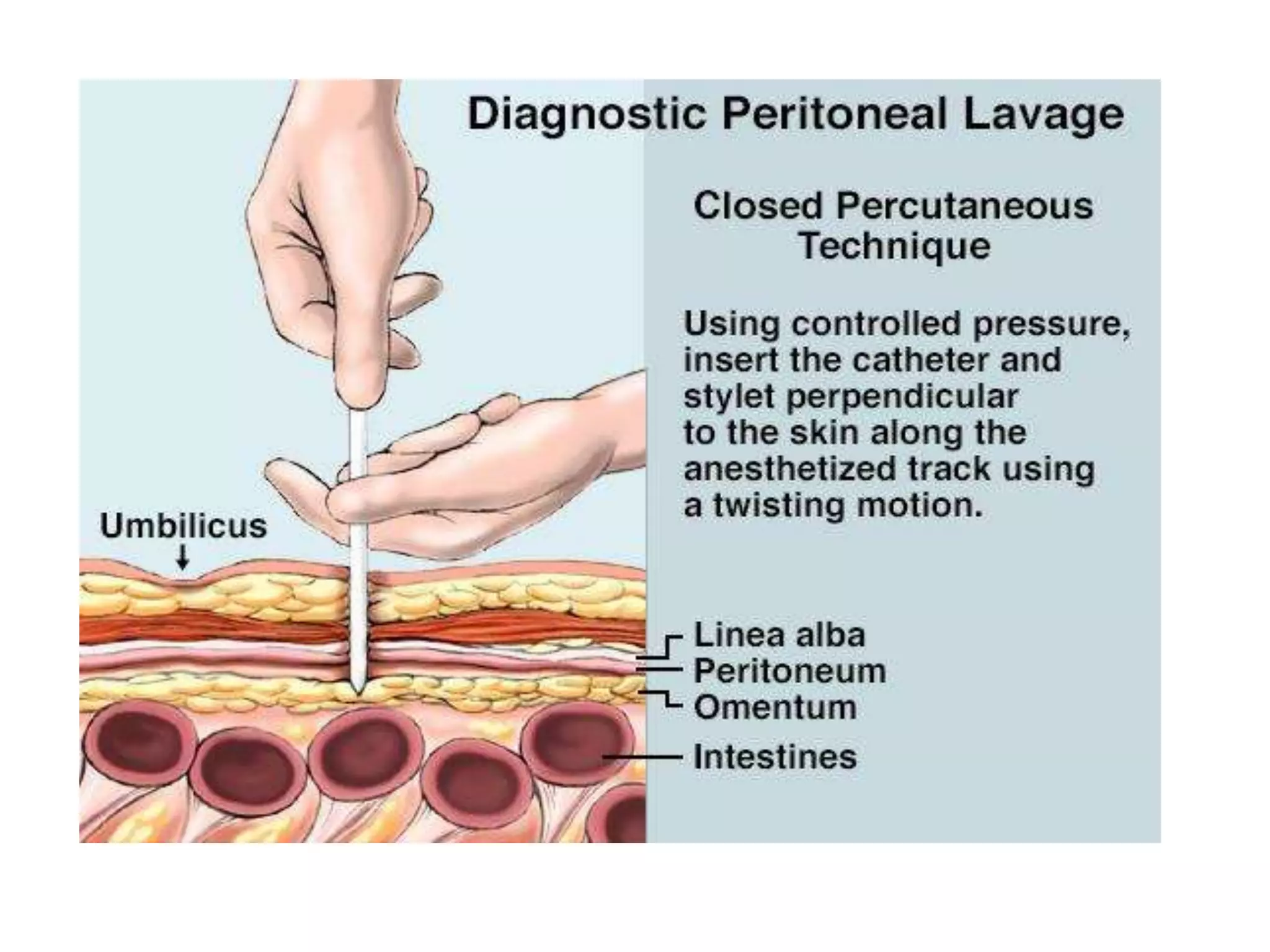
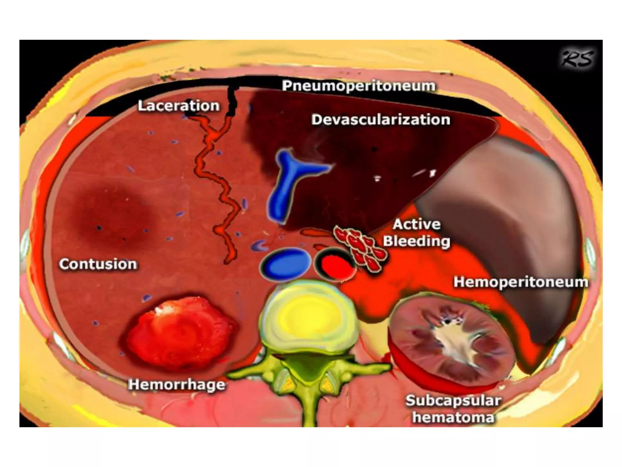
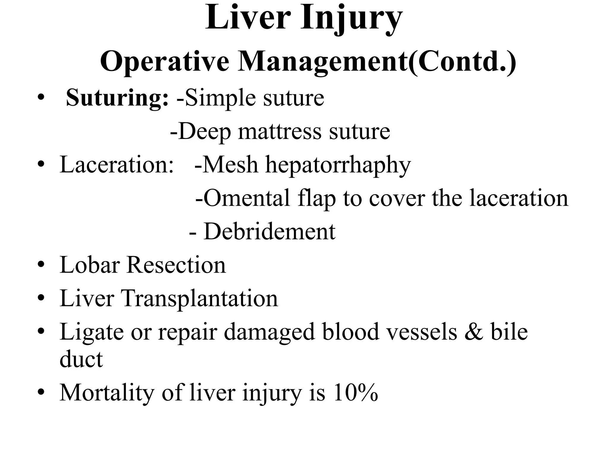
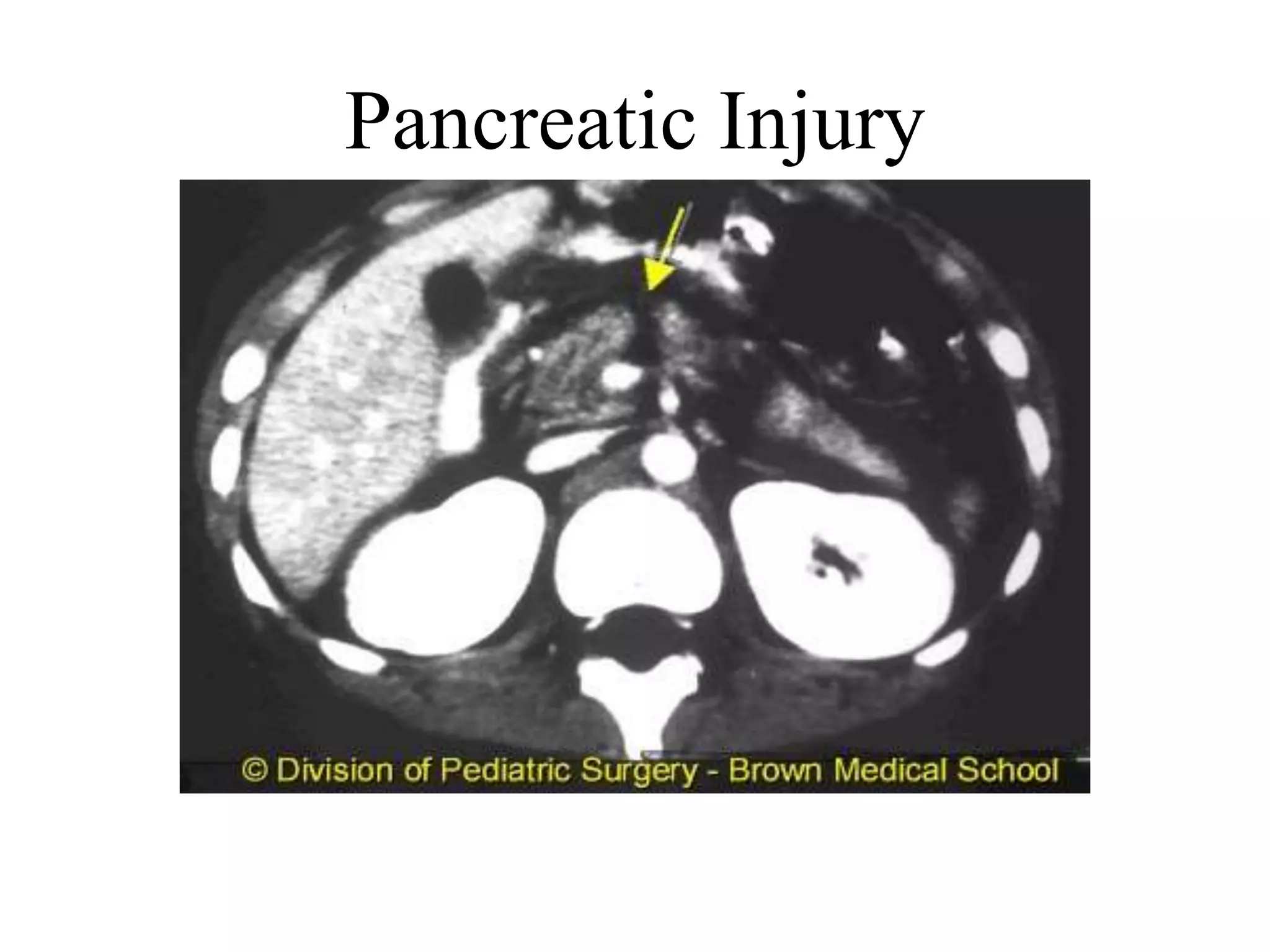
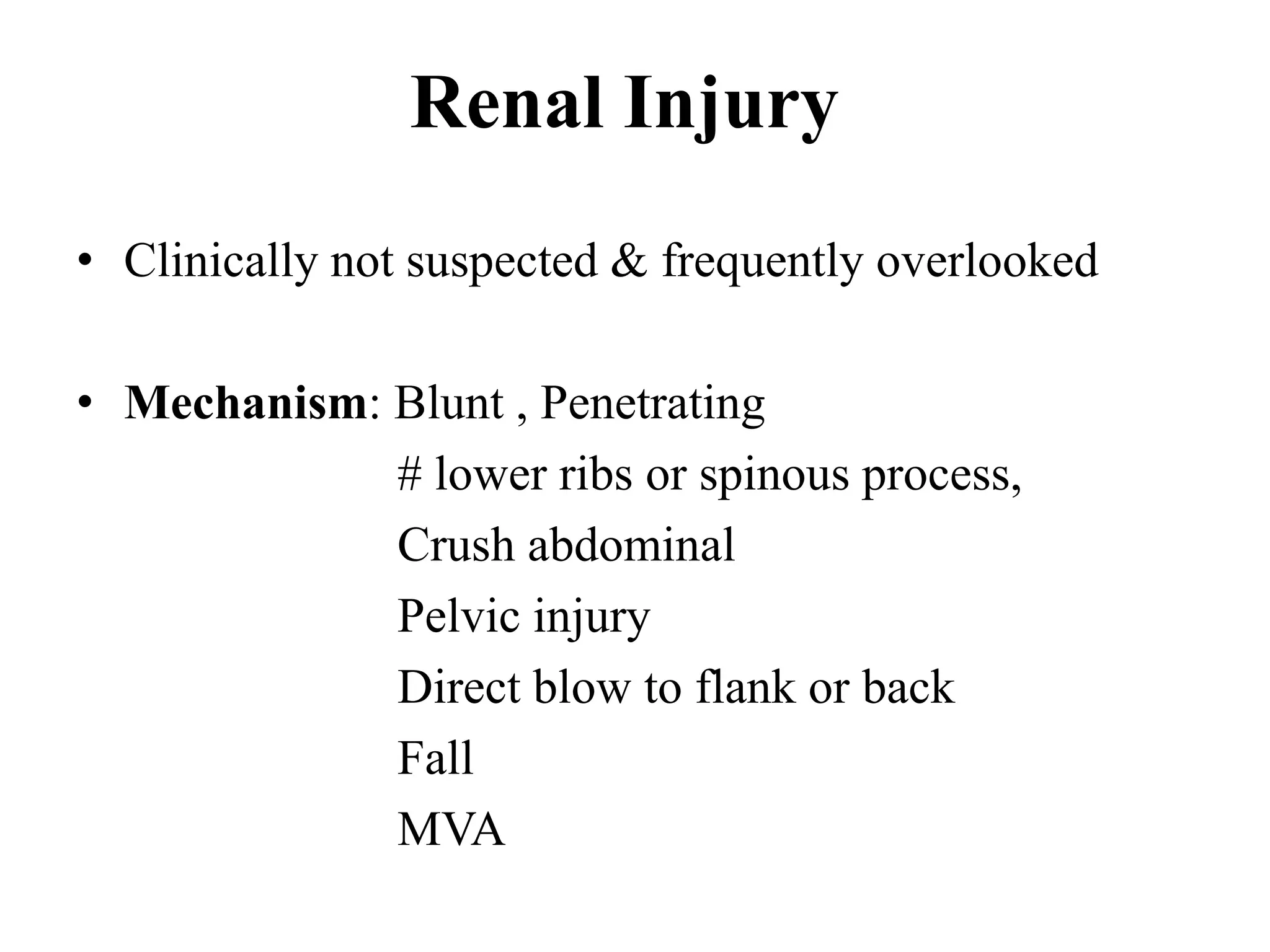
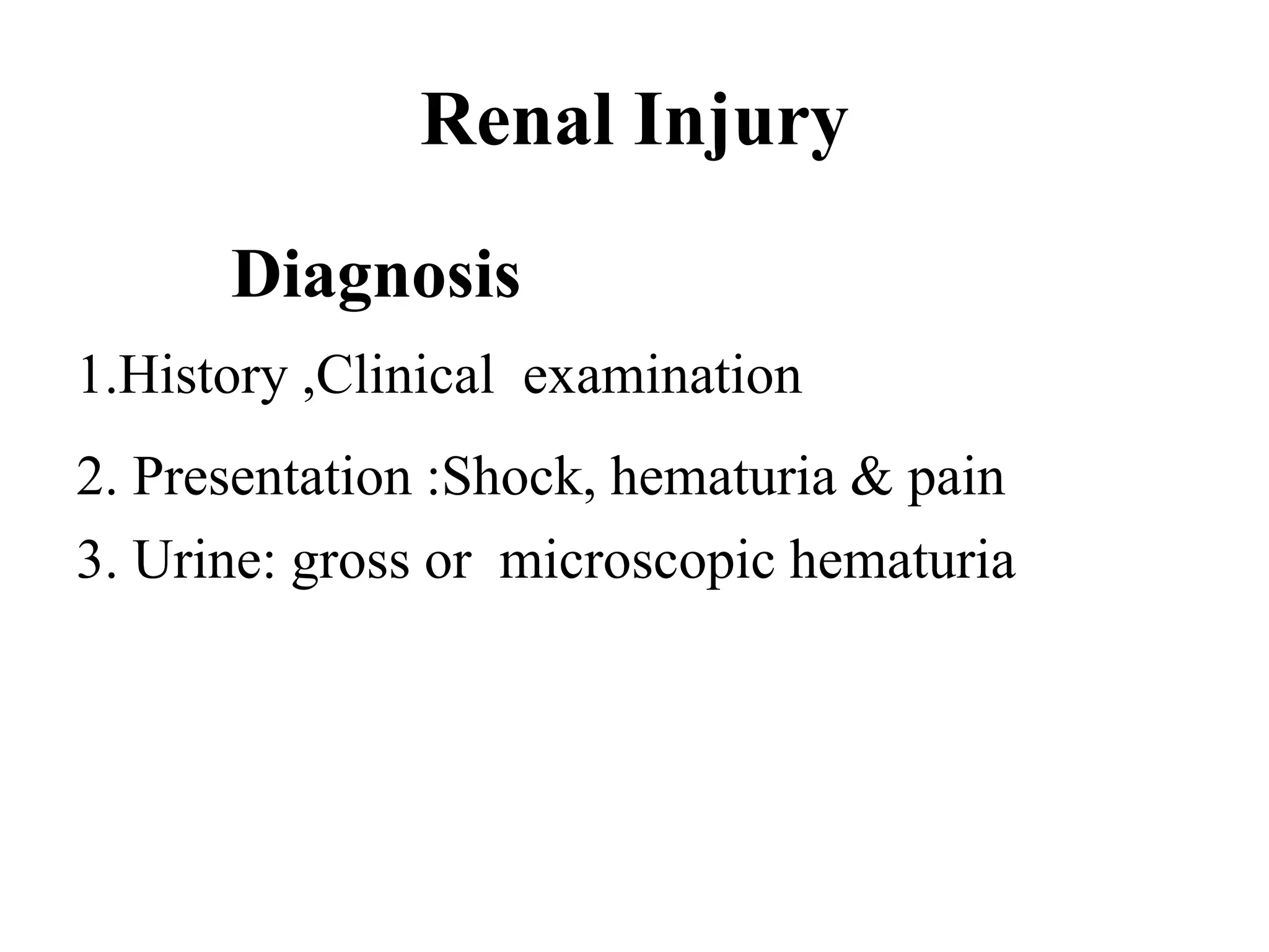
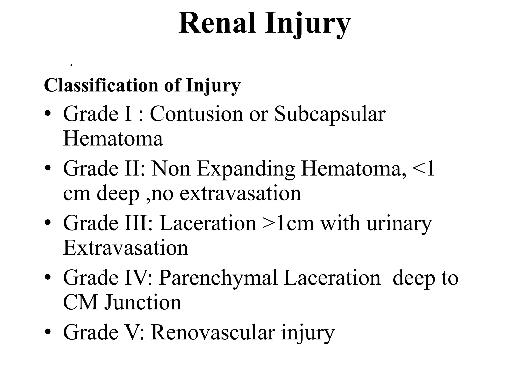
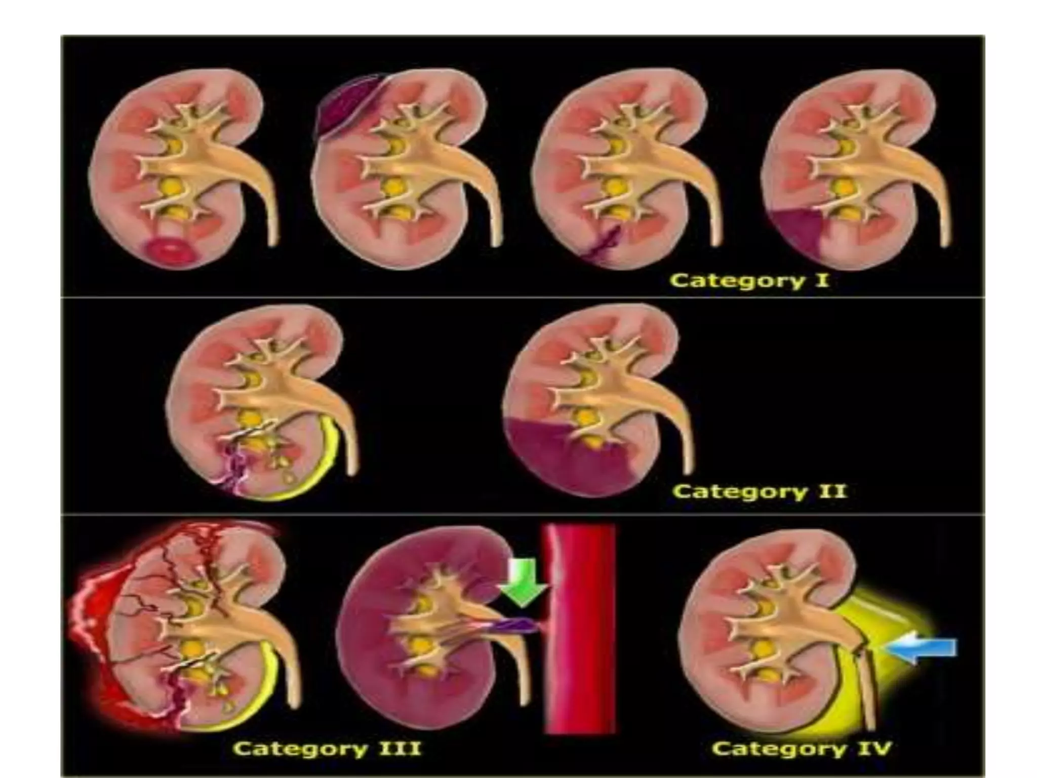
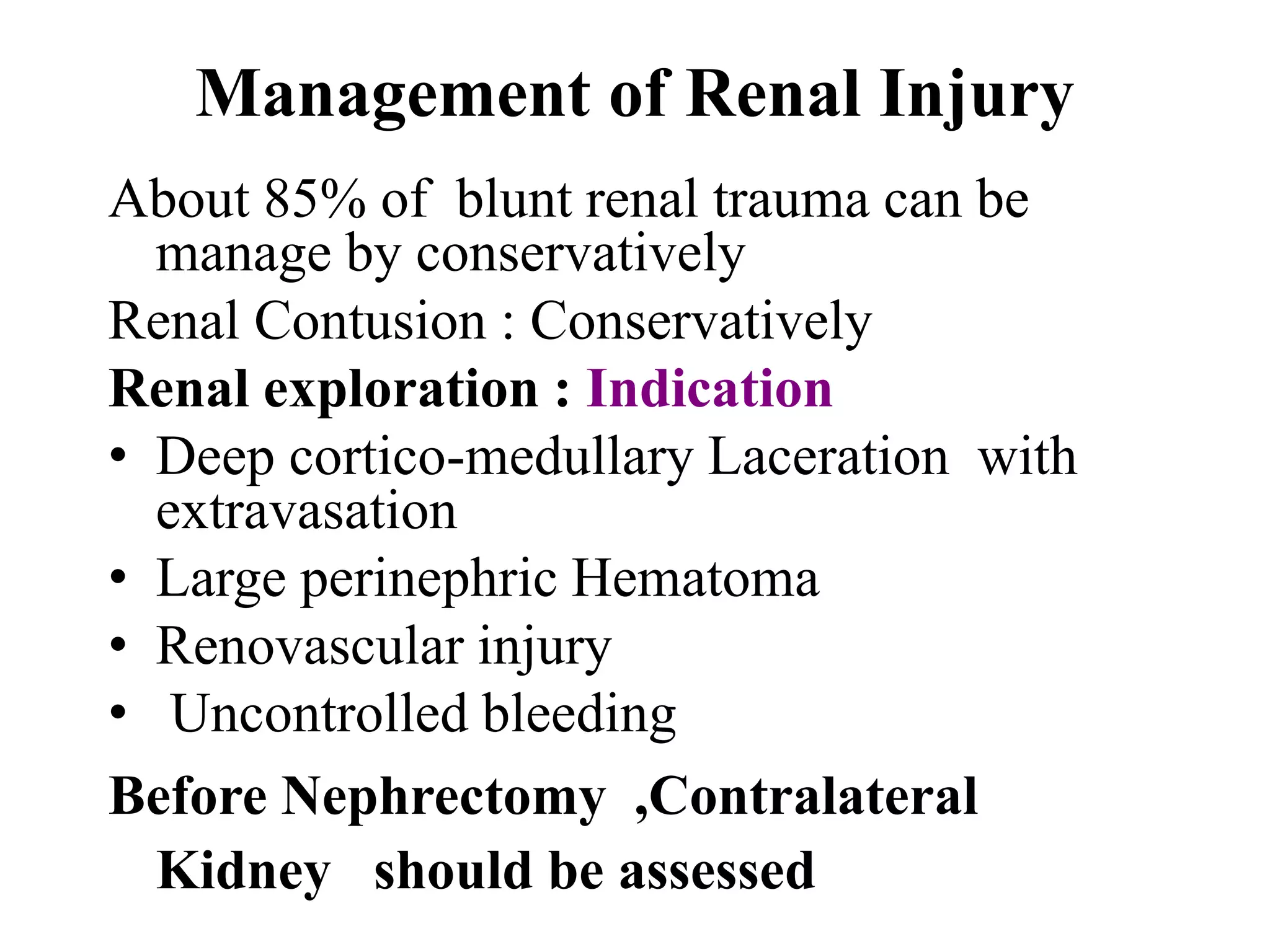
Procedures like paracentesis, diagnostic peritoneal lavage, and the nuances of injuries to solid organs. Injury details focusing on spleen, liver, pancreas, and renal injuries, including diagnosis and management strategies.
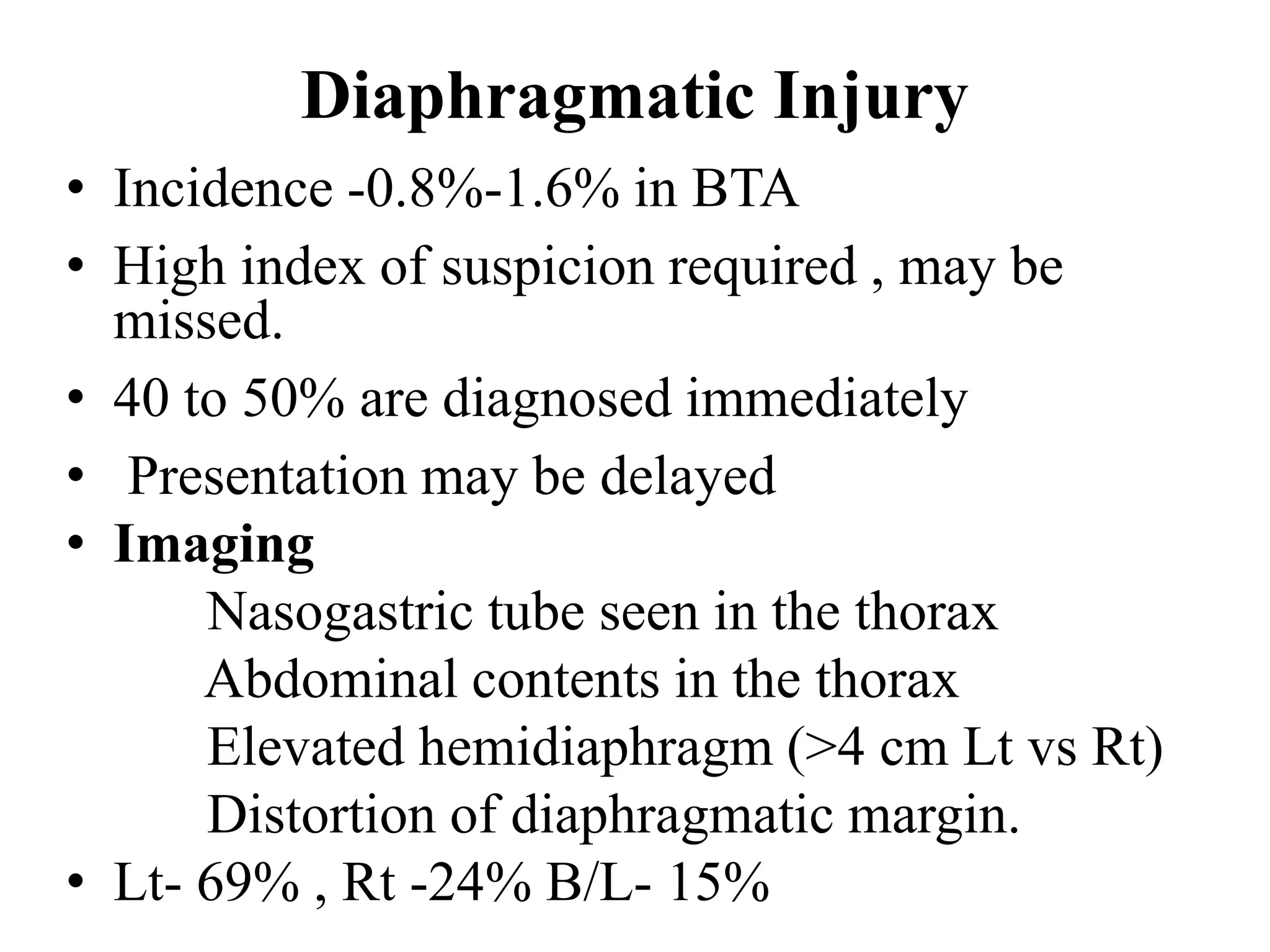
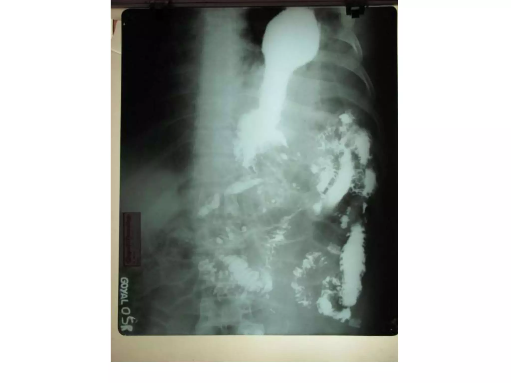
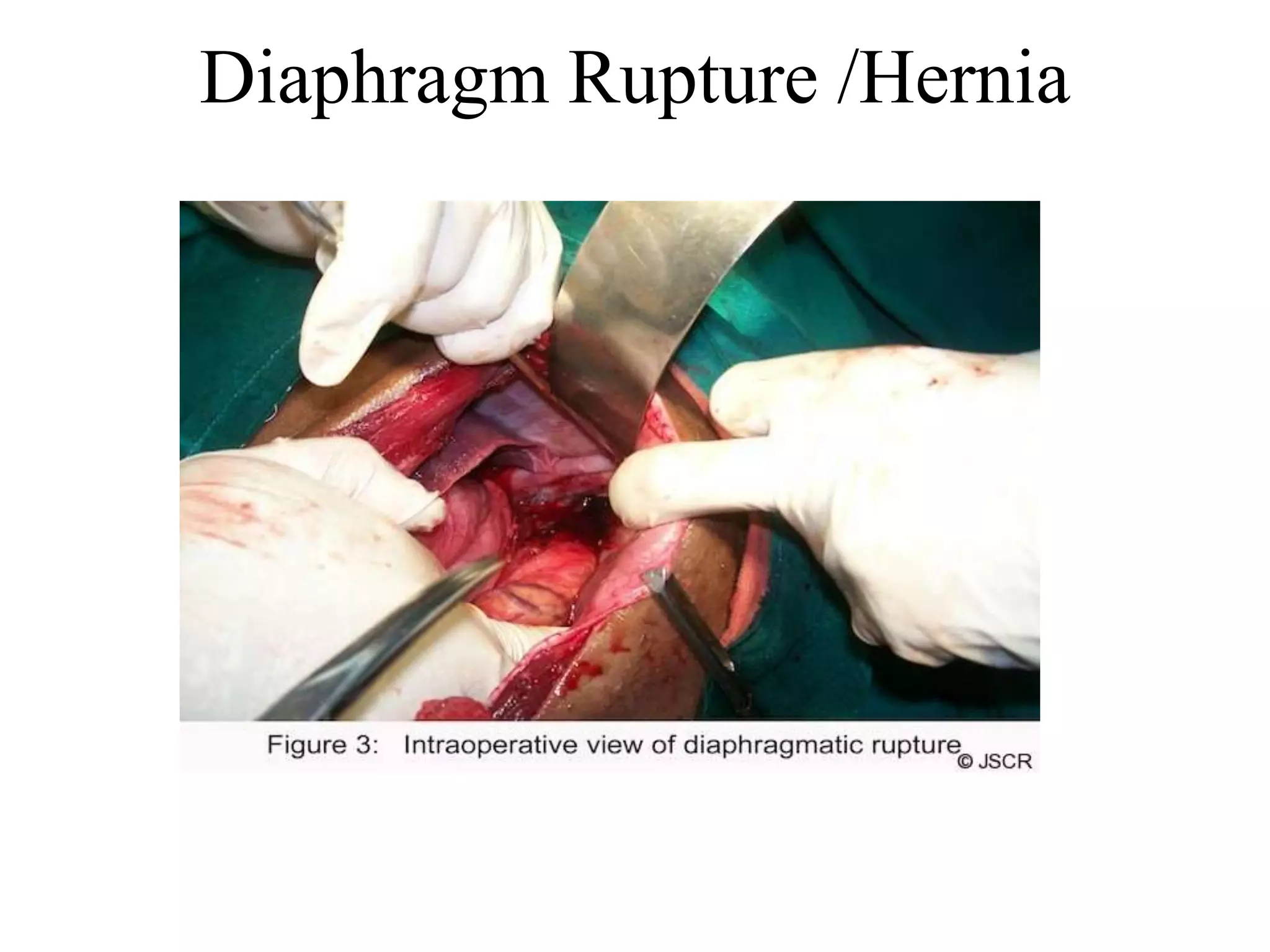
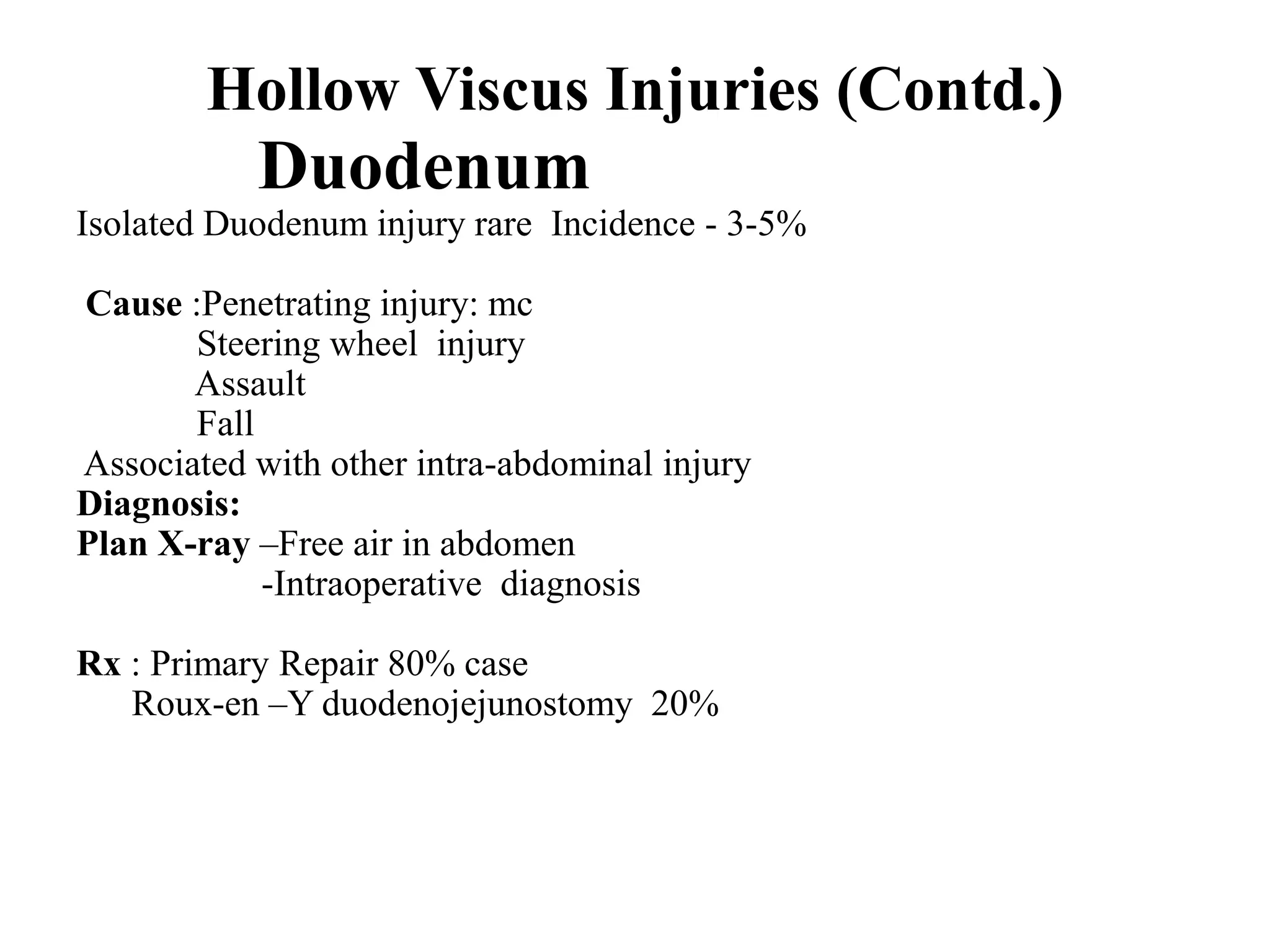
Challenges in diagnosing diaphragm injuries and hollow viscus injuries, including strategies for treatment.
Bladder and ureteral injuries, diagnosis methods, and management practices.
Incidence of trauma in pregnant women, causes, and the multidisciplinary approach needed for management.
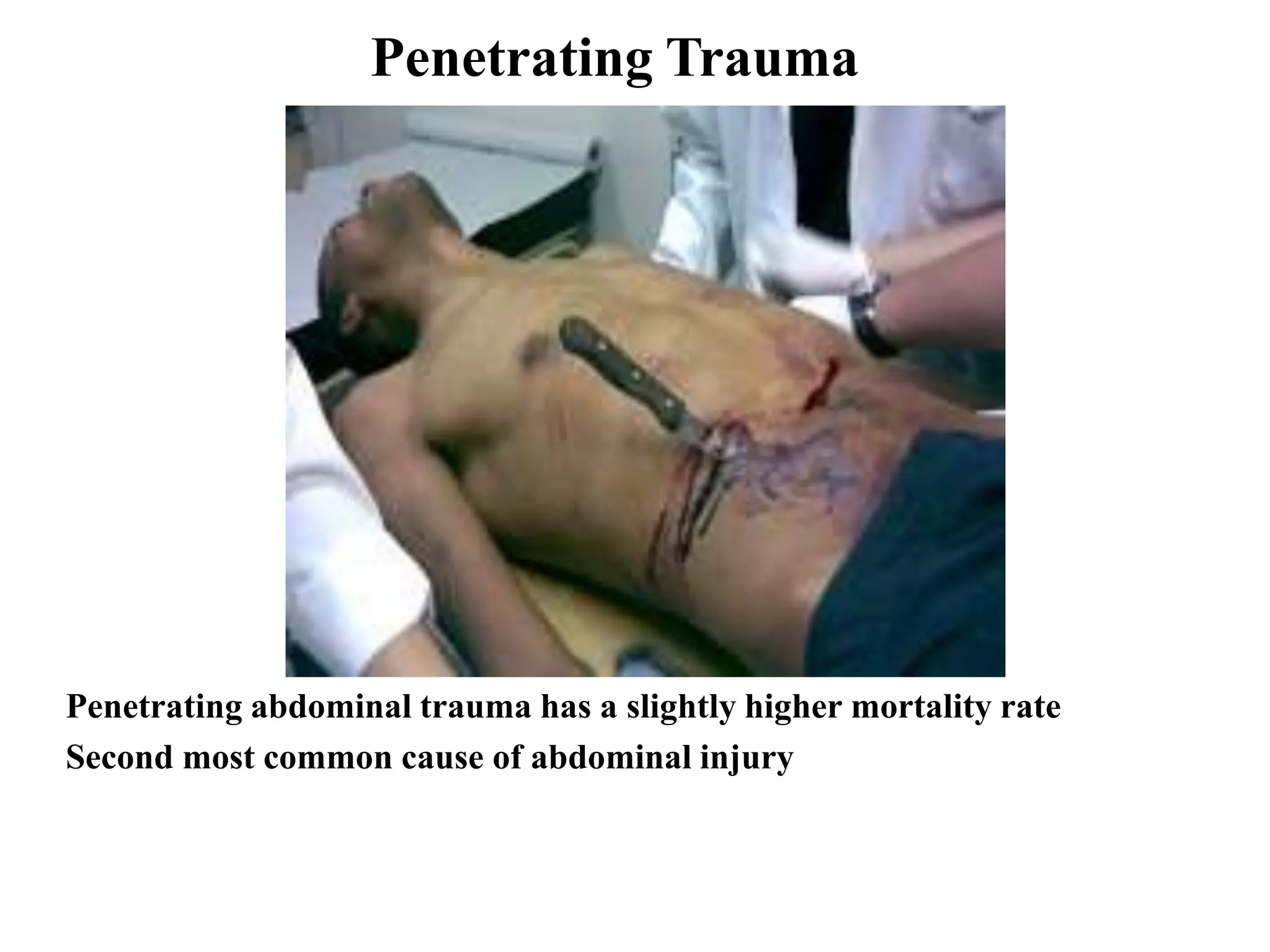
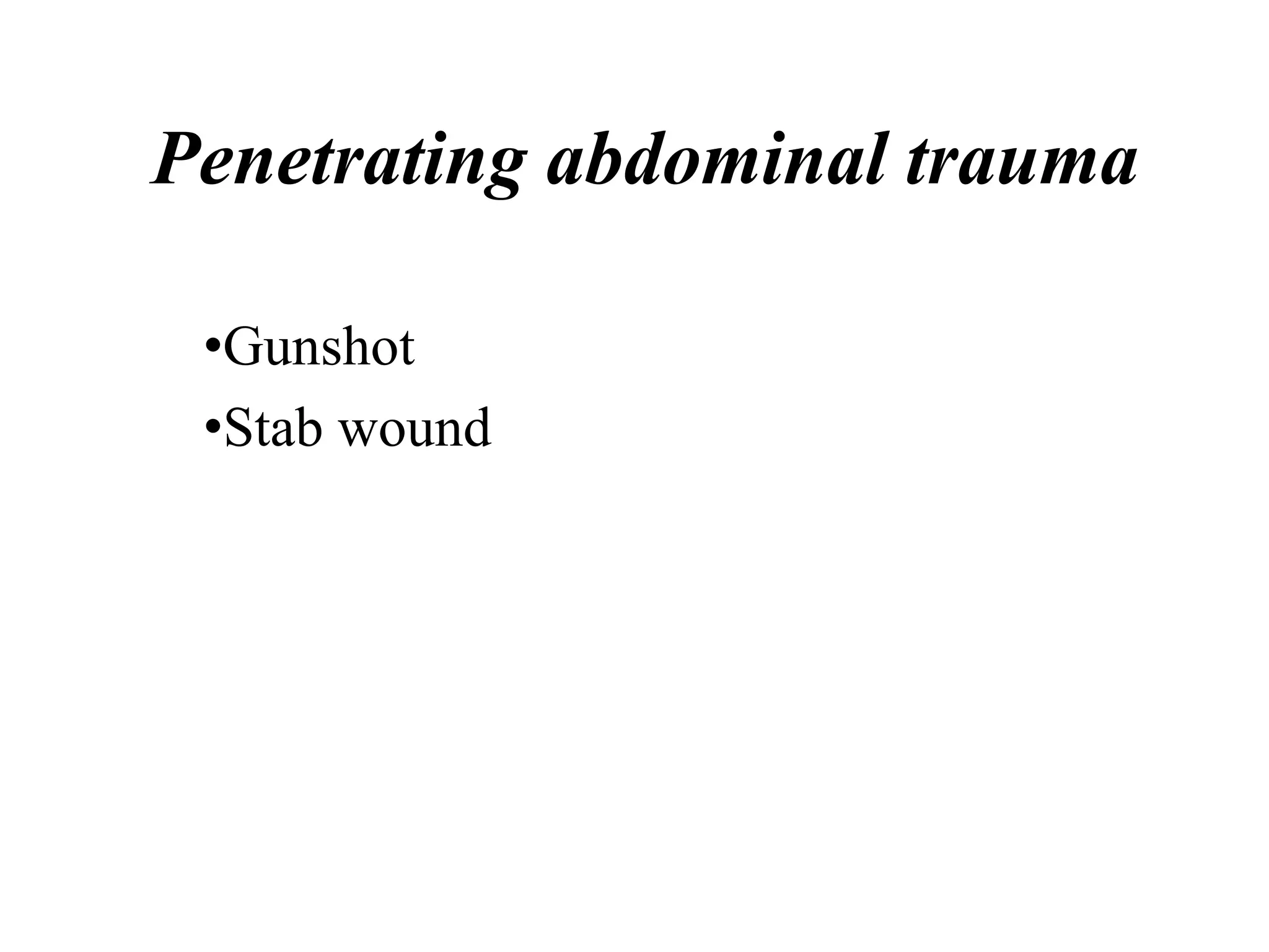
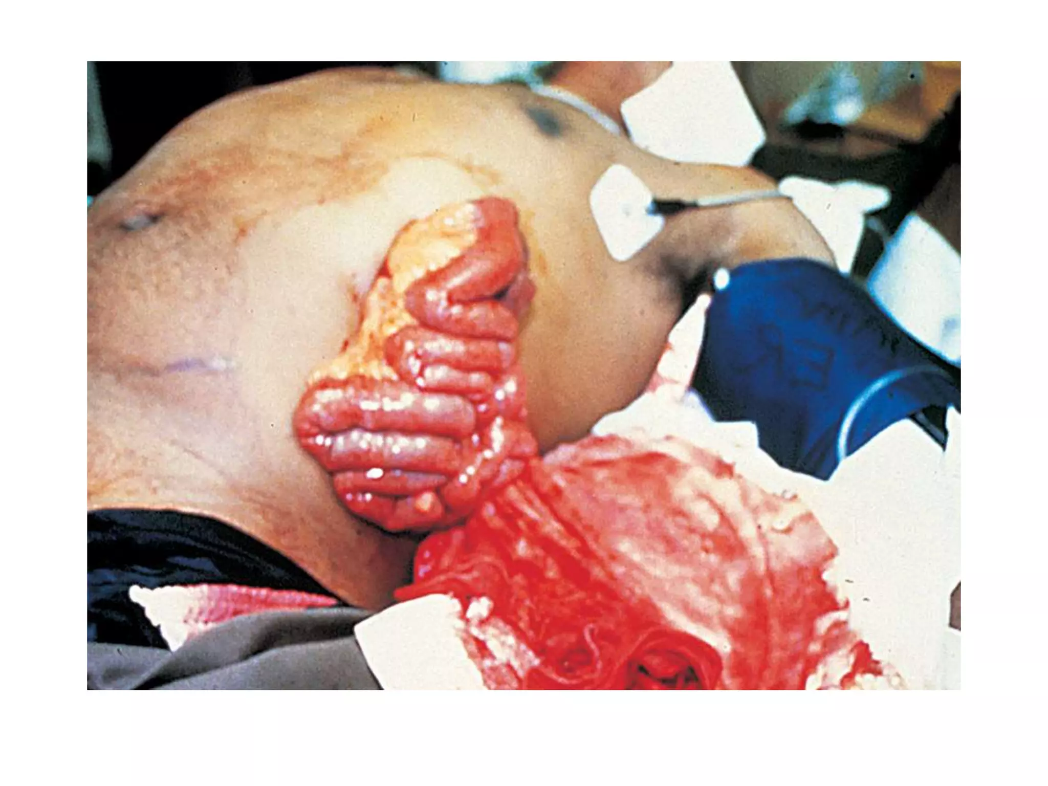
Characteristics of penetrating injuries, specifics about surgery requirements, and associated risks.
Injury prevention strategies focusing on education, safety measures, and statistical impact on injury rates.
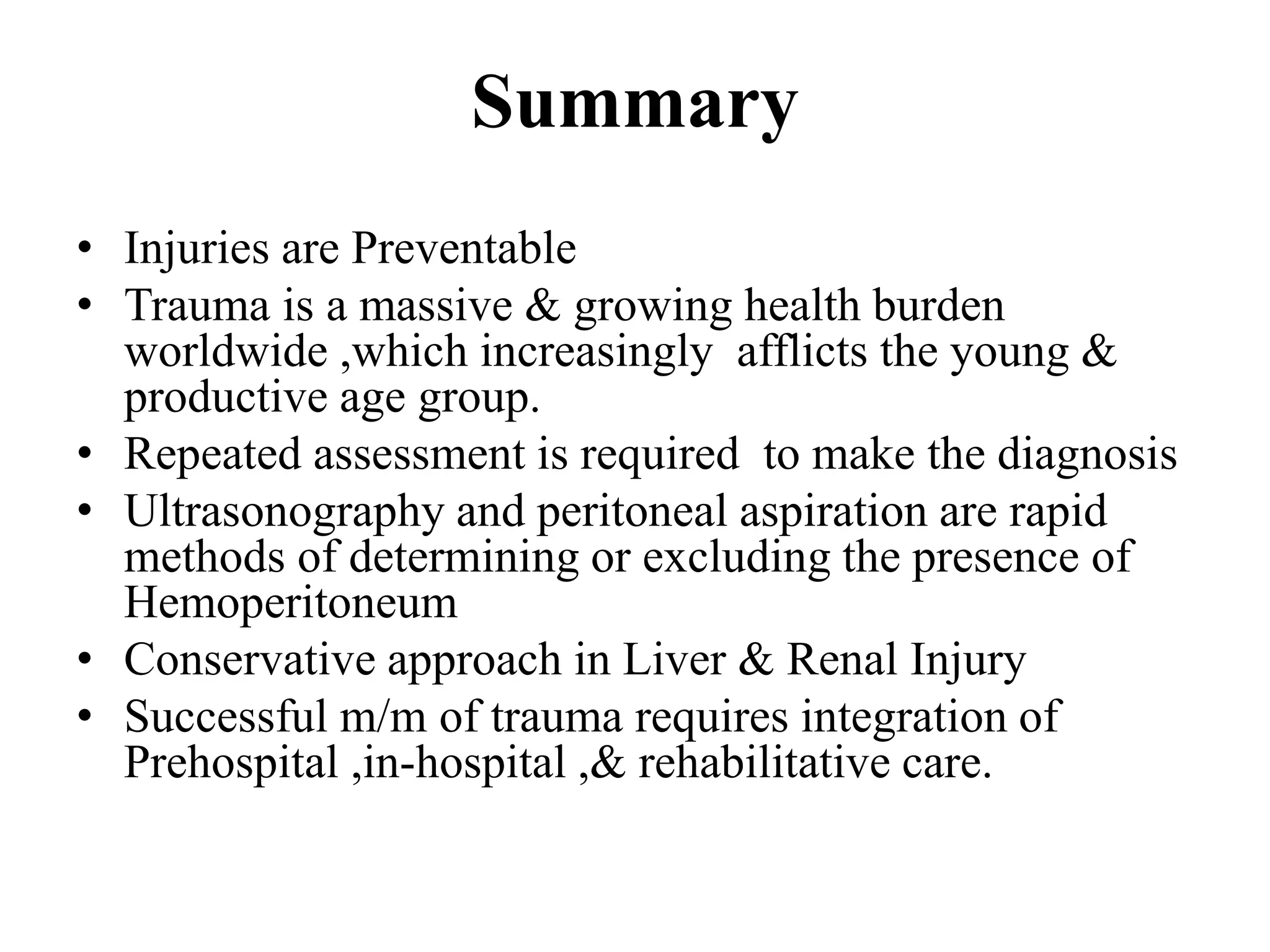
Overall insights on injury preventability, trauma health burden, and the importance of integrated care.
ABDOMINAL TRAUMA :AN OVERVIEW
Dr Tariq Saeed
a Associate Professor
Department of Surgery
KMC/KTH/ KMU Peshawar
2.
Introduction
• Abdominal traumais regularly
encountered in the emergency department
• One of the leading cause of death and
disability
• Identification of serious intra-abdominal
injuries is often challenging
• Many injuries may not manifest during
the initial assessment and
treatment period
3.
Epidemiology
• Peak incidenceAbdominal Trauma
15 - 30yr
• More than 1.5 Lac people die every year
as a result of injuries by motor vehicle
accident , fall, suicide and homicide
• Injury accounts for 10% of all deaths
• Estimates indicate that by 2020, 8.4
million people will die yearly.
• Prevalence: 13%
Prehospital Care
• Thegoal of prehospital is to deliver the pt
to hospital for definitive care as rapidly
as possible. ‘Scoop and Run’
• Maintain airway & start I V line
• Care of spinal cord
• Communicate to medical control
• Rapid transport of patient to trauma
centre
11.
Initial Assessment andResuscitation
Primary survey
Identification & treatment of life threatening
conditions
• Airway , with cervical spine precautions
• Breathing
• Circulation
• Disability
• Exposure
12.
Emergency Care
• IV fluids
• Control external bleeding
• Dressing of wounds
• Protect eviscerated organs with a sterile
dressing
• Stabilize an impaled object in place
• Give high flow oxygen
• Immobilize the patient with a fractured pelvis
• Keep the patient warm
• Analgesics
13.
Secondary Survey
• General&Systemic Examination-to identify
all occult injuries .
• Special attention to Back, Axilla , Perineum
• PR - sphincter tone ,bleeding ,perforation ,
high riding prostate
• Foley’s catheter- monitor urine out put
• Nasogastric tube
Examination
Grey-Turner’s Sign: (1877-1951)
Bluishdiscoloration of the flanks
Retroperitoneal Hematoma
hemorrhagic pancreatitis.
Kehr’s sign (1862-1916).
Referred pain, Right shoulder
irritation of the diaphragm
(Splenic injury, free air,
intra-abdominal bleeding)
18.
Examination
Balance’s Sign
Dullness onpercussion of the left upper quadrant
ruptured spleen
Labia and Scrotum : Pooling of blood from
abdominal and pelvic cavities.
19.
Examination
Auscultation :1. Bowelsounds in the thoracic
cavity (Diaphragmatic rupture)
2. Haemothorax
Palpation: -Mass
-Tenderness
-Signs of peritonitis
-# Ribs
-Chest & Pelvic compression test
Focused Assessment withSonography in
Trauma (FAST)
• First used in 1996
• Rapid , Accurate
• Sensitivity 86- 99%
• Can detect 100 mL of blood
• Cost effective
• Four different views- Pericardiac
Perihepatic
Perisplenic
Peripelvic space
• Eliminates unnecessary CT scans
• Helps in management plan
22.
Plain X-Ray Chest& Abdomen
• Pneumotharax, Haemothorax
• Free air under diaphragm
• Nasogastric tube, bowel loops in the chest
• Elevation of the both /Single diaphragm
• Lower Ribs # -Liver /Spleen Injury
• Ground Glass Appearance –
Massive Hemoperitoneum
• Obliteration of Psoas Shadow –Retroperitoneal
Bleeding
• #vertebra
23.
USG
Advantage
• Easy &Early to Diagnose
• Noninvasive
• No Radiation Exposure
Resuscitation/Emergency
room
Used in initial Evaluation
Low cost
Disadvantage
. Examiner Dependent
• Obesity
• Gas interposition
• Low Sensitivity for free fluid
less 500 mL
• False –Negative
retroperitoneal & Hallow
viscus injury
25.
Paracentasis
• Four quadrantaspiration of abdomen
• A Positive tap – blood , air , bile
stained fluid
• Negative tap doesn’t rule out injury.
• False negatives are as high as 22-60%
26.
Diagnostic Peritoneal Lavage
•First described in 1965
• Rapid & Accurate test used to identify
intra-abdominal injuries
• Predictive value of greater than 90%
• The RBC count for lavage fluid is >
1,00,000/cu m.m.
• A WBC count > 500/cu m.m.
• Test is highly sensitive to presence of
intraperitoneal blood
• However specificity is low
CT Scan
•Gold Standard
•HaemodynamicallyStable
• Provides excellent imaging of pancreas,
duodenum and Genitourinary system
•Standard for detection of solid organs injury.
• Determines the source and amount of bleeding
• Can reveal other associated injuries e.g.
Vertebral & Pelvic # & injury in the thoracic
cavity .
•High Specificity-95%
Solid Organ Injuries
•Grading of injured solid organs such as Spleen, Liver &
Kidneys are on the basis of subcapsular hematoma ,capsular
tear, parenchymal lacerations & avulsion of vascular pedicle
• Bleeds significantly and cause rapid blood loss
• Difficult to identify injury by physical exam
• Repeated assessment is required to make the diagnosis
• Slowly oozing blood into peritoneal cavity
35.
SPLENIC INJURY
• Mostcommon intra- abdominal organ to injured (40-55%)
• 20% of splenic injuries due to left lower rib fractures
• Commonly arterial hemorrhage
• Conservative management :
-Hemodynamic stability
- Negative abdominal examination
-Absence of contrast extravasation in CT
- Absence of other indication of Laprotomy
-Grade 1to 3 (Subcapsular Hematoma ,Laceration <3 cm)
Monitoring
• Serial abdo. Examinations & Haematocrit are essential
• Success rate of conservative m/m is >80%
36.
Splenic Injuries
Operative Management
Capsulartears (I)- Compression & topical haemostatic
agent
Deep Laceration (II)- Horizontal mattress suture
or Splenorrhaphy
Major Laceration not involving hilum (IV)-
Partial Splenectomy
Hillar injury (V)–Total Splenectomy
Grade IV-V: almost invariably require operative
intervention
Success rate of Splenic salvage procedure is 40-60%
37.
Liver injury
• Liveris the largest organ in abdomen
• 2nd most common organ injured (35-
45%) in BTA
• Driving and fighting responsible for
50% of deaths due to liver injury
• Usually venous bleeding
• 85% of all patients with blunt hepatic
trauma are stable
• CT is the mainstay of diagnosis in stable
pt.
38.
Liver Injury
• 50%liver injury have stop bleeding
spontaneously by the time of surgery
Non Operative m/m
• Haemodynamically Stable
• No other intra-abdominal injury require surgery
• < 2 units of BT required
• Hemoperitoneum <500 ml on CT
• Grade I-III(subcapsular & intr-perenchymal hematoma)
39.
Liver Injury
Operative m/m
•Packing
- Bleeding can be stopped by
packing of abdomen
-Pack removed after 48 hr
-haemostatic agents
-34 % survival in packing only
40.
• Suturing: -Simplesuture
-Deep mattress suture
• Laceration: -Mesh hepatorrhaphy
-Omental flap to cover the laceration
- Debridement
• Lobar Resection
• Liver Transplantation
• Ligate or repair damaged blood vessels & bile
duct
• Mortality of liver injury is 10%
Liver Injury
Operative Management(Contd.)
43.
Pancreatic Injury
• Rare10-20% of all abdominal injury
• Crush , Direct blow to abdo & Seat belt injury
• Associated with abdo. Duodenal injury, Vascular
injury & liver injury
• Diagnosis – Difficult, High index of suspicion
• CECT Scan is helpful
• Serum amylase is a poor indicator
• Usually diagnose on Laparotomy
• Distal Pancreatic injury - Distal resection
• Pancreaticojejunostomy – Injury to Ampulla of
Vater, Head & Body of Pancreas
Renal Injury
Diagnosis (contd.)
5.X-rayKUB
IVP
7. USG
6.CT Scan abdomen
8. Radionuclide Scan
The degree of hematuria may not predict the
severity of renal injury
Renal Injury
.
Classification ofInjury
• Grade I : Contusion or Subcapsular
Hematoma
• Grade II: Non Expanding Hematoma, <1
cm deep ,no extravasation
• Grade III: Laceration >1cm with urinary
Extravasation
• Grade IV: Parenchymal Laceration deep to
CM Junction
• Grade V: Renovascular injury
51.
Management of RenalInjury
About 85% of blunt renal trauma can be
manage by conservatively
Renal Contusion : Conservatively
Renal exploration : Indication
• Deep cortico-medullary Laceration with
extravasation
• Large perinephric Hematoma
• Renovascular injury
• Uncontrolled bleeding
Before Nephrectomy ,Contralateral
Kidney should be assessed
52.
Diaphragmatic Injury
• Incidence-0.8%-1.6% in BTA
• High index of suspicion required , may be
missed.
• 40 to 50% are diagnosed immediately
• Presentation may be delayed
• Imaging
Nasogastric tube seen in the thorax
Abdominal contents in the thorax
Elevated hemidiaphragm (>4 cm Lt vs Rt)
Distortion of diaphragmatic margin.
• Lt- 69% , Rt -24% B/L- 15%
53.
Diaphragm Rupture /Hernia
•S Lal, Y Kailasia , S Chouhan , APS Gaharwar, GP Shrivastava . Delayed
presentation of post traumatic diaphragmatic hernia.
Hollow Viscus Injuries
GastricInjury : Penetrating trauma MC
Blunt trauma abdomen 1%
Causes
Penetrating Injury
-Crushing Against the Spine
-CPR
-Vigorous Ventilation with ET Tube in the Esophagus
-Heimlich Maneuver
Diagnosis : X-Ray chest & Abdomen
CT scan
Diagnostic Peritoneal Lavage
During Surgical Exploration
T/t : Expl. Laparotomy with Primary Repair
57.
Hollow Viscus Injuries(Contd.)
Duodenum
Isolated Duodenum injury rare Incidence - 3-5%
Cause :Penetrating injury: mc
Steering wheel injury
Assault
Fall
Associated with other intra-abdominal injury
Diagnosis:
Plan X-ray –Free air in abdomen
-Intraoperative diagnosis
Rx : Primary Repair 80% case
Roux-en –Y duodenojejunostomy 20%
58.
Hollow Viscus Injuries
SmallIntestine& Colonic Injuries
Commonly Injured in Penetrating injury
Blunt Trauma -Incidence 5% -20%
Mechanism : -Crush Injury
-At Fixed point DJ & IC Junction
Rx : Exploratory Laprotomy
59.
Bladder Injury
• Commonlyin BTA
• 70% of bladder Injury are associated with pelvic fracture .
• Hematuria
Type 1.Extraperitoneal Rupture-by bony fragment
• 2. Intraperitoneal Rupture- at dome
when blow in distended bladder
• Diagnosis -1. Clinical 2. Cystography
T/t 1. Intraperitoneal –trans-peritoneal - closure +SPC
2:Extraperitoneal Rupture : Foley’s catheter -10 -14 days
60.
Ureteral Injury
• Uncommon
•Mostly occur after penetrating trauma
• Associated with concomitant intra-abdominal or
genitourinary injury
• Diagnosis
-IVP
-15-20% Retrograde ureteroscopy
- At the time of Laparotomy
• Operative procedure
Proximal & mid ureter -End to end Anastomosis over
DJ Stent
Distal –Ureteric Reimplantaion
61.
Vascular Injury
• Incidence5-10%
• Highly lethal.
• Associated with extremely rapid rates of blood
loss
• Exposure is difficult in Laparotomy
• Initial Control by digital pressure
• Heparinized saline (50U/ml) injected in both end
of vessel
• Rx Lateral suture ,End to end Anastomosis &
Interposition graft
• Mortality rate is very high
62.
Trauma in Pregnancy
•Incidence- 10-20%
• Causes: 1.Domestic violence
2.Sexual Assault 3. Accident
• Third trimester- mc- balance & coordination disturbed
• Multidisciplinary team- Obstetrician, surgeon, and
neonatologist
• Peritoneal sign are delayed
• “Supine hypotensive syndrome” > 20 weeks’ gestation.
COMPLICATIONS
• Fetal Injury & Death –fetoplacental injury, maternal shock,
• Placental Abruption
• Rupture of Uterus
Penetrating Abdominal Trauma(Contd.)
•Multiple in 20% of cases
• Most stab wounds do not cause an
intraperitoneal injury
• A complete Laparotomy is
mandatory
Gunshot Injury
• Handguns,Rifles, and Shotgun
• More dangerous than penetrating injury
• The degree of injury depends .
Amount of kinetic energy imparted by the bullet to the
victim
Mass of the bullet and the square of its velocity
Distance .
• Injury multiple organ
72.
Injury Prevention
1.Primary: Preventan injury from its occurrence in
the first place: Educational activity such as anti-
drink-driving campaigns , speed limit rule
-Children should accompanied with parent
2.Secondary: Attempts to lesson the consequences
of injury – making road & safer car, anti-locking
brakes, air bags , helmets, seat belt
3. Tertiary: Minimize the effect of injury by health
care by individuals & system.
73.
Injury Prevention (Contd.)
•Speed is a critical factor ; a 10% increase
speed translate into a 40% rise in the case
fatality rate.
• Use of seat belt reduces the risk of death or
serious injury by 45%.
• Air Bags reduces the risk of fatal injury by 30%
& deaths by 11 %.
• Children Below 12yrs should be properly
restraints in the back seat.
• Motorcycle experience death rate 35 time
greater than car.
74.
Summary
• Injuries arePreventable
• Trauma is a massive & growing health burden
worldwide ,which increasingly afflicts the young &
productive age group.
• Repeated assessment is required to make the diagnosis
• Ultrasonography and peritoneal aspiration are rapid
methods of determining or excluding the presence of
Hemoperitoneum
• Conservative approach in Liver & Renal Injury
• Successful m/m of trauma requires integration of
Prehospital ,in-hospital ,& rehabilitative care.