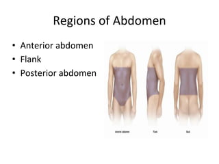
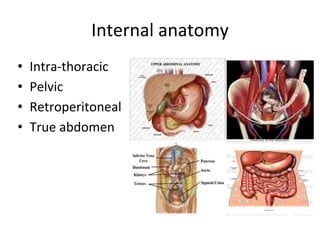
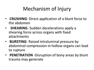
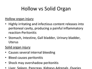
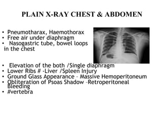
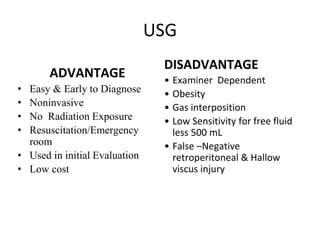
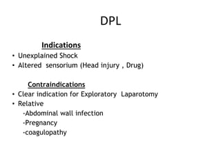
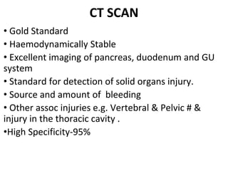
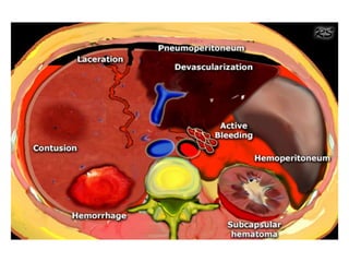
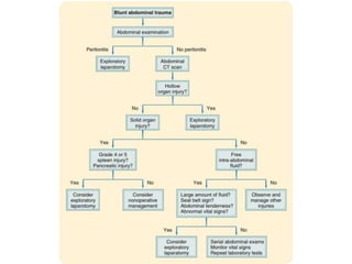
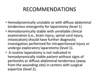
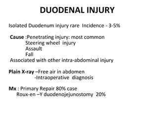
SlideShare a Scribd company logo
BLUNT TRAUMA ABDOMEN
SURG LT CDR K S PATEL
Scope
• Types
• Regional anatomy
• Causes
• Mechanism
• Examination
• Investigation
• Management
Types of Abdominal Trauma
1.Blunt Trauma- Any force to abdomen which
does not leave open wound.
2.Penetrating Trauma
-Stab
-Gun shot Injury
Regions of Abdomen
• Anterior abdomen
• Flank
• Posterior abdomen
Internal anatomy
• Intra-thoracic
• Pelvic
• Retroperitoneal
• True abdomen
Mechanism of Injury
• CRUSHING- Direct application of a blunt force to
the abdomen
• SHEARING- Sudden decelerations apply a
shearing force across organs with fixed
attachments
• BURSTING- Raised intraluminal pressure by
abdominal compression in hollow organs can lead
to rupture
• PENETRATION -Disruption of bony areas by blunt
trauma may generate
Hollow vs Solid Organ
Hollow organ injury
• Highly irritating and infectious content releases into
peritoneal cavity, producing a painful inflammatory
reaction-Peritonitis
• Stomach, Intestine, Gall bladder, Urinary bladder,
Uterus
Solid organ injury
• Causes several internal bleeding
• Bleed causes peritonitis
• Shock may overshadow peritonitis
CAUSES
• RTA with motor vehicle accident 60%
• Direct blow to abdomen 15%
• Fall 9%
• Domestic violence
PRIMARY SURVEY
• Identification & treatment of life threatening
conditions
• Airway , with cervical spine precautions
• Breathing
• Circulation
• Disability-AVPU (neurological status)
• Exposure-remove all clothes
EMERGENCY CARE
• I V fluids
• Control external bleeding
• Dressing of wounds
• Protect eviscerated organs with a sterile
dressing
• Stabilize an impaled object in place
• Give high flow oxygen
• Keep the patient warm
• Analgesics
SECONDARY SURVEY
• General &Systemic Examination-to identify
all occult injuries .
• Special attention to Back, Axilla , Perineum
• P/R - sphincter tone ,bleeding ,perforation ,
high riding prostate
• Foley’s catheter- monitor urine out put
• Nasogastric tube
SECONDARY SURVEY
AMPLE History
A: Allergy
M: Medications
P: Past medical history
L: Last meal
E: Event
EXAMINATION
Cullen’s Sign: Bluish discoloration around umbilicus.
Diffusion of blood along periumbilical tissues
Hemoperitoneum
Severe pancreatitis
13
EXAMINATION
Grey-Turner’s Sign: Bluish discoloration of the
flanks.Retroperitoneal Hematoma hemorrhagic
pancreatitis.
Kehr’s sign: Referred pain, Right shoulder
irritation of the diaphragm (Splenic injury, free
air,intra-abdominal bleeding)
EXAMINATION
Balance’s Sign: Dullness on percussion of the left
upper quadrant ruptured spleen
Labia and Scrotum : Pooling of blood from
abdominal and pelvic cavities.
• Seat belt sign
London sign
Examination
Palpation: -Mass
-Tenderness
-Rebound tenderness
- Guarding /Rigidity
-Signs of peritonitis
-# Ribs
-Chest & Pelvic compression test
Auscultation :
1. Bowel sounds in the thoracic cavity
(Diaphragmatic rupture)
2. Haemothorax
INVESTIGATIONS
• FAST
• X-Ray Chest & Abdomen
• USG
• CT Scan
• Diagnostic Peritoneal Lavage
• Diagnostic Laparoscopy
Focussed Assessment with Sonography in
Trauma (FAST)
• Rapid , Accurate
• Sensitivity 86- 99%
• Can detect 100 mL of blood
• Cost effective
• Four different views- Pericardiac
Perihepatic
Perisplenic
Peripelvic space
• Eliminates unnecessary CT scans
• Helps in management plan
PLAIN X-RAY CHEST & ABDOMEN
• Pneumotharax, Haemothorax
• Free air under diaphragm
• Nasogastric tube, bowel loops
in the chest
• Elevation of the both /Single diaphragm
• Lower Ribs # -Liver /Spleen Injury
• Ground Glass Appearance – Massive Hemoperitoneum
• Obliteration of Psoas Shadow –Retroperitoneal
Bleeding
• #vertebra
USG
ADVANTAGE
• Easy & Early to Diagnose
• Noninvasive
• No Radiation Exposure
• Resuscitation/Emergency
room
• Used in initial Evaluation
• Low cost
DISADVANTAGE
• Examiner Dependent
• Obesity
• Gas interposition
• Low Sensitivity for free fluid
less 500 mL
• False –Negative
retroperitoneal & Hallow
viscus injury
DIAGNOSTIC PERITONEAL LAVAGE
• Rapid & Accurate test used to identify intra-
abdominal injuries
• Predictive value of greater than 90%
• The RBC count for lavage fluid is >
1,00,000/cu m.m.
• A WBC count > 500/cu m.m.
• Test is highly sensitive to presence of
intraperitoneal blood
• However specificity is low
DPL
Indications
• Unexplained Shock
• Altered sensorium (Head injury , Drug)
Contraindications
• Clear indication for Exploratory Laparotomy
• Relative
-Abdominal wall infection
-Pregnancy
-coagulopathy
CT SCAN
• Gold Standard
• Haemodynamically Stable
• Excellent imaging of pancreas, duodenum and GU
system
• Standard for detection of solid organs injury.
• Source and amount of bleeding
• Other assoc injuries e.g. Vertebral & Pelvic # &
injury in the thoracic cavity .
•High Specificity-95%
CT SCAN
Contraindication:
• Clear indication for Laparotomy
• Haemodynamically Unstable
• Allergy to contrast media
RECOMMENDATIONS
• Hemodynamically unstable or with diffuse abdominal
tenderness emergently for laparotomy (level 1)
• Hemodynamically stable with unreliable clinical
examination (i.e., brain injury, spinal cord injury,
intoxication) should have further diagnostic
investigation performed for intraperitoneal injury or
undergo exploratory laparotomy (level 1).
• A routine laparotomy is not indicated in
hemodynamically stable patient without signs of
peritonitis or diffuse abdominal tenderness (away
from the wounding site) in centers with surgical
expertise (level 2).
• A routine laparotomy is not indicated in
hemodynamically stable patients with abdominal
GSWs if the wounds are tangential and there are no
peritoneal signs (level 2).
• In patients selected for initial NOM, abdomino-pelvic
CT should be strongly considered as a diagnostic tool
to facilitate initial management decisions (level 2).
• Diagnostic laparoscopy may be considered as a tool to
evaluate diaphragmatic lacerations and peritoneal
penetration (level 2)
SOLID ORGAN INJURIES
• Grading of injured solid organs such as Spleen, Liver &
Kidneys are on the basis of
• subcapsular hematoma
• capsular tear
• parenchymal lacerations
• avulsion of vascular pedicle
• Bleeds significantly and cause rapid blood loss
• Difficult to identify injury by physical exam
• Repeated assessment is required to make the diagnosis
SPLENIC INJURY
• Most common intra- abdominal organ to injured (40-55%)
• 20% due to left lower rib fractures
• Arterial hemorrhage
Splenic injury
• Conservative management :
-Hemodynamic stability
- Negative abdominal examination
-Absence of contrast extravasation in CT
- Absence of other indication of Laprotomy
-Grade 1to 3 (Subcapsular Hematoma ,Laceration <3
cm)
Monitoring
• Serial abdo examinations & Haematocrit are essential
• Success rate is >80%
SPLENIC INJURIES
Operative Management
Capsular tears (I)- Compression & topical haemostatic agent
Deep Laceration (II) and (III) - Horizontal mattress suture
or Splenorrhaphy
Major Laceration not involving hilum (IV)-
Partial Splenectomy
Hillar injury (V)–Total Splenectomy
Grade IV-V: almost invariably require operative intervention
Success rate of Splenic salvage procedure is 40-60%
LIVER INJURY
• 2nd most common organ injured (35-45%) in BTA
• RTA and fights are responsible for 50% of liver
injury
• Venous bleeding
• Around 85% of all patients remain stable
• CT mainstay in stable pt.
Liver injury
LIVER INJURY
• 50% stop bleeding spontaneously by the time of surgery
Non Operative Mx
• Haemodynamically Stable
• No other intra-abdominal injury require surgery
• < 2 units of Blood Transfusion required
• Hemoperitoneum <500 ml on CT
• Grade I-III(subcapsular & intr-perenchymal hematoma)
38
LIVER INJURY
OPERATIVE MANAGEMENT
• Packing
- Bleeding can be stopped by
packing
-Pack removed after 48 hr
-Haemostatic agents
-34 % survival in packing only
• Suturing: -Simple suture
-Deep mattress suture
• Laceration: -Mesh hepatorrhaphy
-Omental flap to cover the laceration
• Lobar Resection
• Ligate or repair damaged blood vessels & bile duct
• Mortality due to liver injury is 10%
LIVER INJURY
OPERATIVE MANAGEMENT
PANCREATIC INJURY
• 10-20% of abdomen injury.
• Crush, direct blow to abdo & Seat belt injury
• Associated with duodenal injury, Vascular injury &
liver injury
• Pancreatic enzymes are caustic, source of massive
systemic inflammation and subsequent poor
outcomes.
• Diagnosis – Difficult, CECT :- malperfusion of the
pancreatic parenchyma surrounding fluid
• Hematoma
• Extent of parenchymal injury and degree of ductal
involvement poorly characterize
• High index of suspicion –rib/ vertebra fracture
Pancreatic Injury
The injury involves the pancreatic neck and appears as a 2-cm segment of
nonperfused pancreas tissue with surrounding edema as identified by the arrow
PANCREATIC INJURY
Management
• Exposure of the entire pancreas is required
• Mobilization:- hepatic flexure gastrocolic
ligament to retract the transverse colon and
mesocolon inferiorly.
• Ext drainage imp component
• Distal feeding access provide early enteral
nutrition
RENAL INJURY
• Frequently overlooked
• Mechanism: Blunt , Penetrating
# lower ribs or spinous process,
Crush abdominal
Pelvic injury
Direct blow to flank or back
Fall
RTA
RENAL INJURY
Diagnosis
1.History ,Clinical examination
2. Presentation :Shock, hematuria & pain
3. Urine: gross or microscopic hematuria
5.X-ray KUB
IVP
6.CT Scan abdomen
7. USG
The degree of hematuria may not predict the
severity of renal injury
RENAL INJURY
Classification of Injury
• Grade I : Contusion or Subcapsular Hematoma
• Grade II: Non Expanding Hematoma, <1 cm deep
,no extravasation
• Grade III: Laceration >1cm with urinary
Extravasation
• Grade IV: Parenchymal Laceration deep to CM
Junction
• Grade V: Renovascular injury
MANAGEMENT OF RENAL INJURY
About 85% of trauma can be managed conservatively
Renal Contusion : Conservatively
Renal exploration : Indication
• Deep cortico-medullary Laceration with extravasation
• Large perinephric Hematoma
• Renovascular injury
• Uncontrolled bleeding
DIAPHRAGMATIC INJURY
• Incidence -0.8%-1.6% in BTA
• High index of suspicion required , may be
missed.
• Presentation may be delayed
• Imaging
Nasogastric tube seen in the thorax
Abdominal contents in the thorax
Elevated hemidiaphragm
Distortion of diaphragmatic margin.-
-Primary repair with interrupted horizontal mattress
suture
HOLLOW VISCUS INJURIES
Gastric Injury : Penetrating trauma
Blunt trauma abdomen 1%
Causes
Penetrating Injury
-Crushing Against the Spine
-CPR
-Vigorous Ventilation with ET Tube in the Esophagus
-Heimlich Maneuver
Diagnosis : X-Ray chest & Abdomen
CT scan
Diagnostic Peritoneal Lavage
During Surgical Exploration
T/t : Expl. Laparotomy with Primary Repair
DUODENAL INJURY
Isolated Duodenum injury rare Incidence - 3-5%
Cause :Penetrating injury: most common
Steering wheel injury
Assault
Fall
Associated with other intra-abdominal injury
Plain X-ray –Free air in abdomen
-Intraoperative diagnosis
Mx : Primary Repair 80% case
Roux-en –Y duodenojejunostomy 20%
HOLLOW VISCUS INJURIES
Small Intestine& Colonic Injuries
Commonly Injured in Penetrating injury
Blunt Trauma -Incidence 5% -20%
Mechanism : -Crush Injury
-At Fixed point DJ & IC Junction
Mx : Exploratory Laprotomy
Primary repair
Resection with anastomosis
End colostomy
Primary repair with diverting colostomy
BLADDER INJURY
• 70% of bladder Injury are associated with pelvic fracture .
• Hematuria
Type 1.Extraperitoneal Rupture-by bony fragment
• 2. Intraperitoneal Rupture- at dome when blow in
distended bladder
• Diagnosis -1. Clinical 2. Cystography
T/t 1. Intraperitoneal – closure with absorbable sutures +SPC
2:Extraperitoneal Rupture : Foley’s catheter -10 -14 days
THANK YOU

More Related Content

What's hot

Bile duct injuries.slideshare
Bile duct injuries.slideshareBile duct injuries.slideshare
Bile duct injuries.slideshare
drksreenath
 

What's hot (20)

Abdominal injuries, lecture
Abdominal injuries, lectureAbdominal injuries, lecture
Abdominal injuries, lecture
 
Abdominal trauma
Abdominal traumaAbdominal trauma
Abdominal trauma
 
Damage Control Surgery
Damage Control SurgeryDamage Control Surgery
Damage Control Surgery
 
Penetrating Abdominal Trauma Emergency Management
Penetrating Abdominal Trauma Emergency ManagementPenetrating Abdominal Trauma Emergency Management
Penetrating Abdominal Trauma Emergency Management
 
Traumatic Retroperitoneal Hematoma
Traumatic Retroperitoneal HematomaTraumatic Retroperitoneal Hematoma
Traumatic Retroperitoneal Hematoma
 
Abdominal trauma
Abdominal traumaAbdominal trauma
Abdominal trauma
 
Abdominal truma 2007
Abdominal truma 2007Abdominal truma 2007
Abdominal truma 2007
 
Liver trauma
Liver traumaLiver trauma
Liver trauma
 
Abdominal trauma
Abdominal traumaAbdominal trauma
Abdominal trauma
 
Lt hemicolectomy - Surgical Approach, Complications.
Lt hemicolectomy - Surgical Approach, Complications.Lt hemicolectomy - Surgical Approach, Complications.
Lt hemicolectomy - Surgical Approach, Complications.
 
Spleen Trauma
Spleen TraumaSpleen Trauma
Spleen Trauma
 
Management of duodenal trauma
Management of duodenal traumaManagement of duodenal trauma
Management of duodenal trauma
 
Ventral hernia
Ventral herniaVentral hernia
Ventral hernia
 
Colonic trauma, colon injury, colorectal trauma
Colonic trauma, colon injury, colorectal traumaColonic trauma, colon injury, colorectal trauma
Colonic trauma, colon injury, colorectal trauma
 
Blunt abdominal trauma
Blunt abdominal traumaBlunt abdominal trauma
Blunt abdominal trauma
 
Traumatic retroperitoneal injury
Traumatic retroperitoneal injuryTraumatic retroperitoneal injury
Traumatic retroperitoneal injury
 
Bile duct injuries.slideshare
Bile duct injuries.slideshareBile duct injuries.slideshare
Bile duct injuries.slideshare
 
PENETRATING ABDOMINAL TRAUMA
PENETRATING ABDOMINAL TRAUMAPENETRATING ABDOMINAL TRAUMA
PENETRATING ABDOMINAL TRAUMA
 
Abdominal trauma ,an overview
Abdominal trauma ,an overviewAbdominal trauma ,an overview
Abdominal trauma ,an overview
 
Management of abdominal trauma
Management of abdominal traumaManagement of abdominal trauma
Management of abdominal trauma
 

Similar to Blunt trauma abdomen

Abdominal trauma : an overview
Abdominal trauma  : an overviewAbdominal trauma  : an overview
Abdominal trauma : an overview
shyamesic
 
1588832705-abdominal-injuries-in-sports.pptx
1588832705-abdominal-injuries-in-sports.pptx1588832705-abdominal-injuries-in-sports.pptx
1588832705-abdominal-injuries-in-sports.pptx
Aymanshahzad4
 
Pancreatitis DEFINITION, COMPLICATIONS, TREATMENT .pptx
Pancreatitis DEFINITION, COMPLICATIONS, TREATMENT .pptxPancreatitis DEFINITION, COMPLICATIONS, TREATMENT .pptx
Pancreatitis DEFINITION, COMPLICATIONS, TREATMENT .pptx
rohanbijarnia2
 
ABDOMINAL TRAUMA ASSESSMENT AND TREATMENT.pptx
ABDOMINAL TRAUMA ASSESSMENT AND TREATMENT.pptxABDOMINAL TRAUMA ASSESSMENT AND TREATMENT.pptx
ABDOMINAL TRAUMA ASSESSMENT AND TREATMENT.pptx
Nethaji Perumal
 

Similar to Blunt trauma abdomen (20)

Abdominal trauma : an overview
Abdominal trauma  : an overviewAbdominal trauma  : an overview
Abdominal trauma : an overview
 
Abdominal trauma
Abdominal traumaAbdominal trauma
Abdominal trauma
 
Abdominal trauma
Abdominal  traumaAbdominal  trauma
Abdominal trauma
 
AT.pptx
AT.pptxAT.pptx
AT.pptx
 
AT.pptx
AT.pptxAT.pptx
AT.pptx
 
Abdominal-injuries-in-sports.pptx
Abdominal-injuries-in-sports.pptxAbdominal-injuries-in-sports.pptx
Abdominal-injuries-in-sports.pptx
 
Abdominal injuries
Abdominal injuriesAbdominal injuries
Abdominal injuries
 
1588832705-abdominal-injuries-in-sports.pptx
1588832705-abdominal-injuries-in-sports.pptx1588832705-abdominal-injuries-in-sports.pptx
1588832705-abdominal-injuries-in-sports.pptx
 
Abdominal injuries lit review .pptx
Abdominal injuries lit review .pptxAbdominal injuries lit review .pptx
Abdominal injuries lit review .pptx
 
Abdominal trauma 2.pptx
Abdominal trauma 2.pptxAbdominal trauma 2.pptx
Abdominal trauma 2.pptx
 
Trauma I-1.pptx
Trauma I-1.pptxTrauma I-1.pptx
Trauma I-1.pptx
 
abdominal trauma.ppt
abdominal trauma.pptabdominal trauma.ppt
abdominal trauma.ppt
 
Abdominal trauma (1)
Abdominal trauma (1)Abdominal trauma (1)
Abdominal trauma (1)
 
Abdominal trauma.pptx
Abdominal trauma.pptxAbdominal trauma.pptx
Abdominal trauma.pptx
 
Abdominal Trauma 3.pptx
Abdominal Trauma 3.pptxAbdominal Trauma 3.pptx
Abdominal Trauma 3.pptx
 
ABDOMINAL TRAUMA.pptx
ABDOMINAL TRAUMA.pptxABDOMINAL TRAUMA.pptx
ABDOMINAL TRAUMA.pptx
 
Pancreatitis DEFINITION, COMPLICATIONS, TREATMENT .pptx
Pancreatitis DEFINITION, COMPLICATIONS, TREATMENT .pptxPancreatitis DEFINITION, COMPLICATIONS, TREATMENT .pptx
Pancreatitis DEFINITION, COMPLICATIONS, TREATMENT .pptx
 
Abdominal injury
Abdominal injuryAbdominal injury
Abdominal injury
 
ABDOMINAL TRAUMA ASSESSMENT AND TREATMENT.pptx
ABDOMINAL TRAUMA ASSESSMENT AND TREATMENT.pptxABDOMINAL TRAUMA ASSESSMENT AND TREATMENT.pptx
ABDOMINAL TRAUMA ASSESSMENT AND TREATMENT.pptx
 
pancreatic trauma and its management.pptx
pancreatic trauma and its management.pptxpancreatic trauma and its management.pptx
pancreatic trauma and its management.pptx
 

Recently uploaded

Cardiac Impulse: Rhythmical Excitation and Conduction in the Heart
Cardiac Impulse: Rhythmical Excitation and Conduction in the HeartCardiac Impulse: Rhythmical Excitation and Conduction in the Heart
Cardiac Impulse: Rhythmical Excitation and Conduction in the Heart
MedicoseAcademics
 

Recently uploaded (20)

Hemodialysis: Chapter 2, Extracorporeal Blood Circuit - Dr.Gawad
Hemodialysis: Chapter 2, Extracorporeal Blood Circuit - Dr.GawadHemodialysis: Chapter 2, Extracorporeal Blood Circuit - Dr.Gawad
Hemodialysis: Chapter 2, Extracorporeal Blood Circuit - Dr.Gawad
 
The hemodynamic and autonomic determinants of elevated blood pressure in obes...
The hemodynamic and autonomic determinants of elevated blood pressure in obes...The hemodynamic and autonomic determinants of elevated blood pressure in obes...
The hemodynamic and autonomic determinants of elevated blood pressure in obes...
 
Young at heart: Cardiovascular health stations to empower healthy lifestyle b...
Young at heart: Cardiovascular health stations to empower healthy lifestyle b...Young at heart: Cardiovascular health stations to empower healthy lifestyle b...
Young at heart: Cardiovascular health stations to empower healthy lifestyle b...
 
DECIPHERING COMMON ECG FINDINGS IN ED.pptx
DECIPHERING COMMON ECG FINDINGS IN ED.pptxDECIPHERING COMMON ECG FINDINGS IN ED.pptx
DECIPHERING COMMON ECG FINDINGS IN ED.pptx
 
Non-Invasive assessment of arterial stiffness in advanced heart failure patie...
Non-Invasive assessment of arterial stiffness in advanced heart failure patie...Non-Invasive assessment of arterial stiffness in advanced heart failure patie...
Non-Invasive assessment of arterial stiffness in advanced heart failure patie...
 
Vaccines: A Powerful and Cost-Effective Tool Protecting Americans Against Dis...
Vaccines: A Powerful and Cost-Effective Tool Protecting Americans Against Dis...Vaccines: A Powerful and Cost-Effective Tool Protecting Americans Against Dis...
Vaccines: A Powerful and Cost-Effective Tool Protecting Americans Against Dis...
 
Cardiovascular Physiology - Regulation of Cardiac Pumping
Cardiovascular Physiology - Regulation of Cardiac PumpingCardiovascular Physiology - Regulation of Cardiac Pumping
Cardiovascular Physiology - Regulation of Cardiac Pumping
 
Compare home pulse pressure components collected directly from home
Compare home pulse pressure components collected directly from homeCompare home pulse pressure components collected directly from home
Compare home pulse pressure components collected directly from home
 
Arterial health throughout cancer treatment and exercise rehabilitation in wo...
Arterial health throughout cancer treatment and exercise rehabilitation in wo...Arterial health throughout cancer treatment and exercise rehabilitation in wo...
Arterial health throughout cancer treatment and exercise rehabilitation in wo...
 
TEST BANK For Advanced Practice Nursing in the Care of Older Adults, 2nd Edit...
TEST BANK For Advanced Practice Nursing in the Care of Older Adults, 2nd Edit...TEST BANK For Advanced Practice Nursing in the Care of Older Adults, 2nd Edit...
TEST BANK For Advanced Practice Nursing in the Care of Older Adults, 2nd Edit...
 
Factors Affecting child behavior in Pediatric Dentistry
Factors Affecting child behavior in Pediatric DentistryFactors Affecting child behavior in Pediatric Dentistry
Factors Affecting child behavior in Pediatric Dentistry
 
180-hour Power Capsules For Men In Ghana
180-hour Power Capsules For Men In Ghana180-hour Power Capsules For Men In Ghana
180-hour Power Capsules For Men In Ghana
 
Book Trailer: PGMEE in a Nutshell (CEE MD/MS PG Entrance Examination)
Book Trailer: PGMEE in a Nutshell (CEE MD/MS PG Entrance Examination)Book Trailer: PGMEE in a Nutshell (CEE MD/MS PG Entrance Examination)
Book Trailer: PGMEE in a Nutshell (CEE MD/MS PG Entrance Examination)
 
Effects of vaping e-cigarettes on arterial health
Effects of vaping e-cigarettes on arterial healthEffects of vaping e-cigarettes on arterial health
Effects of vaping e-cigarettes on arterial health
 
Multiple sclerosis diet.230524.ppt3.pptx
Multiple sclerosis diet.230524.ppt3.pptxMultiple sclerosis diet.230524.ppt3.pptx
Multiple sclerosis diet.230524.ppt3.pptx
 
DIGITAL RADIOGRAPHY-SABBU KHATOON .pptx
DIGITAL RADIOGRAPHY-SABBU KHATOON  .pptxDIGITAL RADIOGRAPHY-SABBU KHATOON  .pptx
DIGITAL RADIOGRAPHY-SABBU KHATOON .pptx
 
End Feel -joint end feel - Normal and Abnormal end feel
End Feel -joint end feel - Normal and Abnormal end feelEnd Feel -joint end feel - Normal and Abnormal end feel
End Feel -joint end feel - Normal and Abnormal end feel
 
Cardiac Impulse: Rhythmical Excitation and Conduction in the Heart
Cardiac Impulse: Rhythmical Excitation and Conduction in the HeartCardiac Impulse: Rhythmical Excitation and Conduction in the Heart
Cardiac Impulse: Rhythmical Excitation and Conduction in the Heart
 
CURRENT HEALTH PROBLEMS AND ITS SOLUTION BY AYURVEDA.pptx
CURRENT HEALTH PROBLEMS AND ITS SOLUTION BY AYURVEDA.pptxCURRENT HEALTH PROBLEMS AND ITS SOLUTION BY AYURVEDA.pptx
CURRENT HEALTH PROBLEMS AND ITS SOLUTION BY AYURVEDA.pptx
 
Temporal, Infratemporal & Pterygopalatine BY Dr.RIG.pptx
Temporal, Infratemporal & Pterygopalatine BY Dr.RIG.pptxTemporal, Infratemporal & Pterygopalatine BY Dr.RIG.pptx
Temporal, Infratemporal & Pterygopalatine BY Dr.RIG.pptx
 

Blunt trauma abdomen

  • 1. BLUNT TRAUMA ABDOMEN SURG LT CDR K S PATEL
  • 2. Scope • Types • Regional anatomy • Causes • Mechanism • Examination • Investigation • Management
  • 3. Types of Abdominal Trauma 1.Blunt Trauma- Any force to abdomen which does not leave open wound. 2.Penetrating Trauma -Stab -Gun shot Injury
  • 4. Regions of Abdomen • Anterior abdomen • Flank • Posterior abdomen
  • 5. Internal anatomy • Intra-thoracic • Pelvic • Retroperitoneal • True abdomen
  • 6. Mechanism of Injury • CRUSHING- Direct application of a blunt force to the abdomen • SHEARING- Sudden decelerations apply a shearing force across organs with fixed attachments • BURSTING- Raised intraluminal pressure by abdominal compression in hollow organs can lead to rupture • PENETRATION -Disruption of bony areas by blunt trauma may generate
  • 7. Hollow vs Solid Organ Hollow organ injury • Highly irritating and infectious content releases into peritoneal cavity, producing a painful inflammatory reaction-Peritonitis • Stomach, Intestine, Gall bladder, Urinary bladder, Uterus Solid organ injury • Causes several internal bleeding • Bleed causes peritonitis • Shock may overshadow peritonitis
  • 8. CAUSES • RTA with motor vehicle accident 60% • Direct blow to abdomen 15% • Fall 9% • Domestic violence
  • 9. PRIMARY SURVEY • Identification & treatment of life threatening conditions • Airway , with cervical spine precautions • Breathing • Circulation • Disability-AVPU (neurological status) • Exposure-remove all clothes
  • 10. EMERGENCY CARE • I V fluids • Control external bleeding • Dressing of wounds • Protect eviscerated organs with a sterile dressing • Stabilize an impaled object in place • Give high flow oxygen • Keep the patient warm • Analgesics
  • 11. SECONDARY SURVEY • General &Systemic Examination-to identify all occult injuries . • Special attention to Back, Axilla , Perineum • P/R - sphincter tone ,bleeding ,perforation , high riding prostate • Foley’s catheter- monitor urine out put • Nasogastric tube
  • 12. SECONDARY SURVEY AMPLE History A: Allergy M: Medications P: Past medical history L: Last meal E: Event
  • 13. EXAMINATION Cullen’s Sign: Bluish discoloration around umbilicus. Diffusion of blood along periumbilical tissues Hemoperitoneum Severe pancreatitis 13
  • 14. EXAMINATION Grey-Turner’s Sign: Bluish discoloration of the flanks.Retroperitoneal Hematoma hemorrhagic pancreatitis. Kehr’s sign: Referred pain, Right shoulder irritation of the diaphragm (Splenic injury, free air,intra-abdominal bleeding)
  • 15. EXAMINATION Balance’s Sign: Dullness on percussion of the left upper quadrant ruptured spleen Labia and Scrotum : Pooling of blood from abdominal and pelvic cavities.
  • 18. Examination Palpation: -Mass -Tenderness -Rebound tenderness - Guarding /Rigidity -Signs of peritonitis -# Ribs -Chest & Pelvic compression test Auscultation : 1. Bowel sounds in the thoracic cavity (Diaphragmatic rupture) 2. Haemothorax
  • 19. INVESTIGATIONS • FAST • X-Ray Chest & Abdomen • USG • CT Scan • Diagnostic Peritoneal Lavage • Diagnostic Laparoscopy
  • 20. Focussed Assessment with Sonography in Trauma (FAST) • Rapid , Accurate • Sensitivity 86- 99% • Can detect 100 mL of blood • Cost effective • Four different views- Pericardiac Perihepatic Perisplenic Peripelvic space • Eliminates unnecessary CT scans • Helps in management plan
  • 21. PLAIN X-RAY CHEST & ABDOMEN • Pneumotharax, Haemothorax • Free air under diaphragm • Nasogastric tube, bowel loops in the chest • Elevation of the both /Single diaphragm • Lower Ribs # -Liver /Spleen Injury • Ground Glass Appearance – Massive Hemoperitoneum • Obliteration of Psoas Shadow –Retroperitoneal Bleeding • #vertebra
  • 22. USG ADVANTAGE • Easy & Early to Diagnose • Noninvasive • No Radiation Exposure • Resuscitation/Emergency room • Used in initial Evaluation • Low cost DISADVANTAGE • Examiner Dependent • Obesity • Gas interposition • Low Sensitivity for free fluid less 500 mL • False –Negative retroperitoneal & Hallow viscus injury
  • 23.
  • 24. DIAGNOSTIC PERITONEAL LAVAGE • Rapid & Accurate test used to identify intra- abdominal injuries • Predictive value of greater than 90% • The RBC count for lavage fluid is > 1,00,000/cu m.m. • A WBC count > 500/cu m.m. • Test is highly sensitive to presence of intraperitoneal blood • However specificity is low
  • 25. DPL Indications • Unexplained Shock • Altered sensorium (Head injury , Drug) Contraindications • Clear indication for Exploratory Laparotomy • Relative -Abdominal wall infection -Pregnancy -coagulopathy
  • 26. CT SCAN • Gold Standard • Haemodynamically Stable • Excellent imaging of pancreas, duodenum and GU system • Standard for detection of solid organs injury. • Source and amount of bleeding • Other assoc injuries e.g. Vertebral & Pelvic # & injury in the thoracic cavity . •High Specificity-95%
  • 27.
  • 28. CT SCAN Contraindication: • Clear indication for Laparotomy • Haemodynamically Unstable • Allergy to contrast media
  • 29.
  • 30. RECOMMENDATIONS • Hemodynamically unstable or with diffuse abdominal tenderness emergently for laparotomy (level 1) • Hemodynamically stable with unreliable clinical examination (i.e., brain injury, spinal cord injury, intoxication) should have further diagnostic investigation performed for intraperitoneal injury or undergo exploratory laparotomy (level 1). • A routine laparotomy is not indicated in hemodynamically stable patient without signs of peritonitis or diffuse abdominal tenderness (away from the wounding site) in centers with surgical expertise (level 2).
  • 31. • A routine laparotomy is not indicated in hemodynamically stable patients with abdominal GSWs if the wounds are tangential and there are no peritoneal signs (level 2). • In patients selected for initial NOM, abdomino-pelvic CT should be strongly considered as a diagnostic tool to facilitate initial management decisions (level 2). • Diagnostic laparoscopy may be considered as a tool to evaluate diaphragmatic lacerations and peritoneal penetration (level 2)
  • 32. SOLID ORGAN INJURIES • Grading of injured solid organs such as Spleen, Liver & Kidneys are on the basis of • subcapsular hematoma • capsular tear • parenchymal lacerations • avulsion of vascular pedicle • Bleeds significantly and cause rapid blood loss • Difficult to identify injury by physical exam • Repeated assessment is required to make the diagnosis
  • 33. SPLENIC INJURY • Most common intra- abdominal organ to injured (40-55%) • 20% due to left lower rib fractures • Arterial hemorrhage
  • 34. Splenic injury • Conservative management : -Hemodynamic stability - Negative abdominal examination -Absence of contrast extravasation in CT - Absence of other indication of Laprotomy -Grade 1to 3 (Subcapsular Hematoma ,Laceration <3 cm) Monitoring • Serial abdo examinations & Haematocrit are essential • Success rate is >80%
  • 35. SPLENIC INJURIES Operative Management Capsular tears (I)- Compression & topical haemostatic agent Deep Laceration (II) and (III) - Horizontal mattress suture or Splenorrhaphy Major Laceration not involving hilum (IV)- Partial Splenectomy Hillar injury (V)–Total Splenectomy Grade IV-V: almost invariably require operative intervention Success rate of Splenic salvage procedure is 40-60%
  • 36. LIVER INJURY • 2nd most common organ injured (35-45%) in BTA • RTA and fights are responsible for 50% of liver injury • Venous bleeding • Around 85% of all patients remain stable • CT mainstay in stable pt.
  • 38. LIVER INJURY • 50% stop bleeding spontaneously by the time of surgery Non Operative Mx • Haemodynamically Stable • No other intra-abdominal injury require surgery • < 2 units of Blood Transfusion required • Hemoperitoneum <500 ml on CT • Grade I-III(subcapsular & intr-perenchymal hematoma) 38
  • 39.
  • 40. LIVER INJURY OPERATIVE MANAGEMENT • Packing - Bleeding can be stopped by packing -Pack removed after 48 hr -Haemostatic agents -34 % survival in packing only
  • 41. • Suturing: -Simple suture -Deep mattress suture • Laceration: -Mesh hepatorrhaphy -Omental flap to cover the laceration • Lobar Resection • Ligate or repair damaged blood vessels & bile duct • Mortality due to liver injury is 10% LIVER INJURY OPERATIVE MANAGEMENT
  • 42.
  • 43. PANCREATIC INJURY • 10-20% of abdomen injury. • Crush, direct blow to abdo & Seat belt injury • Associated with duodenal injury, Vascular injury & liver injury • Pancreatic enzymes are caustic, source of massive systemic inflammation and subsequent poor outcomes. • Diagnosis – Difficult, CECT :- malperfusion of the pancreatic parenchyma surrounding fluid • Hematoma • Extent of parenchymal injury and degree of ductal involvement poorly characterize • High index of suspicion –rib/ vertebra fracture
  • 44. Pancreatic Injury The injury involves the pancreatic neck and appears as a 2-cm segment of nonperfused pancreas tissue with surrounding edema as identified by the arrow
  • 45. PANCREATIC INJURY Management • Exposure of the entire pancreas is required • Mobilization:- hepatic flexure gastrocolic ligament to retract the transverse colon and mesocolon inferiorly. • Ext drainage imp component • Distal feeding access provide early enteral nutrition
  • 46.
  • 47. RENAL INJURY • Frequently overlooked • Mechanism: Blunt , Penetrating # lower ribs or spinous process, Crush abdominal Pelvic injury Direct blow to flank or back Fall RTA
  • 48. RENAL INJURY Diagnosis 1.History ,Clinical examination 2. Presentation :Shock, hematuria & pain 3. Urine: gross or microscopic hematuria 5.X-ray KUB IVP 6.CT Scan abdomen 7. USG The degree of hematuria may not predict the severity of renal injury
  • 49. RENAL INJURY Classification of Injury • Grade I : Contusion or Subcapsular Hematoma • Grade II: Non Expanding Hematoma, <1 cm deep ,no extravasation • Grade III: Laceration >1cm with urinary Extravasation • Grade IV: Parenchymal Laceration deep to CM Junction • Grade V: Renovascular injury
  • 50. MANAGEMENT OF RENAL INJURY About 85% of trauma can be managed conservatively Renal Contusion : Conservatively Renal exploration : Indication • Deep cortico-medullary Laceration with extravasation • Large perinephric Hematoma • Renovascular injury • Uncontrolled bleeding
  • 51. DIAPHRAGMATIC INJURY • Incidence -0.8%-1.6% in BTA • High index of suspicion required , may be missed. • Presentation may be delayed • Imaging Nasogastric tube seen in the thorax Abdominal contents in the thorax Elevated hemidiaphragm Distortion of diaphragmatic margin.- -Primary repair with interrupted horizontal mattress suture
  • 52.
  • 53. HOLLOW VISCUS INJURIES Gastric Injury : Penetrating trauma Blunt trauma abdomen 1% Causes Penetrating Injury -Crushing Against the Spine -CPR -Vigorous Ventilation with ET Tube in the Esophagus -Heimlich Maneuver Diagnosis : X-Ray chest & Abdomen CT scan Diagnostic Peritoneal Lavage During Surgical Exploration T/t : Expl. Laparotomy with Primary Repair
  • 54. DUODENAL INJURY Isolated Duodenum injury rare Incidence - 3-5% Cause :Penetrating injury: most common Steering wheel injury Assault Fall Associated with other intra-abdominal injury Plain X-ray –Free air in abdomen -Intraoperative diagnosis Mx : Primary Repair 80% case Roux-en –Y duodenojejunostomy 20%
  • 55. HOLLOW VISCUS INJURIES Small Intestine& Colonic Injuries Commonly Injured in Penetrating injury Blunt Trauma -Incidence 5% -20% Mechanism : -Crush Injury -At Fixed point DJ & IC Junction Mx : Exploratory Laprotomy Primary repair Resection with anastomosis End colostomy Primary repair with diverting colostomy
  • 56. BLADDER INJURY • 70% of bladder Injury are associated with pelvic fracture . • Hematuria Type 1.Extraperitoneal Rupture-by bony fragment • 2. Intraperitoneal Rupture- at dome when blow in distended bladder • Diagnosis -1. Clinical 2. Cystography T/t 1. Intraperitoneal – closure with absorbable sutures +SPC 2:Extraperitoneal Rupture : Foley’s catheter -10 -14 days