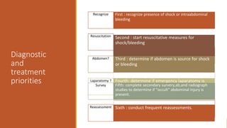
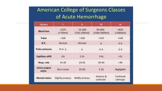
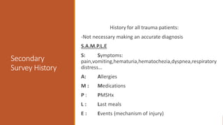
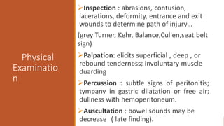
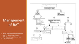
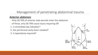
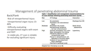
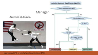
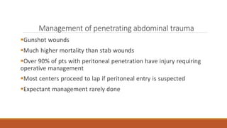
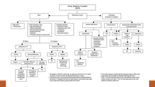
Management
Of
Abdominal
Trauma
BY : CHONG LIH YIN
Index
1. classification of abdominal injury
2. Pathophysiology of abdominal injury -PAT
, BAT
3. Primary Survery
4. Secondary Survey- Physical examination,Lab Test
5. Imaging –Plain radiography , FAST scan, CT
6. Other diagnosis method -DPL,LWE,,Laparascopy, Exploratory Laparatomy
7. Management of BAT and PAT
8. Specific Organ injury
9. -Spleen , Diaphragm, stomach , small intestine, Colorectal injury,
10. Damage control resuscitation
11. Abdominal compartment syndrome
12. Reference
Abdominal Trauma
Blunt Abdominal Trauma
◦Greater mortality than PAT (more difficult to diagnose, commonly associated with
trauma to multiple organs/systems)
◦Most commonly injured organs?
- spleen > liver, intestine is the most likely hollow viscus.
◦Most common causes?
- MVA (50 - 75% of cases) > blows to abdomen (15%) > falls (6 - 9%)
Penetrating Abdominal Trauma
◦Stabbing 3x more common than firearm wounds
◦Gun shot wound cause 90% of the deaths
◦Most commonly injured organs?
- small intestine > colon > liver
Pathophysiology of injury
Penetrating Abdominal
Trauma
Stab Wounds
◦Knives, ice picks, pens, coat
hangers, broken bottles
◦Liver, small bowel, spleen
Gunshot wounds
◦small bowel, colon and liver
◦Often multiple organ injuries,
bowel perforations
Rosen’s Emergency Medicine, 7th ed. 2009
Pathophysiology of injury
Rosen’s Emergency Medicine, 7th ed. 2009
Pathophysiology of injury
Blunt Abdominal Trauma
• Rupture or burst injury of a hollow organ by sudden rises in intra-abdominal
pressures
• Crushing effect
• Acceleration and deceleration forces → shear injury
• Seat belt injuries
◦ “seat belt sign” = highly correlated with intraperitoneal injury
Rosen’s Emergency Medicine, 7th ed. 2009
Primary Survey –ATLS approach
ABCDE pattern: Airway, Breathing, Circulation, Disability (neurologic status), and
Exposure.
A - intubation may be required if patient is shocked, hypotensive or unconscious or in need
for ventilation. *with cervical precaution.
B - watch for hemothorax in both blunt and penetrating thoracoabdominal injuries.
C - start with 2 L crystalloid (If active bleeding you must find source and stop the bleeding)
D – May seen associated with thorocolumbar #
E - Watch for other injury
Diagnostic
and
treatment
priorities
Recognize First : recognize presence of shock or intraabdominal
bleeding
Resuscitation Second : start resuscitative measures for
shock/bleeding
Abdomen? Third : determine if abdomen is source for shock
or bleeding
Laparatomy ? Fourth: determine if emergency laparatomy is
needed
Survey Fifth: complete secondary survery,ab,and radiograph
studies to determine if “occult” abdominal injury is
present.
Reassessment Sixth : conduct frequent reassessments.
Secondary
Survey History
History for all trauma patients:
-Not necessary making an accurate diagnosis
S.A.M.P.L.E
S: Symptoms:
pain,vomiting,hematuria,hematochezia,dyspnea,respiratory
distress…
A: Allergies
M : Medications
P : PMSHx
L : Last meals
E : Events (mechanism of injury)
Physical
Examinatio
n
Inspection : abrasions, contusion,
lacerations, deformity, entrance and exit
wounds to determine path of injury…
(grey Turner, Kehr, Balance,Cullen,seat belt
sign)
Palpation: elicits superficial , deep , or
rebound tenderness; involuntary muscle
duarding
Percussion : subtle signs of peritonitis;
tympany in gastric dilatation or free air;
dullness with hemoperitoneum.
Auscultation : bowel sounds may be
decrease ( late finding).
Physical examination
Grey-Turner sign : bluid discoloration of
lower flanks, lower back; associated with
retroperitoneal bleeding of
pancrease,kidney or pelvic fracture.
Cullen sign : bluish discoloration around
umbilicus, indicates peritoneal bleeding,
often pancreatic hemorrhage.
Kehr sign: shoulder pain while supine
;caused by diaphragmatic irritation(splenic
injury, free air, intra-abdominal bleeding)
Balance sign : dull percussion in
LUQ.Sign of splenic injury; blood
accumulation in subcapsular or
extracapsular spleen
In the trauma patient, a ‘normal’ physical exam of the abdomen
doesn’t equate to much. You NEED to do further testing.
Laboratory tests
- limited
-Hematocrit – below 30% increases the likelihood of intra-
abdominal injury.
-Leukocyte count – In BAT, the white blood cell (WBC) count is
nonspecific and of little value. Catecholamine release due to
trauma can cause demargination and may elevate the WBC to
12,000 to 20,000/mm3 with a moderate left shift. Solid or
hollow viscus injury can cause comparable elevations
-Pancreatic enzymes – Normal serum amylase and lipase
concentrations cannot exclude significant pancreatic injury . And
while elevated concentrations raise the possibility of pancreatic
injury,
-Liver function tests – Hepatic injury is associated with
elevations in liver transaminase concentrations
-Urinalysis – Gross hematuria suggests serious renal injury and
mandates further investigation
-Base deficit and lactate - Base deficit less than -6 was
associated with intra-abdominal hemorrhage and the need for
laparotomy and blood transfusion
• FAST ultrasound
• Diagnostic Peritoneal Tap
• CT Scan, contrast study
• Local wound exploration
• Angiography
• Urethrocystography
• IVU
Imaging in Abdominal Trauma
Plain films generally have NO
ROLE in acute abdominal
trauma
What else do we have?
Plain radiograph
Findings on chest radiograph that suggest
intra- abdominal injury include:
Lower rib fracture
•Diaphragmatic hernia
•Free air under the diaphragm
(FAST) Focused assessment with
sonography for trauma
- To diagnosed free intraperitoneal fluid.
- evaluate solid organ hematoma
- Four areas:
1. Pericardium (subxiphoid)
2.
3.
4.
Perihepatic &hepatorenal space (morrison’s pouch)
Perisplenic
Pelvis (pouch of Douglas /rectovesical pouch)
Sensitivity 60-95% for detecting 100ml -500 ml of fluid
E-fast(extended)
-add thoracic windows to look for pneumothorax.
Sensitivity 59%,specificity,specificity up to (99% for
pneumothorax. )
1 3
2
4
FAST Ultrasound
Advantages
• Sensitivity at detecting 100cc fluid is 60-
95%
• Portable(bedside),fast(<5 min) and ability
to repeat
• No radiation or contrast
• Noninvasive
• Rapid results, hemodynamically unstable
patient that unable to go for CT scan
• Less expensive
Disadvantages
• -Injury to solid parenchyma, the retroperitoneum,
or the diaphragm is not well seen.
• -Uncooperative patients, obesity, bowel gas, and
subcutaneous air interfere with image quality.
• -Low sensitivity in comparison to CT, particularly for
non-hypotensive patients. Cannot reliably exclude
clinically significant injuries
• -Blood cannot be distinguished from ascites or
urine.
• -Subcapsular injuries cannot be detected.
• -Insensitive for detecting bowel injury
• -Limited in detecting<200cc intraperitoneal fluid
Pericardium
(subxiphoid)
FAST-Morrison’s pouch (hepato-renal space)
Rosen’s Emergency Medicine, 7th ed. 2009
FAST Perisplenic view
FAST-Retrovesicle (Pouch of Douglas)
trauma.org
th
CT Imaging
◦ Accurate for solid visceral lesions and intraperitoneal hemorrhage
◦ guide nonoperative management of solid organ damage
◦ IV not oral contrast
◦ Disadvantages : insensitive for injury of the pancreas, diaphragm, small
bowel, and mesentery
Rosen’s Emergency Medicine, 7th ed. 2009
Diagnostic Peritoneal Taps
DPA - The recovery of 10 cc of frank blood (or more) from the
peritoneum is a strong predictor (90% PPV in blunt trauma) of
intraperitoneal injury, and the procedure is then terminated.
DPL - If aspiration findings are negative, lavage is conducted in which the
peritoneal cavity is washed with saline. RBC count exceeding
100,000/cc is considered positive and generally specific for injury.
Sensitivity 90%.
Diagnositic Peritoneal ‘Lavage’
Is actually a 2 Step Process.
Step 1. DPA (closed).
◦ Patient supine
◦ Landmark is 2 finger widths below umbilicus
◦ Local freezing, puncture skin 30-degrees to the head
◦ Seldinger technique to introduce a DPL catheter
◦ Aspirate using 30cc syringe
DPA
Advantages
◦ Highly accurate for hemoperitoneum (SENS 90-100%)
◦ Most sensitive test for hollow viscus injury
Disadvantages
◦ Invasive (complication rate 1-5%)
◦ Time consuming (20 minutes)
◦ False positives. Up to 25% non-therapeutic laparotomies
DPA
•If 10cc frank blood or more is aspirated, you are
done, patient needs to go to the OR.
If the DPA is negative, you proceed to Step 2…
Diagnostic Peritoneal Lavage
Step 2. DPL.
◦Hook up 1L of Ringer’s to the peritoneal catheter, and
squeeze into the abdomen.
◦Once infused, put the empty Ringer’s bag on the floor,
and let it back-fill via gravity
◦Send off 10cc for analysis, if 100,000 RBC/cc it is positive
Is there still a role for DPA?
FAST has largely replaced DPA, likely due to
ease of use.
However, 2 areas where still is warranted:
◦Hemodynamically unstable and an equivocal FAST
◦No FAST available
“DPL is safe, sensitive, and reduces the use of
CT” (Journal of Trauma 2007)
Local Wound Exploration
To determine the depth of penetration in stab wounds
If peritoneum is violated, must do more diagnostics
Prep, extend wound, carefully examine (No blind probing)
Indicated for anterior abdominal stab wounds, less clear for
other areas
Rosen’s Emergency Medicine, 7th ed. 2009
Laparoscopy
Most useful to eval penetrating wounds to thoracoabdominal
region in stable pt
esp for diaphragm injury: Sens 87.5%, specificity 100%
Can repair organs via the laparoscope
diaphragm, solid viscera, stomach, small bowel.
Disadvantages:
poor sensitivity for hollow visceral injury, retroperitoneum
Complications from trocar misplacement.
If diaphragm injury, PTX during insufflation
Exploratory laparatomy
Potential indications include the following:
Haemodynamic instability
 Evidence of Peritonitis to achieve control of haemorrhage and control of spillage
Traumatic diaphragmatic injury with herniation
 Severe solid organ injury (e.g. kidney and spleen)
 Infarction due to post traumatic occlusion of the blood supply
 Mesenteric tear/s
 Unexplained Moderate to large amounts of free fluid (200-≥500mls)
 Failed non-operative management
Management
of BAT
• NOM: nonoperative management
• Abd CT: abdominal CT scan;
• DPT: diagnostic peritoneal tap;
• LAP: laparotomy
Management of penetrating abdominal trauma
Mandatory laparotomy
vs
Selective nonoperative management
Management of penetrating abdominal trauma
Mandatory laparotomy
◦ standard of care for abdominal stab wounds until 1960s, for GSWs until
recently
◦ Now thought unnecessary in 70% of abdominal stab wounds
◦ Increased complication rates, length of stay, costs
◦ Immediate laparotomy indicated for shock, evisceration, and peritonitis
Management of penetrating abdominal trauma
Selective management used to reduce unnecessary
laparotomies
Diagnostic studies to determine if there is intraperitoneal injury
requiring operative repair
Strategy depends on abdominal region:
Thoracoabdomen
Nipple line to costal margin
Anterior abdomen
Xiphoid to pubis
Flank and back
Posterior to anterior axillary line
Management of penetrating abdominal trauma
Thoracoabdomen
Big concern is diaphragmatic injury
◦ 7% of thoracoabdominal wounds
Diagnostic evaluation:
◦ CXR (hemothorax or pneumothorax)
◦ Diagnostic peritoneal lavage
◦ FAST
◦ Thoracoscopy
Biffl et al. Management guidelines for penetrating abdominal trauma. Curr Opin Crit Care 2010;16:609-617
Thoracoabdomen
Management of penetrating abdominal trauma
Anterior abdomen
◦ Only 50-70% of anterior stab wounds enter the abdomen
◦ of these, only 50-70% cause injury requiring OR
1. is immediate lap indicated ?
2. Has peritoneal cavity been violated?
3. Is laparotomy required?
Management of penetrating abdominal trauma
Back/Flank
◦Risk of retroperitoneal injury
◦Intraperitoneal organ injury 15-
40%
◦Difficulty evaluating
retroperitoneal organs with exam
and FAST
◦In stable pts, CT scan is reliable
for excluding significant injury:
Biffl et al. Management guidelines for penetrating abdominal trauma. Curr Opin Crit Care 2010;16:609-617
Management of PAT
Anterior abdomen
laparoscopy (LPY), or serial physical examinations (SPEs)
Management of penetrating abdominal trauma
Gunshot wounds
Much higher mortality than stab wounds
Over 90% of pts with peritoneal penetration have injury requiring
operative management
Most centers proceed to lap if peritoneal entry is suspected
Expectant management rarely done
Management of PAT
Gunshot wounds
-assess peritoneal entry by missile path,
LWE, CT
, US, laparoscopy (all limited)
laparoscopy (LPY), or serial physical examinations (SPEs)
Specific Organ Injury
Specific organ
trauma:
1.peritoneal
2.retroperitoneal
3.diaphragm
-Treatment of an organ injury is
similar whether the injury
mechanism is penetrating or
blunt
-An exception to the rule is a
retroperitoneal hematoma
-explore all retroperitoneal
hematoma caused by
penetrating injury.
Splenic Injury - Grading System(AAST)
I.- Hematoma, subcapsular <10% SA Capsular Laceration <1cm
II.- Hematoma, subcapsular 10-50% SA; intraparenchymal
<5cm Capsular Laceration 1-3cm
III.- Hematoma, subcapsular >50% SA; intraparenchymal >5cm
Capsular Laceration >3cm (or parenchymal depth)
IV.- Hematoma ruptured into parenchyma Hilar Injury
devascularizing spleen >25%
V.- Vascular hilar injury devascularing spleen 100%, or
‘Shattered’
WSES classification
Minor spleen injuries:
WSES class I includes hemodynamically stable AAST-OIS grade I–II blunt and penetrating lesions.
Moderate spleen injuries:
WSES class II includes hemodynamically stable AAST-OIS grade III blunt and penetrating lesions.
WSES class III includes hemodynamically stable AAST-OIS grade IV–V blunt and penetrating
lesions.
Severe spleen injuries:
WSES class IV includes hemodynamically unstable AAST-OIS grade I–V blunt and penetrating
lesions.
Diaphragmatic injury
Its possible in injuries to the thoracoabdominal region
Can be due to blunt(>85%) or penetrating injury and is larger in the blunt
Possible cardiac injury if the penetrating wound is more central
The weakest point of diaphragm is the left posteriorlateral(80%)
Often missed in multitrauma
In isolated injury it may go unnoticed and there is often a delay between the injury and the diagnosis
Patients present with non specific symptoms and may complain of chest pain,abdominal pain,dyspnea
,tachypnea and cough
Rupture with herniation is diagnosed by CXR or CT but without herniation is difficult to diagnose
Thoracoscopy or laparascopy is diagnostic
Treatment
Once identified must be repaired because it will not close spontaneously regardless the size.
Early diagnosis needs abdominal approach using the interrupted nonabsorbable suture and
the large defect(>25cm2) may need nonabsorbable mesh.
In the event of a gross contamination, endogenous tissue can be utilized for a definitive
repair as latissimus dorsi flap, tensor fascia lata or omentum.
There are some who advocate using biologic tissue grafts, such as AlloDerm(human acellular
tissue matrix).The durability of such a repair is questionable.
Place chest tube on the surgery side at the time of repair
Stomach
More common in penetrating trauma than blunt &
its about 10% of penetrating injuries of the abdomen
Diagnosis:
Physical exam:
-epigastric tenderness,
-peritoneal signs,
-bloody gastric aspirate.
Plain radiography in <50%:
-free air under diaphragm
• FAST examination:-
unreliable
• DPL: WBC, RBC < Gross
contamination
• CT scan:
pneumoperitoneum
• Laparoscopy:-operator
dependent
Stomach treatment is according to the
severity
administer preop abx
Hematoma is evacuated ,hemostasis and closure with nonabsorbable suture.
Small perforation can be closed in one or two layered
Large injuries near the greater curvature can be closed by suture or GIA stapler
Certain defects may be closed using a TA stapler
A pyloric wound may be converted to pyloroplasty
Destructive wound may need proximal or distal gastrectomy
In rare cases a total gastrectomy and Roux-en –y esophagojejunostomy are necessary for severe
cases.
Small intestine
The small bowel is the mc injured intraabdominal organ in penetrating
tauma, a blunt trauma cause is less common,but not rare(10%)
Small isolated perforation probably result from blowout of
pseudoclosed loops(seatbelt related injuries)
Larger perforation, complete disruptions and injuries associated with
large mesenteric hematoma or laceration are caused by direct blows or
shearing injury or contusion
Perforation from blunt injury is the mc at the ligament of triez,ileocecal
valve,midjejunum or in the areas of adhesion
Small intestinee
•CT has a significant false negative rate in the diagnosis of small-
bowel injury.
•CT findings in small-bowel injury include:
Fluid collections without solid viscus injury
Bowel wall thickening
Mesenteric infiltration
Free intraperitoneal air
Oral contrast extravasation
Colon and rectum
-Diagnosis
• Peritoneal signs or free intraperitoneal air.
• At laparotomy, small injuries in the wall of the colon can be missed so explore all blood staining
or hematomas of the colonic wall.
• Consider proctoscopy or proctosigmoidescopy in :
- Gross blood on PR in the presence of a pelvic fracture
- Penetrating abdominal, buttock, thigh or pelvic wound.
- Any patient with a major pelvic fracture if the patient is stable.
• The location of the injury can be important in planning the operation. Even if the hole cannot
be visualized on proctoscopy, assume the patient has a colorectal injury, if there is intraluminal
blood.
• In hemodynamically unstable patients, proceed with laparotomy first.
Colon
and
rectum
Current operative options include :
-Primary repair of the injury,
-.Resection and anastomosis, and
-Colostomy..
Traditional contraindications to primary repair include :
• Patients with shock, underlying disease, significant associated injuries, or peritonitis
• Extensive intraperitoneal spillage of feces,
• Multisegmental or extensive colonic injury requiring resection, and
• Major loss of the abdominal wall or mesh repair of the abdominal wall; Colon and rectum
Treatment is operative
If a primary repair cannot be
performed safely for anatomic
reasons (bowel wall edema,
vascular compromise), a
colostomy may be a safer option.
The guidelines for primary repair include :
• Minimal fecal spillage,
• No shock (defined as systolic blood pressure <90 mmHg),
• Minimal associated intraabdominal injuries,
• <8-hour delay in diagnosis and treatment, and
• <1-L blood transfusion.
Colon and rectum
Rectum -intraperitoneal or extraperitoneal
1.Often, intraperitoneal rectal injuries can be managed as in colonic injury (primarily
repaired). 2.Treat extraperitoneal rectal tears by diverting sigmoid colostomy. Acceptable
options include:
• Hartmann resection with end colostomy,
•End colostomy with a mucus fistula, or
•Loop colostomy with a stapled distal end.
3.If the defect is not readily identified on proctoscopy…..
4.Presacral drainage and irrigation of the distal rectal stump…..
5.If a colostomy is necessary in a patient with a pelvic fracture requiring fixation……
6.Perioperative broad-spectrum antibiotics should be administered for colon and rectal
wounds
Damage
control
Resuscitatio
n
-can be applied to unstable
patient who are with life
threatening hemorrhage &
going to need massive
transfusion.
It’s an alternative resuscitation approach to hemmorhagic
shock which involves:
1.rapid control of surgical bleeding
2.Early and increased use of RBC, plasma and platelets in
a 1:1:1 ratio.
3.limitation of excessive crystalloid use
4.prevention and treatment of
hypothermia,hypocalcemia and acidosis.
5.Permissive hypotension. (hypotensive resuscitation
strategies).
Approach
Before:
ER->OR ->death
Now:
ER->OR->DCS->ICU->OR->ICU
Major complication of abdominal
trauma- Abdominal Compartment
Syndrome
Common problem with abdominal trauma
Definition: elevated intraabdominal pressure (IAP) of ≥20 mm Hg,
with single or multiple organ system failure
◦± APP below 50 mm Hg
Primary ACS: associated with injury/disease in abdomen
Secondary (“medical”) ACS: due to problems outside the abdomen
(eg sepsis, capillary leak)
Sugrue M. Abdominal compartment syndrome. Curr Opin Crit Care 2005; 11:333-338
Abdominal Compartment Syndrome
Bailey J, Shapiro M. Abdominal compartment syndrome. Crit Care 2000, 4:23–29
Abdominal Compartment Syndrome
Effects of elevated IAP
◦Renal dysfunction
◦ Decreased cardiac output
◦Increased airway
pressures and decreased
compliance
◦ Visceral hypoperfusion
Sugrue M. Abdominal compartment syndrome. Curr Opin Crit Care 2005; 11:333-338
Abdominal Compartment Syndrome
Management
◦Surgical abdominal decompression
◦Nonsurgical: paracentesis, NGT,
sedation
◦Staged approach to abdominal
repair
◦Temporary abdominal closure
Conclusions
Watch out for implements and missiles violating the abdomen
Laparotomy is mandatory if shock, evisceration, or peritonitis
Diagnostic studies used to determine need for laparotomy in PAT and BAT
FAST is noninvasive, quick and accurate way to evaluate for intraperitoneal
blood
Damage Control is a principle of staged operative management with
control and resuscitation prior to definitive repair
Abdominal compartment syndrome is a common problem in abdominal
trauma
Reference
Biffl et al. Management guidelines for penetrating abdominal trauma. Curr Opin Crit Care
2010;16:609-617
https://wjes.biomedcentral.com/articles/10.1186/s13017-017-0151-4#
http://www.aast.org/library/traumatools/injuryscoringscales.aspx#pancreas
https://www.uptodate.com/contents/initial-evaluation-and-management-of-blunt-abdominal-trauma-
in-
adults?search=abdominal%20trauma&source=search_result&selectedTitle=1~150&usage_type=d
efault&display_rank=1#subscribeMessage

abdominal trauma.ppt

  • 1.
  • 2.
    Index 1. classification ofabdominal injury 2. Pathophysiology of abdominal injury -PAT , BAT 3. Primary Survery 4. Secondary Survey- Physical examination,Lab Test 5. Imaging –Plain radiography , FAST scan, CT 6. Other diagnosis method -DPL,LWE,,Laparascopy, Exploratory Laparatomy 7. Management of BAT and PAT 8. Specific Organ injury 9. -Spleen , Diaphragm, stomach , small intestine, Colorectal injury, 10. Damage control resuscitation 11. Abdominal compartment syndrome 12. Reference
  • 4.
    Abdominal Trauma Blunt AbdominalTrauma ◦Greater mortality than PAT (more difficult to diagnose, commonly associated with trauma to multiple organs/systems) ◦Most commonly injured organs? - spleen > liver, intestine is the most likely hollow viscus. ◦Most common causes? - MVA (50 - 75% of cases) > blows to abdomen (15%) > falls (6 - 9%) Penetrating Abdominal Trauma ◦Stabbing 3x more common than firearm wounds ◦Gun shot wound cause 90% of the deaths ◦Most commonly injured organs? - small intestine > colon > liver
  • 5.
    Pathophysiology of injury PenetratingAbdominal Trauma Stab Wounds ◦Knives, ice picks, pens, coat hangers, broken bottles ◦Liver, small bowel, spleen Gunshot wounds ◦small bowel, colon and liver ◦Often multiple organ injuries, bowel perforations Rosen’s Emergency Medicine, 7th ed. 2009
  • 6.
    Pathophysiology of injury Rosen’sEmergency Medicine, 7th ed. 2009
  • 7.
    Pathophysiology of injury BluntAbdominal Trauma • Rupture or burst injury of a hollow organ by sudden rises in intra-abdominal pressures • Crushing effect • Acceleration and deceleration forces → shear injury • Seat belt injuries ◦ “seat belt sign” = highly correlated with intraperitoneal injury Rosen’s Emergency Medicine, 7th ed. 2009
  • 8.
    Primary Survey –ATLSapproach ABCDE pattern: Airway, Breathing, Circulation, Disability (neurologic status), and Exposure. A - intubation may be required if patient is shocked, hypotensive or unconscious or in need for ventilation. *with cervical precaution. B - watch for hemothorax in both blunt and penetrating thoracoabdominal injuries. C - start with 2 L crystalloid (If active bleeding you must find source and stop the bleeding) D – May seen associated with thorocolumbar # E - Watch for other injury
  • 9.
    Diagnostic and treatment priorities Recognize First :recognize presence of shock or intraabdominal bleeding Resuscitation Second : start resuscitative measures for shock/bleeding Abdomen? Third : determine if abdomen is source for shock or bleeding Laparatomy ? Fourth: determine if emergency laparatomy is needed Survey Fifth: complete secondary survery,ab,and radiograph studies to determine if “occult” abdominal injury is present. Reassessment Sixth : conduct frequent reassessments.
  • 11.
    Secondary Survey History History forall trauma patients: -Not necessary making an accurate diagnosis S.A.M.P.L.E S: Symptoms: pain,vomiting,hematuria,hematochezia,dyspnea,respiratory distress… A: Allergies M : Medications P : PMSHx L : Last meals E : Events (mechanism of injury)
  • 12.
    Physical Examinatio n Inspection : abrasions,contusion, lacerations, deformity, entrance and exit wounds to determine path of injury… (grey Turner, Kehr, Balance,Cullen,seat belt sign) Palpation: elicits superficial , deep , or rebound tenderness; involuntary muscle duarding Percussion : subtle signs of peritonitis; tympany in gastric dilatation or free air; dullness with hemoperitoneum. Auscultation : bowel sounds may be decrease ( late finding).
  • 13.
    Physical examination Grey-Turner sign: bluid discoloration of lower flanks, lower back; associated with retroperitoneal bleeding of pancrease,kidney or pelvic fracture. Cullen sign : bluish discoloration around umbilicus, indicates peritoneal bleeding, often pancreatic hemorrhage. Kehr sign: shoulder pain while supine ;caused by diaphragmatic irritation(splenic injury, free air, intra-abdominal bleeding) Balance sign : dull percussion in LUQ.Sign of splenic injury; blood accumulation in subcapsular or extracapsular spleen In the trauma patient, a ‘normal’ physical exam of the abdomen doesn’t equate to much. You NEED to do further testing.
  • 14.
    Laboratory tests - limited -Hematocrit– below 30% increases the likelihood of intra- abdominal injury. -Leukocyte count – In BAT, the white blood cell (WBC) count is nonspecific and of little value. Catecholamine release due to trauma can cause demargination and may elevate the WBC to 12,000 to 20,000/mm3 with a moderate left shift. Solid or hollow viscus injury can cause comparable elevations -Pancreatic enzymes – Normal serum amylase and lipase concentrations cannot exclude significant pancreatic injury . And while elevated concentrations raise the possibility of pancreatic injury, -Liver function tests – Hepatic injury is associated with elevations in liver transaminase concentrations -Urinalysis – Gross hematuria suggests serious renal injury and mandates further investigation -Base deficit and lactate - Base deficit less than -6 was associated with intra-abdominal hemorrhage and the need for laparotomy and blood transfusion
  • 15.
    • FAST ultrasound •Diagnostic Peritoneal Tap • CT Scan, contrast study • Local wound exploration • Angiography • Urethrocystography • IVU Imaging in Abdominal Trauma Plain films generally have NO ROLE in acute abdominal trauma What else do we have?
  • 16.
    Plain radiograph Findings onchest radiograph that suggest intra- abdominal injury include: Lower rib fracture •Diaphragmatic hernia •Free air under the diaphragm
  • 17.
    (FAST) Focused assessmentwith sonography for trauma - To diagnosed free intraperitoneal fluid. - evaluate solid organ hematoma - Four areas: 1. Pericardium (subxiphoid) 2. 3. 4. Perihepatic &hepatorenal space (morrison’s pouch) Perisplenic Pelvis (pouch of Douglas /rectovesical pouch) Sensitivity 60-95% for detecting 100ml -500 ml of fluid E-fast(extended) -add thoracic windows to look for pneumothorax. Sensitivity 59%,specificity,specificity up to (99% for pneumothorax. ) 1 3 2 4
  • 18.
    FAST Ultrasound Advantages • Sensitivityat detecting 100cc fluid is 60- 95% • Portable(bedside),fast(<5 min) and ability to repeat • No radiation or contrast • Noninvasive • Rapid results, hemodynamically unstable patient that unable to go for CT scan • Less expensive Disadvantages • -Injury to solid parenchyma, the retroperitoneum, or the diaphragm is not well seen. • -Uncooperative patients, obesity, bowel gas, and subcutaneous air interfere with image quality. • -Low sensitivity in comparison to CT, particularly for non-hypotensive patients. Cannot reliably exclude clinically significant injuries • -Blood cannot be distinguished from ascites or urine. • -Subcapsular injuries cannot be detected. • -Insensitive for detecting bowel injury • -Limited in detecting<200cc intraperitoneal fluid
  • 19.
  • 20.
    FAST-Morrison’s pouch (hepato-renalspace) Rosen’s Emergency Medicine, 7th ed. 2009
  • 21.
  • 22.
    FAST-Retrovesicle (Pouch ofDouglas) trauma.org th
  • 23.
    CT Imaging ◦ Accuratefor solid visceral lesions and intraperitoneal hemorrhage ◦ guide nonoperative management of solid organ damage ◦ IV not oral contrast ◦ Disadvantages : insensitive for injury of the pancreas, diaphragm, small bowel, and mesentery Rosen’s Emergency Medicine, 7th ed. 2009
  • 24.
    Diagnostic Peritoneal Taps DPA- The recovery of 10 cc of frank blood (or more) from the peritoneum is a strong predictor (90% PPV in blunt trauma) of intraperitoneal injury, and the procedure is then terminated. DPL - If aspiration findings are negative, lavage is conducted in which the peritoneal cavity is washed with saline. RBC count exceeding 100,000/cc is considered positive and generally specific for injury. Sensitivity 90%.
  • 25.
    Diagnositic Peritoneal ‘Lavage’ Isactually a 2 Step Process. Step 1. DPA (closed). ◦ Patient supine ◦ Landmark is 2 finger widths below umbilicus ◦ Local freezing, puncture skin 30-degrees to the head ◦ Seldinger technique to introduce a DPL catheter ◦ Aspirate using 30cc syringe
  • 28.
    DPA Advantages ◦ Highly accuratefor hemoperitoneum (SENS 90-100%) ◦ Most sensitive test for hollow viscus injury Disadvantages ◦ Invasive (complication rate 1-5%) ◦ Time consuming (20 minutes) ◦ False positives. Up to 25% non-therapeutic laparotomies
  • 29.
    DPA •If 10cc frankblood or more is aspirated, you are done, patient needs to go to the OR. If the DPA is negative, you proceed to Step 2…
  • 30.
    Diagnostic Peritoneal Lavage Step2. DPL. ◦Hook up 1L of Ringer’s to the peritoneal catheter, and squeeze into the abdomen. ◦Once infused, put the empty Ringer’s bag on the floor, and let it back-fill via gravity ◦Send off 10cc for analysis, if 100,000 RBC/cc it is positive
  • 31.
    Is there stilla role for DPA? FAST has largely replaced DPA, likely due to ease of use. However, 2 areas where still is warranted: ◦Hemodynamically unstable and an equivocal FAST ◦No FAST available “DPL is safe, sensitive, and reduces the use of CT” (Journal of Trauma 2007)
  • 32.
    Local Wound Exploration Todetermine the depth of penetration in stab wounds If peritoneum is violated, must do more diagnostics Prep, extend wound, carefully examine (No blind probing) Indicated for anterior abdominal stab wounds, less clear for other areas Rosen’s Emergency Medicine, 7th ed. 2009
  • 33.
    Laparoscopy Most useful toeval penetrating wounds to thoracoabdominal region in stable pt esp for diaphragm injury: Sens 87.5%, specificity 100% Can repair organs via the laparoscope diaphragm, solid viscera, stomach, small bowel. Disadvantages: poor sensitivity for hollow visceral injury, retroperitoneum Complications from trocar misplacement. If diaphragm injury, PTX during insufflation
  • 34.
    Exploratory laparatomy Potential indicationsinclude the following: Haemodynamic instability  Evidence of Peritonitis to achieve control of haemorrhage and control of spillage Traumatic diaphragmatic injury with herniation  Severe solid organ injury (e.g. kidney and spleen)  Infarction due to post traumatic occlusion of the blood supply  Mesenteric tear/s  Unexplained Moderate to large amounts of free fluid (200-≥500mls)  Failed non-operative management
  • 35.
    Management of BAT • NOM:nonoperative management • Abd CT: abdominal CT scan; • DPT: diagnostic peritoneal tap; • LAP: laparotomy
  • 36.
    Management of penetratingabdominal trauma Mandatory laparotomy vs Selective nonoperative management
  • 37.
    Management of penetratingabdominal trauma Mandatory laparotomy ◦ standard of care for abdominal stab wounds until 1960s, for GSWs until recently ◦ Now thought unnecessary in 70% of abdominal stab wounds ◦ Increased complication rates, length of stay, costs ◦ Immediate laparotomy indicated for shock, evisceration, and peritonitis
  • 38.
    Management of penetratingabdominal trauma Selective management used to reduce unnecessary laparotomies Diagnostic studies to determine if there is intraperitoneal injury requiring operative repair Strategy depends on abdominal region: Thoracoabdomen Nipple line to costal margin Anterior abdomen Xiphoid to pubis Flank and back Posterior to anterior axillary line
  • 39.
    Management of penetratingabdominal trauma Thoracoabdomen Big concern is diaphragmatic injury ◦ 7% of thoracoabdominal wounds Diagnostic evaluation: ◦ CXR (hemothorax or pneumothorax) ◦ Diagnostic peritoneal lavage ◦ FAST ◦ Thoracoscopy Biffl et al. Management guidelines for penetrating abdominal trauma. Curr Opin Crit Care 2010;16:609-617
  • 40.
  • 41.
    Management of penetratingabdominal trauma Anterior abdomen ◦ Only 50-70% of anterior stab wounds enter the abdomen ◦ of these, only 50-70% cause injury requiring OR 1. is immediate lap indicated ? 2. Has peritoneal cavity been violated? 3. Is laparotomy required?
  • 42.
    Management of penetratingabdominal trauma Back/Flank ◦Risk of retroperitoneal injury ◦Intraperitoneal organ injury 15- 40% ◦Difficulty evaluating retroperitoneal organs with exam and FAST ◦In stable pts, CT scan is reliable for excluding significant injury: Biffl et al. Management guidelines for penetrating abdominal trauma. Curr Opin Crit Care 2010;16:609-617
  • 43.
    Management of PAT Anteriorabdomen laparoscopy (LPY), or serial physical examinations (SPEs)
  • 44.
    Management of penetratingabdominal trauma Gunshot wounds Much higher mortality than stab wounds Over 90% of pts with peritoneal penetration have injury requiring operative management Most centers proceed to lap if peritoneal entry is suspected Expectant management rarely done
  • 45.
    Management of PAT Gunshotwounds -assess peritoneal entry by missile path, LWE, CT , US, laparoscopy (all limited) laparoscopy (LPY), or serial physical examinations (SPEs)
  • 46.
    Specific Organ Injury Specificorgan trauma: 1.peritoneal 2.retroperitoneal 3.diaphragm -Treatment of an organ injury is similar whether the injury mechanism is penetrating or blunt -An exception to the rule is a retroperitoneal hematoma -explore all retroperitoneal hematoma caused by penetrating injury.
  • 47.
    Splenic Injury -Grading System(AAST) I.- Hematoma, subcapsular <10% SA Capsular Laceration <1cm II.- Hematoma, subcapsular 10-50% SA; intraparenchymal <5cm Capsular Laceration 1-3cm III.- Hematoma, subcapsular >50% SA; intraparenchymal >5cm Capsular Laceration >3cm (or parenchymal depth) IV.- Hematoma ruptured into parenchyma Hilar Injury devascularizing spleen >25% V.- Vascular hilar injury devascularing spleen 100%, or ‘Shattered’
  • 48.
    WSES classification Minor spleeninjuries: WSES class I includes hemodynamically stable AAST-OIS grade I–II blunt and penetrating lesions. Moderate spleen injuries: WSES class II includes hemodynamically stable AAST-OIS grade III blunt and penetrating lesions. WSES class III includes hemodynamically stable AAST-OIS grade IV–V blunt and penetrating lesions. Severe spleen injuries: WSES class IV includes hemodynamically unstable AAST-OIS grade I–V blunt and penetrating lesions.
  • 50.
    Diaphragmatic injury Its possiblein injuries to the thoracoabdominal region Can be due to blunt(>85%) or penetrating injury and is larger in the blunt Possible cardiac injury if the penetrating wound is more central The weakest point of diaphragm is the left posteriorlateral(80%) Often missed in multitrauma In isolated injury it may go unnoticed and there is often a delay between the injury and the diagnosis Patients present with non specific symptoms and may complain of chest pain,abdominal pain,dyspnea ,tachypnea and cough Rupture with herniation is diagnosed by CXR or CT but without herniation is difficult to diagnose Thoracoscopy or laparascopy is diagnostic
  • 51.
    Treatment Once identified mustbe repaired because it will not close spontaneously regardless the size. Early diagnosis needs abdominal approach using the interrupted nonabsorbable suture and the large defect(>25cm2) may need nonabsorbable mesh. In the event of a gross contamination, endogenous tissue can be utilized for a definitive repair as latissimus dorsi flap, tensor fascia lata or omentum. There are some who advocate using biologic tissue grafts, such as AlloDerm(human acellular tissue matrix).The durability of such a repair is questionable. Place chest tube on the surgery side at the time of repair
  • 52.
    Stomach More common inpenetrating trauma than blunt & its about 10% of penetrating injuries of the abdomen Diagnosis: Physical exam: -epigastric tenderness, -peritoneal signs, -bloody gastric aspirate. Plain radiography in <50%: -free air under diaphragm • FAST examination:- unreliable • DPL: WBC, RBC < Gross contamination • CT scan: pneumoperitoneum • Laparoscopy:-operator dependent
  • 53.
    Stomach treatment isaccording to the severity administer preop abx Hematoma is evacuated ,hemostasis and closure with nonabsorbable suture. Small perforation can be closed in one or two layered Large injuries near the greater curvature can be closed by suture or GIA stapler Certain defects may be closed using a TA stapler A pyloric wound may be converted to pyloroplasty Destructive wound may need proximal or distal gastrectomy In rare cases a total gastrectomy and Roux-en –y esophagojejunostomy are necessary for severe cases.
  • 54.
    Small intestine The smallbowel is the mc injured intraabdominal organ in penetrating tauma, a blunt trauma cause is less common,but not rare(10%) Small isolated perforation probably result from blowout of pseudoclosed loops(seatbelt related injuries) Larger perforation, complete disruptions and injuries associated with large mesenteric hematoma or laceration are caused by direct blows or shearing injury or contusion Perforation from blunt injury is the mc at the ligament of triez,ileocecal valve,midjejunum or in the areas of adhesion
  • 55.
    Small intestinee •CT hasa significant false negative rate in the diagnosis of small- bowel injury. •CT findings in small-bowel injury include: Fluid collections without solid viscus injury Bowel wall thickening Mesenteric infiltration Free intraperitoneal air Oral contrast extravasation
  • 57.
    Colon and rectum -Diagnosis •Peritoneal signs or free intraperitoneal air. • At laparotomy, small injuries in the wall of the colon can be missed so explore all blood staining or hematomas of the colonic wall. • Consider proctoscopy or proctosigmoidescopy in : - Gross blood on PR in the presence of a pelvic fracture - Penetrating abdominal, buttock, thigh or pelvic wound. - Any patient with a major pelvic fracture if the patient is stable. • The location of the injury can be important in planning the operation. Even if the hole cannot be visualized on proctoscopy, assume the patient has a colorectal injury, if there is intraluminal blood. • In hemodynamically unstable patients, proceed with laparotomy first.
  • 58.
    Colon and rectum Current operative optionsinclude : -Primary repair of the injury, -.Resection and anastomosis, and -Colostomy..
  • 59.
    Traditional contraindications toprimary repair include : • Patients with shock, underlying disease, significant associated injuries, or peritonitis • Extensive intraperitoneal spillage of feces, • Multisegmental or extensive colonic injury requiring resection, and • Major loss of the abdominal wall or mesh repair of the abdominal wall; Colon and rectum Treatment is operative If a primary repair cannot be performed safely for anatomic reasons (bowel wall edema, vascular compromise), a colostomy may be a safer option. The guidelines for primary repair include : • Minimal fecal spillage, • No shock (defined as systolic blood pressure <90 mmHg), • Minimal associated intraabdominal injuries, • <8-hour delay in diagnosis and treatment, and • <1-L blood transfusion. Colon and rectum
  • 60.
    Rectum -intraperitoneal orextraperitoneal 1.Often, intraperitoneal rectal injuries can be managed as in colonic injury (primarily repaired). 2.Treat extraperitoneal rectal tears by diverting sigmoid colostomy. Acceptable options include: • Hartmann resection with end colostomy, •End colostomy with a mucus fistula, or •Loop colostomy with a stapled distal end. 3.If the defect is not readily identified on proctoscopy….. 4.Presacral drainage and irrigation of the distal rectal stump….. 5.If a colostomy is necessary in a patient with a pelvic fracture requiring fixation…… 6.Perioperative broad-spectrum antibiotics should be administered for colon and rectal wounds
  • 61.
    Damage control Resuscitatio n -can be appliedto unstable patient who are with life threatening hemorrhage & going to need massive transfusion. It’s an alternative resuscitation approach to hemmorhagic shock which involves: 1.rapid control of surgical bleeding 2.Early and increased use of RBC, plasma and platelets in a 1:1:1 ratio. 3.limitation of excessive crystalloid use 4.prevention and treatment of hypothermia,hypocalcemia and acidosis. 5.Permissive hypotension. (hypotensive resuscitation strategies).
  • 63.
  • 65.
    Major complication ofabdominal trauma- Abdominal Compartment Syndrome Common problem with abdominal trauma Definition: elevated intraabdominal pressure (IAP) of ≥20 mm Hg, with single or multiple organ system failure ◦± APP below 50 mm Hg Primary ACS: associated with injury/disease in abdomen Secondary (“medical”) ACS: due to problems outside the abdomen (eg sepsis, capillary leak) Sugrue M. Abdominal compartment syndrome. Curr Opin Crit Care 2005; 11:333-338
  • 66.
    Abdominal Compartment Syndrome BaileyJ, Shapiro M. Abdominal compartment syndrome. Crit Care 2000, 4:23–29
  • 67.
    Abdominal Compartment Syndrome Effectsof elevated IAP ◦Renal dysfunction ◦ Decreased cardiac output ◦Increased airway pressures and decreased compliance ◦ Visceral hypoperfusion Sugrue M. Abdominal compartment syndrome. Curr Opin Crit Care 2005; 11:333-338
  • 68.
    Abdominal Compartment Syndrome Management ◦Surgicalabdominal decompression ◦Nonsurgical: paracentesis, NGT, sedation ◦Staged approach to abdominal repair ◦Temporary abdominal closure
  • 69.
    Conclusions Watch out forimplements and missiles violating the abdomen Laparotomy is mandatory if shock, evisceration, or peritonitis Diagnostic studies used to determine need for laparotomy in PAT and BAT FAST is noninvasive, quick and accurate way to evaluate for intraperitoneal blood Damage Control is a principle of staged operative management with control and resuscitation prior to definitive repair Abdominal compartment syndrome is a common problem in abdominal trauma
  • 71.
    Reference Biffl et al.Management guidelines for penetrating abdominal trauma. Curr Opin Crit Care 2010;16:609-617 https://wjes.biomedcentral.com/articles/10.1186/s13017-017-0151-4# http://www.aast.org/library/traumatools/injuryscoringscales.aspx#pancreas https://www.uptodate.com/contents/initial-evaluation-and-management-of-blunt-abdominal-trauma- in- adults?search=abdominal%20trauma&source=search_result&selectedTitle=1~150&usage_type=d efault&display_rank=1#subscribeMessage