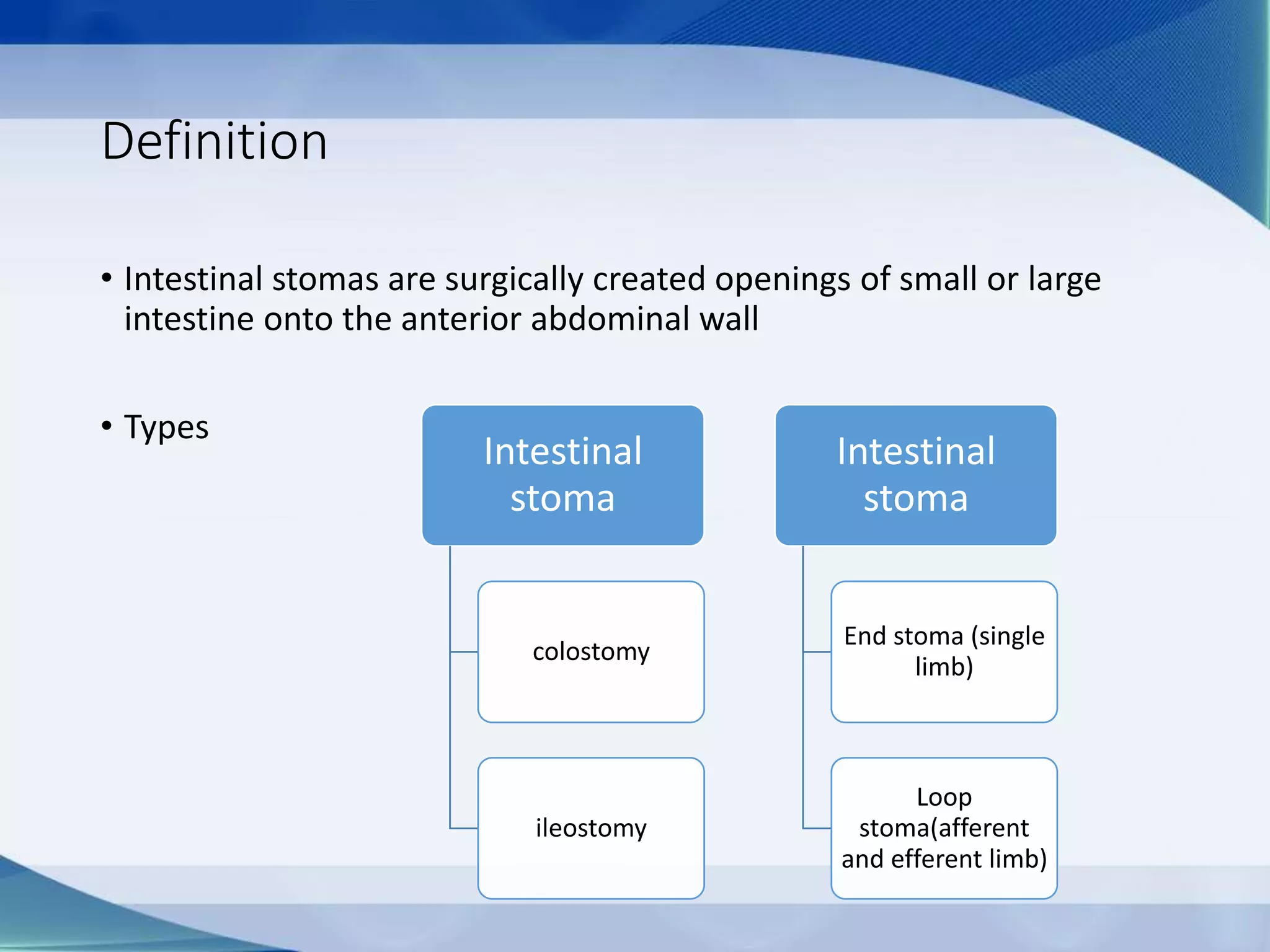
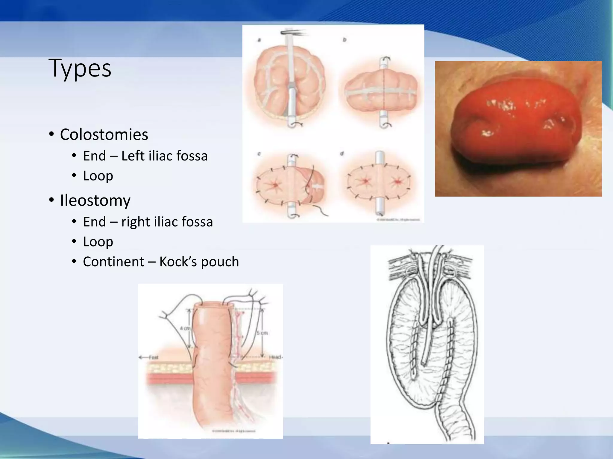
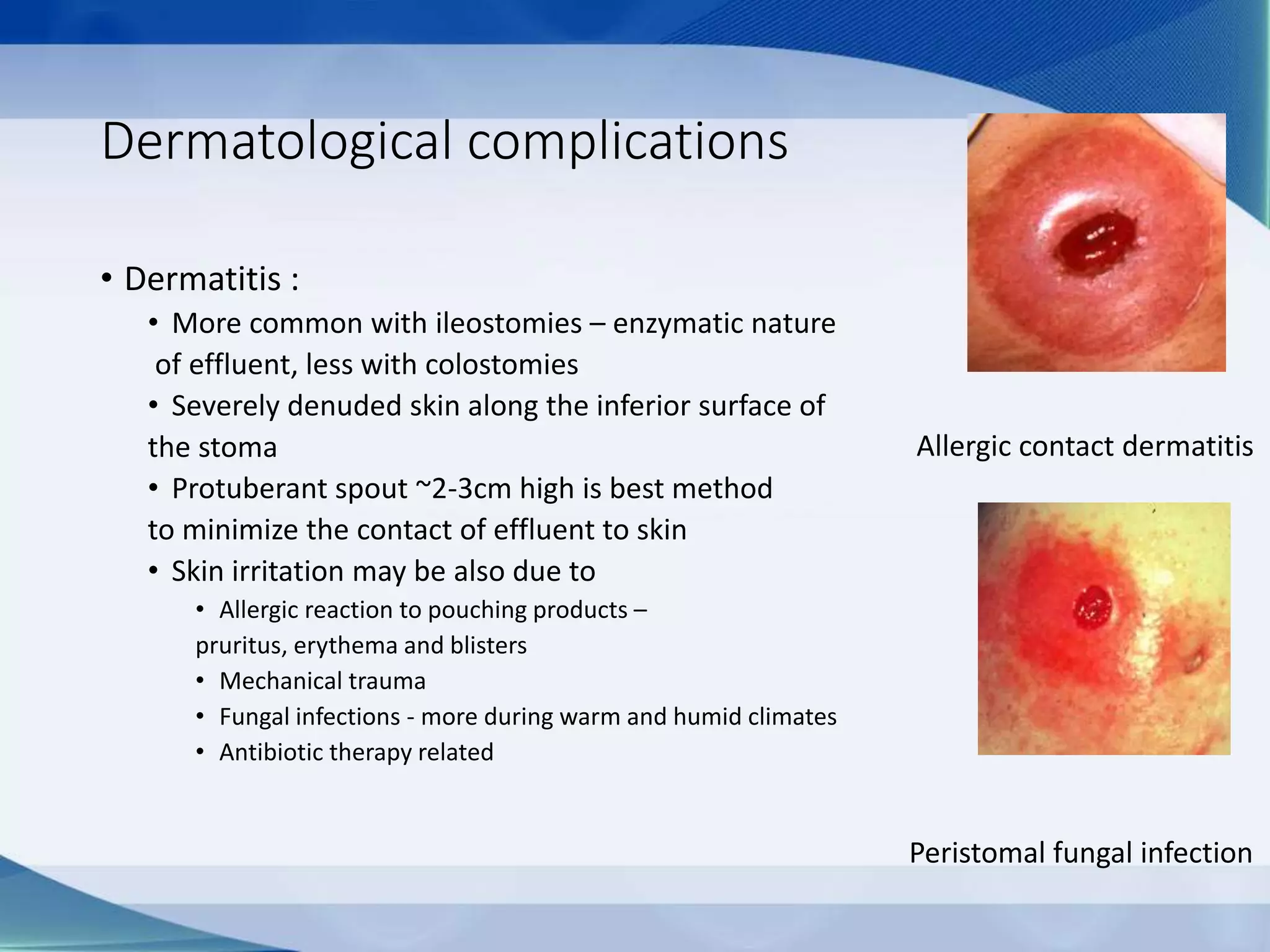
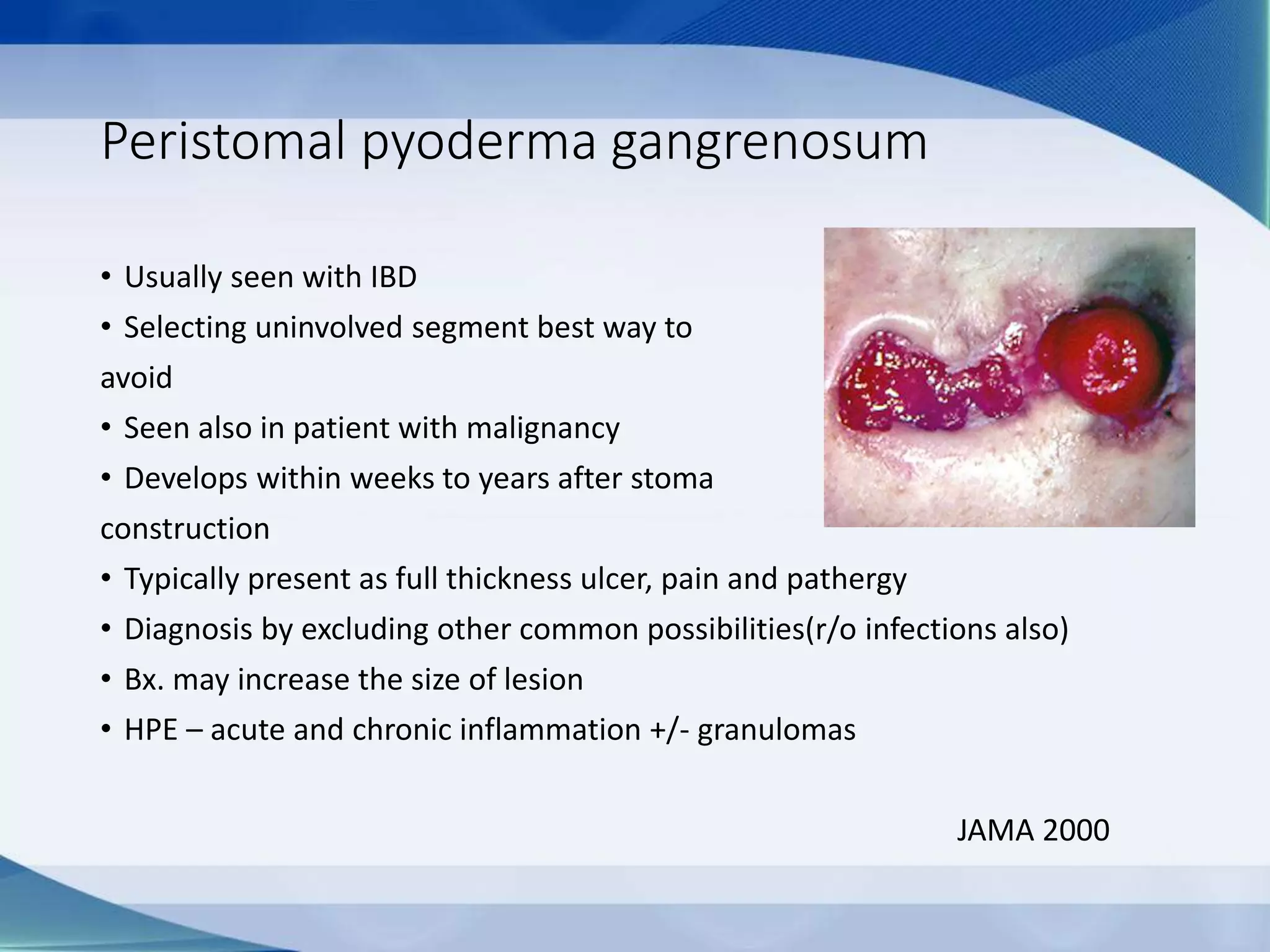
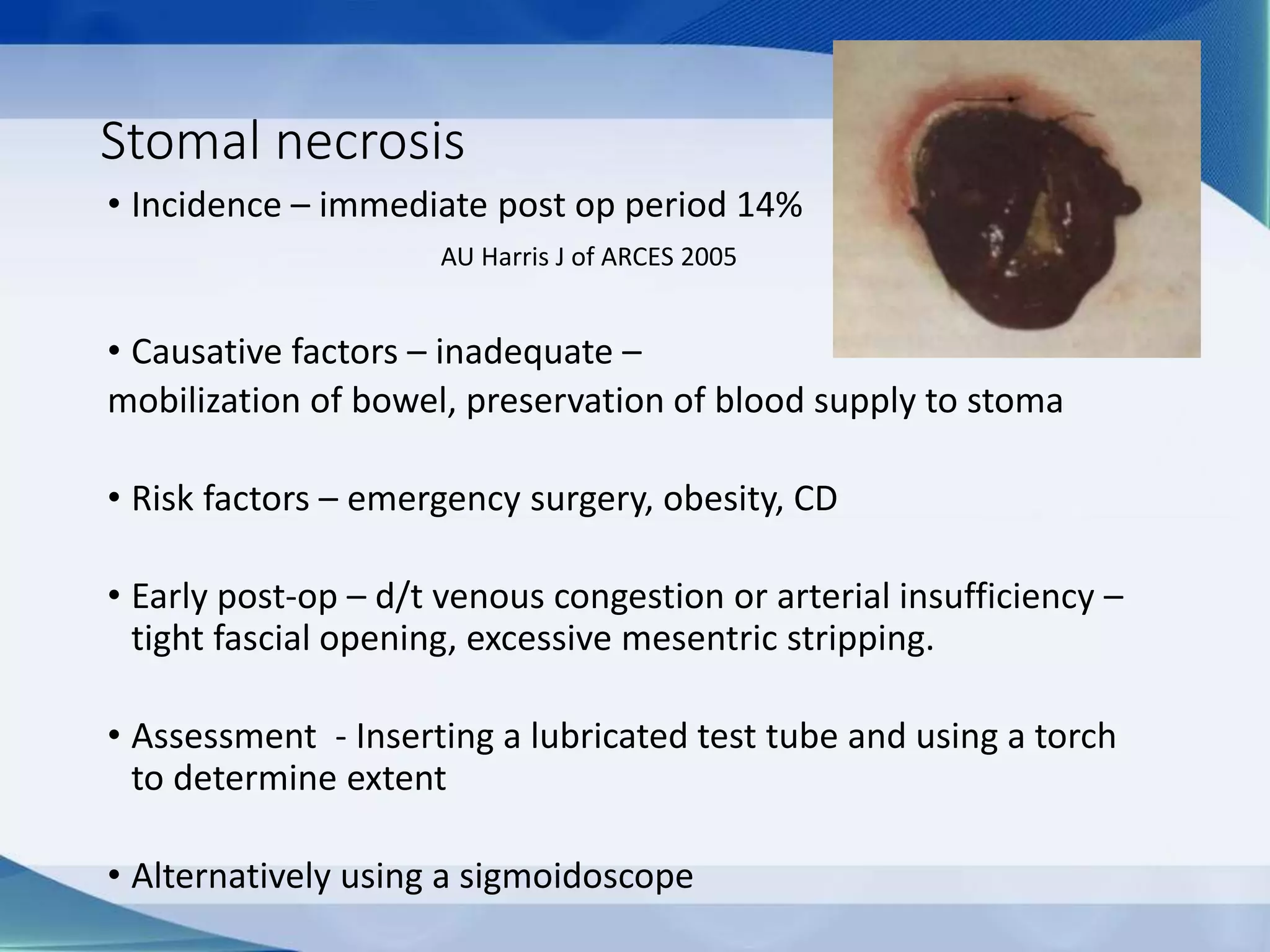
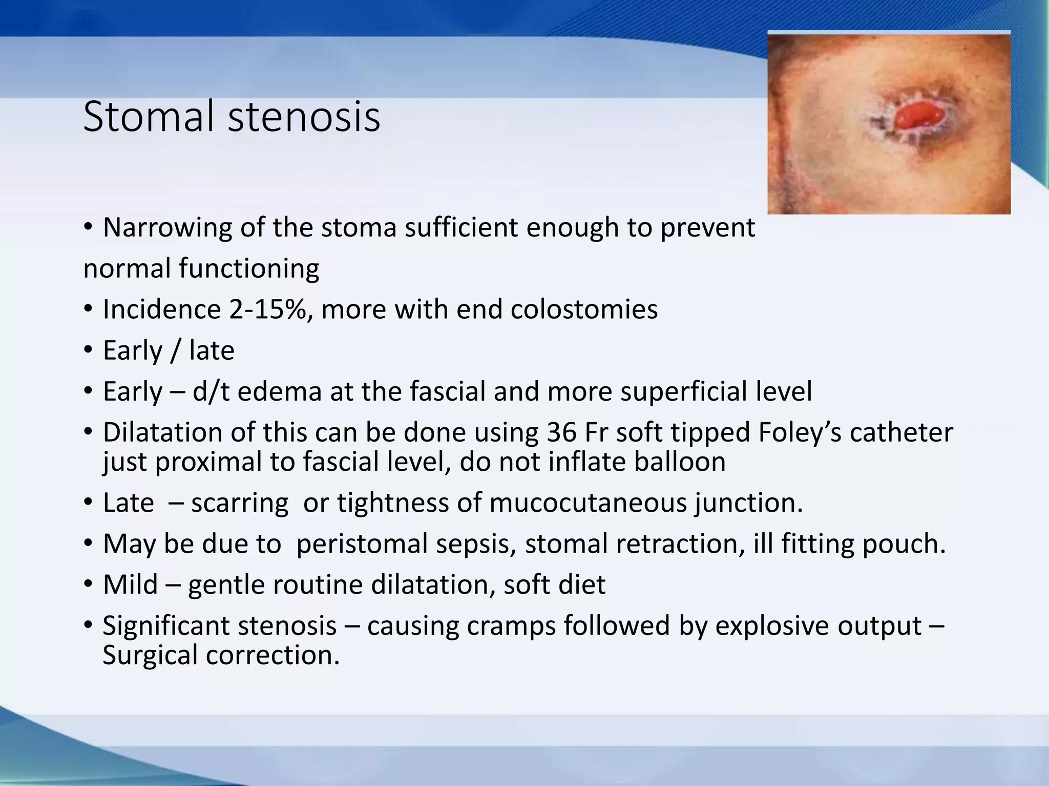
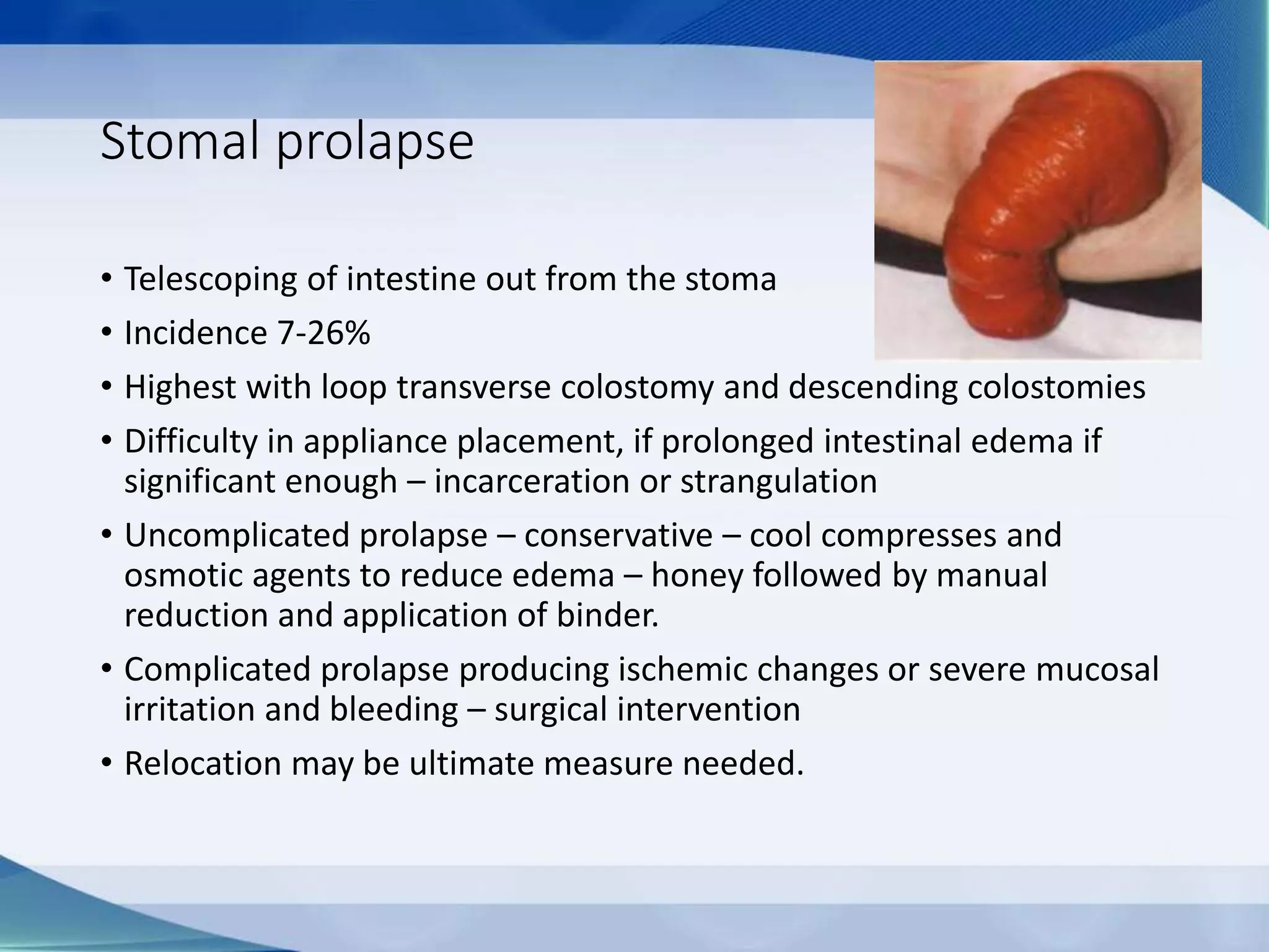
Stoma management
Shankar Zanwar
Definition
• Intestinal stomas are surgically created openings of small or large
intestine onto the anterior abdominal wall
• Types
Intestinal
stoma
colostomy
ileostomy
Intestinal
stoma
End stoma (single
limb)
Loop
stoma(afferent
and efferent limb)
Types
• Colostomies
• End – Left iliac fossa
• Loop
• Ileostomy
• End – right iliac fossa
• Loop
• Continent – Kock’s pouch
Indications of ostomies
• Large bowel obstruction
• Colonic stricture
• Rectovaginal fistula
• Fecal incontinence
• Penetrating colonic injuries
• Complex perianal fistulas
• Necrotising enterocolitis
• High risk of anastomotic leaks
• Total procto-colectomy in in UC/FAP
Stoma site
• Flat area of skin – adequate of adhesion of appliance
• Pt. should be able to see
• To avoid – skin creases, previous scars, umbilicus, bony prominences,
at the belt line.
• Site should be identified with patient lying, sitting and standing
• Usual sites
• Ileostomies – right iliac fossa
• Sigmoid colostomies – Left iliac fossa
• Transverse colostomies – right/left upper quadrant
Basic principles of stoma surgeries
• Midline vertical incision
• Adequate blood supply on either side(skin and bowel)
• Without tension on mesentry or skin
• Avoiding pre-existing infection site
• Avoiding too small hole at fascial level
• No twist
Conventional vs Brooke’s stomies
• Before Brooke, ileostomies were
made by exteriorizing the intestine
through the abdominal wall and
suturing the serosa to skin
• Exposure of ileal serosa to alkaline
stomal effluent resulted in serositis
and ileostomy dysfunction
• Brooke introduced technique of
eversion of the full thickness of the
mucosa and suturing it to the
adjacent dermis.
Pathophysiologic consequences after
ileostomy
• Colonic diversion – absorptive function of colon is lost
• Normal colon absorbs – 1-1.5L of water and 100 mEq/L of sodium
and if need be it can increase to 5 liter.
• Patients with ileostomy obligatory sodium loss is 30-40mEq/L
• A well functioning ileostomies discharges – 500ml to 1.2L of fluid
daily
• Consequently patients are in state of chronic oliguria
• Also they have lower urinary Na/K+
• Changes in urinary composition – increase chances of urolithiasis –
calcium and urate crystals
• Decreased vitamin B-12 absorption and bile acid reabsorption.
Routine care of ostomies
• Pouch placement – types
• Closed end pouches – needs to be removed and replaced
with new pouch every time
• Open end pouches – have reuseable end that can be
opened to drain the content of pouch
• Pouch emptying and care – odor and gas common
concerns of patient
• Assure ostomy bags are odor proof
• Empty the pouch when 1/3rd full prevent pouch seal
from excess weight
• Changing the pouch 1-2/wk and sos
• For foul odor if at all
• Chlorine tabs in bag 1-2 tabs
• Bismuth sub gallate 200 – 400mg
• Cholorophylline complex can be taken orally
Closed end pouch
Open end pouch
Protective skin barrier
• Diet – minimal modification needed, avoid unchewed nuts,
fruits with skin, popcorns that can obstruct stoma
• If gas is bothering patient needs to be given list of gas
producing diet, in short to avoid beans, cabbage, cauliflower,
brussels, broccoli, asparagus.
• Low carb diet with less of potatoes, corn, noodles and wheat
products.
•SOS use of simethicone can be done
•Adequate fluid intake – increase by 500ml -750ml
even more during high output states like sweating
•Should be taught signs of dehydration and fluid
electrolyte imbalance –dry mouth, decreased urine,
marked fatigue, abdominal cramps.
• Physical activity – all usual activites can be performed
without any restriction, bathing can be done with pouch on
or off the stoma,.
• Most sports can be performed even with stoma in place
except for extreme contact sports.
• Sexual activity – does not affect organic function,
dysfunction if at all occurs is due to autonomic denervation
after proctocolectomy
Complications of ostomies
• Over all complications rate of 14 to 79%
• Risk in life time
• Colostomy – 25%
• End –ileostomy – 57%
• Loop ileostomy -75%
Arumugam PJ Colorectal Dis 2003
Complications
• Skin – excoriation, dermatitis, ulcerations
• Ischemia
• Obstruction
• Retraction
• Para- stomal hernia
• Prolapse
• Fistula
• Stenosis
• Bleeding
• Perforation
• Timing of complications – early - are related to technical
issues, defined to be occurring within 3 months
• Usually these are due suboptimal stoma site selection -
necrosis, bleeds, retraction.
• Late – May be related to delayed healing – parastomal
hernia, prolapse, stomal stenosis.
• Risk factors
• Height of stoma <10mm
• Co-morbid medical illness –
• obesity, Crohn’s, diabetes
• Tobacco use
• Obesity
• Emergency surgery
• Malnutrition
• Advanced age
• Malignancy
• Use of steroids
• Inexperience
Orkin B: J of Reoperative Pelvic Surgery 2009
Dermatological complications
• Dermatitis :
• More common with ileostomies – enzymatic nature
of effluent, less with colostomies
• Severely denuded skin along the inferior surface of
the stoma
• Protuberant spout ~2-3cm high is best method
to minimize the contact of effluent to skin
• Skin irritation may be also due to
• Allergic reaction to pouching products –
pruritus, erythema and blisters
• Mechanical trauma
• Fungal infections - more during warm and humid climates
• Antibiotic therapy related
Allergic contact dermatitis
Peristomal fungal infection
•Treatment
•Correction of causative factors
•Elimination of allergens
•Denuded areas – use skin barriers
•Antifungals – Nystatin/ miconazole
•Topical steroids in case of severe allergic reaction
Peristomal pyoderma gangrenosum
• Usually seen with IBD
• Selecting uninvolved segment best way to
avoid
• Seen also in patient with malignancy
• Develops within weeks to years after stoma
construction
• Typically present as full thickness ulcer, pain and pathergy
• Diagnosis by excluding other common possibilities(r/o infections also)
• Bx. may increase the size of lesion
• HPE – acute and chronic inflammation +/- granulomas
JAMA 2000
• Frequent misdiagnosis – stitch abscess, contact
dermatitis, extension of Crohn’s
• Treatment –
• Systemic, intralesional, and or topical anti-inflammatory drugs
- Steroids
• When associated with Crohn’s or in other refractory cases,
tacrolimus 0.1%topical solution daily till healing
• Severely refractory – Infliximab or other biologicals
• Minimize trauma since pathergy is prominent
Stomal necrosis
• Incidence – immediate post op period 14%
AU Harris J of ARCES 2005
• Causative factors – inadequate –
mobilization of bowel, preservation of blood supply to stoma
• Risk factors – emergency surgery, obesity, CD
• Early post-op – d/t venous congestion or arterial insufficiency –
tight fascial opening, excessive mesentric stripping.
• Assessment - Inserting a lubricated test tube and using a torch
to determine extent
• Alternatively using a sigmoidoscope
• Treatment
• If necrosis extends to proximal bowel below the anterior
fascia – immediate revision
• If proximal - bowel viable, limited necrosis – superficial to
ant. fascia – observation
• If sloughing occurs only gentle debridement
Stomal bleeding
• Major bleeding – uncommon
• Usually due to stomal laceration d/t poor fitting appliance or due to peristomal
varices in UC with PSC.
• Initial management –
• direct pressure
• Local cauterisation – hand held cautery/ silver nitrate
• Suturing of bleeding vessel if identified
• Peristomal varices – direct pressure, sclerotherapy or direct suturing
• Beta-blockers may help
• Recurrence are frequent in varices if so – TIPSS
Eur J Gastro –Hep 2006
Stomal retraction
• Defined as stoma that is 0.5cm or more below
the skin surface within six months of construction,
typically as result of tension on stoma.
• Incidence 1-40% of all ostomies
Arumugam Colorectal dis 2005
• It causes – leakages and difficulty with pouch adherence – skin
irritation
• Proper ostomy height(>1cm) and minimising tension at stoma site
prevent retraction
• Management – use of convex pouching system
• If fails surgical correction
• If retracted mobile – non cutting linear stapling
• If immobile - laparotomy
Mucocutnaeous separation
• Separation of ostomy and peristomal skin
• Incidence 12 -24%
• Leads to leakage and skin irritation
• Prevention – meticulous skin approximation
• Partial or circumferential
• If circumferential stomal stenosis may occur as tissue heals
• Circumferential separation – immediate surgical revision
• Partial – fill absorptive material – calcium alginate, skin barrier
powder, hydrofiber
• Covering area with appliance wafer protects the wound from effluent
– promotes healing.
Parastomal hernia
• Type of incisional hernia
• Incidence from – 0-50%
• Unsightly bulge at stoma site
• Leakage around the stoma site
• Skin rashes, non fitting appliances
• Obstruction and strangulation
• Surgical repair –
• local repair, - 100% recurrence
• stoma relocation – 76% recurrence
• Mesh repair – 10% recurrence
Janes, Arch Surg 2004
Stomal stenosis
• Narrowing of the stoma sufficient enough to prevent
normal functioning
• Incidence 2-15%, more with end colostomies
• Early / late
• Early – d/t edema at the fascial and more superficial level
• Dilatation of this can be done using 36 Fr soft tipped Foley’s catheter
just proximal to fascial level, do not inflate balloon
• Late – scarring or tightness of mucocutaneous junction.
• May be due to peristomal sepsis, stomal retraction, ill fitting pouch.
• Mild – gentle routine dilatation, soft diet
• Significant stenosis – causing cramps followed by explosive output –
Surgical correction.
Stomal prolapse
• Telescoping of intestine out from the stoma
• Incidence 7-26%
• Highest with loop transverse colostomy and descending colostomies
• Difficulty in appliance placement, if prolonged intestinal edema if
significant enough – incarceration or strangulation
• Uncomplicated prolapse – conservative – cool compresses and
osmotic agents to reduce edema – honey followed by manual
reduction and application of binder.
• Complicated prolapse producing ischemic changes or severe mucosal
irritation and bleeding – surgical intervention
• Relocation may be ultimate measure needed.
High output stoma
• Definition - >1500ml/day
• Causes
• Adaptation phase
• Gastric acid hypersecretion
• SIBO
• Prestomal ileitis
• Revealed latent disease (celiac/pancreatic insufficiency/thyrotoxicosis)
• Infections including Cl.difficle
• Partial obstruction – parastomal hernias
• Short bowel syndrome
• Uncontrolled inflammation, sepsis, malnutrition.
Assessment
• Complications
• Dehydration
• Dyselectrolytemia
• Renal oxalate stones
• Psychological morbidity
• Death
• Assessment
• Examination of stoma
• Review op notes amount of bowel length remaining
• Small bowel radiology
• Ileostogram
Treatment
•NBM 48hours (on IV fluids) to assess baseline output
•Hyper caloric nutritional supplement
•Reduce oral hypotonic fluids to <500ml use ORS instead
•Reduce oral fluids and increase IV fluids
•Monitor electrolytes frequently including Mg
supplement Mg and vit. D
• Omeprazole – 80mg/d PO, if <50cm of jejunum left - IV
• Antibiotics trial for SIBO
• Megadose loperamide 16-40-100mg/d
• Additional codeine can be used up to 60mg 4X a day
• Octreotide may be used in cases of refractory high outputs upto 100mcg TID
• If baseline output >1500ml/d then consider long term IV fluids/TPN who are
nutritionally deprived with a central line
Guidelines for management of patients with a
short bowel, GUT 2006
Thank you
A well managed stoma
after procto-colectomy for UC.

Stoma management

  • 1.
  • 2.
    Definition • Intestinal stomasare surgically created openings of small or large intestine onto the anterior abdominal wall • Types Intestinal stoma colostomy ileostomy Intestinal stoma End stoma (single limb) Loop stoma(afferent and efferent limb)
  • 3.
    Types • Colostomies • End– Left iliac fossa • Loop • Ileostomy • End – right iliac fossa • Loop • Continent – Kock’s pouch
  • 4.
    Indications of ostomies •Large bowel obstruction • Colonic stricture • Rectovaginal fistula • Fecal incontinence • Penetrating colonic injuries • Complex perianal fistulas • Necrotising enterocolitis • High risk of anastomotic leaks • Total procto-colectomy in in UC/FAP
  • 5.
    Stoma site • Flatarea of skin – adequate of adhesion of appliance • Pt. should be able to see • To avoid – skin creases, previous scars, umbilicus, bony prominences, at the belt line. • Site should be identified with patient lying, sitting and standing • Usual sites • Ileostomies – right iliac fossa • Sigmoid colostomies – Left iliac fossa • Transverse colostomies – right/left upper quadrant
  • 6.
    Basic principles ofstoma surgeries • Midline vertical incision • Adequate blood supply on either side(skin and bowel) • Without tension on mesentry or skin • Avoiding pre-existing infection site • Avoiding too small hole at fascial level • No twist
  • 7.
    Conventional vs Brooke’sstomies • Before Brooke, ileostomies were made by exteriorizing the intestine through the abdominal wall and suturing the serosa to skin • Exposure of ileal serosa to alkaline stomal effluent resulted in serositis and ileostomy dysfunction • Brooke introduced technique of eversion of the full thickness of the mucosa and suturing it to the adjacent dermis.
  • 8.
    Pathophysiologic consequences after ileostomy •Colonic diversion – absorptive function of colon is lost • Normal colon absorbs – 1-1.5L of water and 100 mEq/L of sodium and if need be it can increase to 5 liter. • Patients with ileostomy obligatory sodium loss is 30-40mEq/L • A well functioning ileostomies discharges – 500ml to 1.2L of fluid daily • Consequently patients are in state of chronic oliguria • Also they have lower urinary Na/K+ • Changes in urinary composition – increase chances of urolithiasis – calcium and urate crystals • Decreased vitamin B-12 absorption and bile acid reabsorption.
  • 9.
    Routine care ofostomies • Pouch placement – types • Closed end pouches – needs to be removed and replaced with new pouch every time • Open end pouches – have reuseable end that can be opened to drain the content of pouch • Pouch emptying and care – odor and gas common concerns of patient • Assure ostomy bags are odor proof • Empty the pouch when 1/3rd full prevent pouch seal from excess weight • Changing the pouch 1-2/wk and sos • For foul odor if at all • Chlorine tabs in bag 1-2 tabs • Bismuth sub gallate 200 – 400mg • Cholorophylline complex can be taken orally Closed end pouch Open end pouch Protective skin barrier
  • 10.
    • Diet –minimal modification needed, avoid unchewed nuts, fruits with skin, popcorns that can obstruct stoma • If gas is bothering patient needs to be given list of gas producing diet, in short to avoid beans, cabbage, cauliflower, brussels, broccoli, asparagus. • Low carb diet with less of potatoes, corn, noodles and wheat products.
  • 11.
    •SOS use ofsimethicone can be done •Adequate fluid intake – increase by 500ml -750ml even more during high output states like sweating •Should be taught signs of dehydration and fluid electrolyte imbalance –dry mouth, decreased urine, marked fatigue, abdominal cramps.
  • 12.
    • Physical activity– all usual activites can be performed without any restriction, bathing can be done with pouch on or off the stoma,. • Most sports can be performed even with stoma in place except for extreme contact sports. • Sexual activity – does not affect organic function, dysfunction if at all occurs is due to autonomic denervation after proctocolectomy
  • 13.
    Complications of ostomies •Over all complications rate of 14 to 79% • Risk in life time • Colostomy – 25% • End –ileostomy – 57% • Loop ileostomy -75% Arumugam PJ Colorectal Dis 2003
  • 14.
    Complications • Skin –excoriation, dermatitis, ulcerations • Ischemia • Obstruction • Retraction • Para- stomal hernia • Prolapse • Fistula • Stenosis • Bleeding • Perforation
  • 15.
    • Timing ofcomplications – early - are related to technical issues, defined to be occurring within 3 months • Usually these are due suboptimal stoma site selection - necrosis, bleeds, retraction. • Late – May be related to delayed healing – parastomal hernia, prolapse, stomal stenosis.
  • 16.
    • Risk factors •Height of stoma <10mm • Co-morbid medical illness – • obesity, Crohn’s, diabetes • Tobacco use • Obesity • Emergency surgery • Malnutrition • Advanced age • Malignancy • Use of steroids • Inexperience Orkin B: J of Reoperative Pelvic Surgery 2009
  • 17.
    Dermatological complications • Dermatitis: • More common with ileostomies – enzymatic nature of effluent, less with colostomies • Severely denuded skin along the inferior surface of the stoma • Protuberant spout ~2-3cm high is best method to minimize the contact of effluent to skin • Skin irritation may be also due to • Allergic reaction to pouching products – pruritus, erythema and blisters • Mechanical trauma • Fungal infections - more during warm and humid climates • Antibiotic therapy related Allergic contact dermatitis Peristomal fungal infection
  • 18.
    •Treatment •Correction of causativefactors •Elimination of allergens •Denuded areas – use skin barriers •Antifungals – Nystatin/ miconazole •Topical steroids in case of severe allergic reaction
  • 19.
    Peristomal pyoderma gangrenosum •Usually seen with IBD • Selecting uninvolved segment best way to avoid • Seen also in patient with malignancy • Develops within weeks to years after stoma construction • Typically present as full thickness ulcer, pain and pathergy • Diagnosis by excluding other common possibilities(r/o infections also) • Bx. may increase the size of lesion • HPE – acute and chronic inflammation +/- granulomas JAMA 2000
  • 20.
    • Frequent misdiagnosis– stitch abscess, contact dermatitis, extension of Crohn’s • Treatment – • Systemic, intralesional, and or topical anti-inflammatory drugs - Steroids • When associated with Crohn’s or in other refractory cases, tacrolimus 0.1%topical solution daily till healing • Severely refractory – Infliximab or other biologicals • Minimize trauma since pathergy is prominent
  • 21.
    Stomal necrosis • Incidence– immediate post op period 14% AU Harris J of ARCES 2005 • Causative factors – inadequate – mobilization of bowel, preservation of blood supply to stoma • Risk factors – emergency surgery, obesity, CD • Early post-op – d/t venous congestion or arterial insufficiency – tight fascial opening, excessive mesentric stripping. • Assessment - Inserting a lubricated test tube and using a torch to determine extent • Alternatively using a sigmoidoscope
  • 22.
    • Treatment • Ifnecrosis extends to proximal bowel below the anterior fascia – immediate revision • If proximal - bowel viable, limited necrosis – superficial to ant. fascia – observation • If sloughing occurs only gentle debridement
  • 23.
    Stomal bleeding • Majorbleeding – uncommon • Usually due to stomal laceration d/t poor fitting appliance or due to peristomal varices in UC with PSC. • Initial management – • direct pressure • Local cauterisation – hand held cautery/ silver nitrate • Suturing of bleeding vessel if identified • Peristomal varices – direct pressure, sclerotherapy or direct suturing • Beta-blockers may help • Recurrence are frequent in varices if so – TIPSS Eur J Gastro –Hep 2006
  • 24.
    Stomal retraction • Definedas stoma that is 0.5cm or more below the skin surface within six months of construction, typically as result of tension on stoma. • Incidence 1-40% of all ostomies Arumugam Colorectal dis 2005 • It causes – leakages and difficulty with pouch adherence – skin irritation • Proper ostomy height(>1cm) and minimising tension at stoma site prevent retraction • Management – use of convex pouching system • If fails surgical correction • If retracted mobile – non cutting linear stapling • If immobile - laparotomy
  • 25.
    Mucocutnaeous separation • Separationof ostomy and peristomal skin • Incidence 12 -24% • Leads to leakage and skin irritation • Prevention – meticulous skin approximation • Partial or circumferential • If circumferential stomal stenosis may occur as tissue heals • Circumferential separation – immediate surgical revision • Partial – fill absorptive material – calcium alginate, skin barrier powder, hydrofiber • Covering area with appliance wafer protects the wound from effluent – promotes healing.
  • 26.
    Parastomal hernia • Typeof incisional hernia • Incidence from – 0-50% • Unsightly bulge at stoma site • Leakage around the stoma site • Skin rashes, non fitting appliances • Obstruction and strangulation • Surgical repair – • local repair, - 100% recurrence • stoma relocation – 76% recurrence • Mesh repair – 10% recurrence Janes, Arch Surg 2004
  • 27.
    Stomal stenosis • Narrowingof the stoma sufficient enough to prevent normal functioning • Incidence 2-15%, more with end colostomies • Early / late • Early – d/t edema at the fascial and more superficial level • Dilatation of this can be done using 36 Fr soft tipped Foley’s catheter just proximal to fascial level, do not inflate balloon • Late – scarring or tightness of mucocutaneous junction. • May be due to peristomal sepsis, stomal retraction, ill fitting pouch. • Mild – gentle routine dilatation, soft diet • Significant stenosis – causing cramps followed by explosive output – Surgical correction.
  • 28.
    Stomal prolapse • Telescopingof intestine out from the stoma • Incidence 7-26% • Highest with loop transverse colostomy and descending colostomies • Difficulty in appliance placement, if prolonged intestinal edema if significant enough – incarceration or strangulation • Uncomplicated prolapse – conservative – cool compresses and osmotic agents to reduce edema – honey followed by manual reduction and application of binder. • Complicated prolapse producing ischemic changes or severe mucosal irritation and bleeding – surgical intervention • Relocation may be ultimate measure needed.
  • 29.
    High output stoma •Definition - >1500ml/day • Causes • Adaptation phase • Gastric acid hypersecretion • SIBO • Prestomal ileitis • Revealed latent disease (celiac/pancreatic insufficiency/thyrotoxicosis) • Infections including Cl.difficle • Partial obstruction – parastomal hernias • Short bowel syndrome • Uncontrolled inflammation, sepsis, malnutrition.
  • 30.
    Assessment • Complications • Dehydration •Dyselectrolytemia • Renal oxalate stones • Psychological morbidity • Death • Assessment • Examination of stoma • Review op notes amount of bowel length remaining • Small bowel radiology • Ileostogram
  • 31.
    Treatment •NBM 48hours (onIV fluids) to assess baseline output •Hyper caloric nutritional supplement •Reduce oral hypotonic fluids to <500ml use ORS instead •Reduce oral fluids and increase IV fluids •Monitor electrolytes frequently including Mg supplement Mg and vit. D
  • 32.
    • Omeprazole –80mg/d PO, if <50cm of jejunum left - IV • Antibiotics trial for SIBO • Megadose loperamide 16-40-100mg/d • Additional codeine can be used up to 60mg 4X a day • Octreotide may be used in cases of refractory high outputs upto 100mcg TID • If baseline output >1500ml/d then consider long term IV fluids/TPN who are nutritionally deprived with a central line Guidelines for management of patients with a short bowel, GUT 2006
  • 33.
    Thank you A wellmanaged stoma after procto-colectomy for UC.