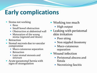
Early complications
Stomanot working
Ileus
Small bowel obstruction
Obstruction at abdominal wall
Maturation of the wrong
stoma (sigmoid and ileum)
Retraction
Stomal necrosis due to vascular
compromise
Muco-cutaneous separation
Retraction
Later stomal stenosis and
stricture
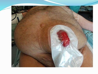
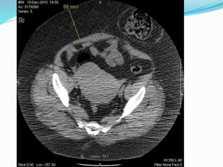
Acute parastomal hernia with
signs of strangulation
Working too much
High output
Leaking with peristomal
skin irritation
Poor siting
Non nippled ileostomy
Muco-cutaneous
separation
Wound infection
Peristomal abscess and
fistula
Necrotizing fascitis
4.
Intermediate or latecomplications
Stenosis
Prolapse
Parastomal herniation
Peristomal varices in patients with portal hypertension
5.
Overall Morbidity
Widelyvaries
21-70% (most 30-50%)
Observer dependent
Stoma type plays a huge role
Likely underestimated by most studies
6.
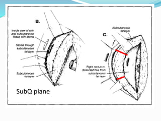
Vascular compromise
Inobese individuals, exteriorizing an ileostomy with an
adequate blood supply can be quite challenging.
The thickened, foreshortened mesentery often does not
have enough length to reach the surface of the
thickened abdominal wall easily, especially when
attempting to create a loop ileostomy.
In these instances, an end-loop configuration may
allow the bowel to more easily reach the abdominal
surface
7.
Vascular compromise
Whenassessing the vascular integrity of a congested
stoma postoperatively, transillumination with a
flashlight will demonstrate viability. A flashlight
placed in direct contact with a viable stoma will still
transilluminate bright red, even in the face of venous
congestion.
Failure to transilluminate the surface of the stoma or
nonviable appearing mucosa beneath the surface
generally indicates that the stoma requires revision.
8.
Vascular compromise
Ifthere is a question regarding viability below the
stomal surface, a well-lubricated blood collection tube
can be carefully passed into the stoma, below the
fascia if possible. When a light is shone into the tube,
viable mucosa will have a healthy, bright-red
appearance.
Darker hues or frank infarction require revision if the
compromise extends below the skin level.
Compromise below the fascia requires relaparotomy.
Questionable stomas can also be evaluated with a
pediatric proctoscope or flexible endoscope
9.
Retraction
Retraction ofa stoma in the immediate postsurgical
period is usually a result of tension on the bowel or its
mesentery due to inadequate mobilization.
Also, in patients who are malnourished, obese, or on
corticosteroid therapy, the stoma may retract due to
poor wound healing and gravity.
10.
Retraction
Mild distalstomal ischemia or stomal necrosis that is
managed expectantly may eventually result in
retraction with or without stenosis.
Complete acute retraction with mucocutaneous
separation can result in subcutaneous or subfascial
contamination, peritonitis, and sepsis. In this case,
immediate laparotomy and revision is advised.
11.
Retraction
More commonly,retraction is seen without complete
mucocutaneous separation.
The most significant problem in this instance is
obtaining a secure seal between the stoma appliance
and the abdominal wall, leading to fecal leakage and
significant peristomal skin irritation.
The majority of these stomas with significant
retraction eventually require revision.
12.
Retraction
The approachto a retracted stoma is similar to distal
ischemia.
If the mucosa is viable and there is no undue tension,
local revision can often be performed by detaching the
mucocutaneous junction, advancing the bowel and
excising devitalized tissue, and resecuring viable
mucosa to the skin using Brooke-type sutures.
If this is not technically feasible, laparotomy and
complete revision is required.
13.
PERISTOMAL SKIN IRRITATION
In most instances, peristomal skin irritation is a direct
result of
(1) chemical dermatitis due to exposure to the stoma
effluent due to leakage, and
(2) desquamation of peristomal skin resulting from
frequent appliance changes. Often, appliance leakage
and local skin irritation result in the need for more
frequent appliance changes, starting a vicious cycle.
14.
PERISTOMAL SKIN IRRITATION
Additionally, allergic reactions due to sensitivity to
skin barriers, adhesives, and tapes are fairly common.
Fungal irritation from Candida albicans colonization
of the peristomal skin also is commonly seen.
Antifungal powders may help alleviate this.
15.
PERISTOMAL INFECTION, ABSCESS,AND FISTULA
FORMATION
In the early postoperative period, parastomal
infections and abscesses are relatively uncommon,
with a reported incidence of 2 to 14.8%.
Peristomal abscesses in the immediate postoperative
period are most commonly seen in the setting of stoma
revision or reconstruction of a stoma at the same site,
mainly due to preoperative colonization of the
peristomal skin and perioperative seeding of the
surgical site.
16.
PERISTOMAL INFECTION, ABSCESS,
ANDFISTULA FORMATION
They may also be seen due to an infected hematoma or
an infected suture granuloma.
In a patient with Crohn's disease, a peristomal fistula
in conjunction with an ileostomy is almost invariably
the result of recurrent Crohn's disease
17.
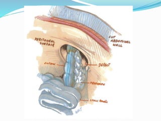
Parastomal hernia (PSH)
Definition
Incisional hernia related to an abdominal wall stoma
Varies in different studies
Palpable defect or bulge adjacent to a stoma
Cough impulse at ostomy site
Radiologic definition-any intra-abdominal content
protruding along an ostomy
Sometimes confused with prolapse
18.
PHS subtypes
Subcutaneous-subcutaneoussac
Interstitial-sac within the muscular or
aponeuroticlayers of the abdomen
Perstomal-the sac is circumferential enclosing the
stoma
Intrastomal-in ileostomies, sac between the
intestinal wall and evertedintestinal layer
NB there may be a diffuse type of hernia due to stretch
and paralysis of abdominal muscles with the stoma on
the summit of this bulge.
21.
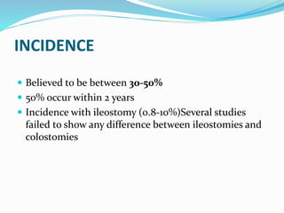
INCIDENCE
Believed tobe between 30-50%
50% occur within 2 years
Incidence with ileostomy (0.8-10%)Several studies
failed to show any difference between ileostomies and
colostomies
22.
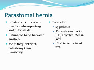
Parastomal hernia
Incidenceis unknown
due to underreporting
and difficult dx.
Estimated to be between
20-80%
More frequent with
colostomy than
ileostomy
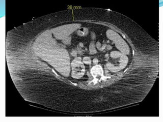
Cingi et al
23 patients
Patient examination
(PE) detected PSH in
52%
CT detected total of
78%
23.
Parastomal Hernia
Early
Presents with acute
pain, mass, obstruction
< 30 days from stoma
Technical failure
Too large of an aperture
in fascia
Late
Inevitable?
Presents with slow
growing mass,
abnormal contour of
tissues around stoma
Consequence of
increasing
intraabdominal tension
“There’s already a hole
there, Doctor.”
R. Schwartz 2008
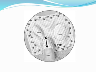
TECHNICAL CONSIDERATIONS IN
STOMAFORMATION
Extraperitoneal vs intrapertioneal(9% vs17%)
Transrectal vs lateral to the rectus (3% vs22%)
Size of the trephine: 2.5cm usually
Todd and Celestine-2cm for ileostomies and 1.5cm
for colostomies with a later retraction of 0.5cm
26.
SYMPTOMS
Asymptomatic +++
Parastomal discomfort with intermittent obstructive
episodes
Stoma appliance issues with leak and skin irritation
Obstruction/strangulation
10-20% have symptoms severe enough to require
surgical repair
27.
SURGICAL MANAGEMENT
Localaponeurotic repair with or without mesh
Relocation of the stoma
Open repair with mesh
Laparoscopic repair
28.
Primary Repair
Justsew the hole around
the stoma
High recurrence rate
historically 50-100%
Add mesh?
Still doesn’t work 50-
88% recurrence
29.
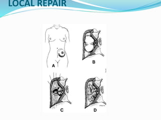
SURGICAL MANAGEMENT
LOCALREPAIR
Aponeurotic repair-primary closure of the defect-
recurrence 50-76% (up to 100%)
Onlay mesh repair-involves applying a non
resorbable mesh on top of the primary repair and
fixing it to the fascia-recurrence 9-10% (small
studies without long follow up)
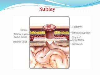
Sublay mesh repair-the mesh is placed in the
properitoneal space after plication of the sac
SURGICAL MANAGEMENT
RELOCATION
Risk of recurrence at least as high as the primary
site
Recurrence rates as high as 24-86%
Higher if relocated on the same side
The primary site should be treated as an
incisional hernia and repaired with mesh
placement-recurrence rate 26-48%
33.
Re-Siting of Stoma
Traditional boards answer for symptomatic PSH
Has expected high recurrence rate
Baig et al. 4/27 recurrences at 56 months
3/16 with laparotomy
1/11 without laparotomy
Historically has rates up to 50-68% (essentially the
same as hernia rate for each new stoma)
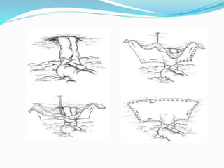
SURGICAL MANAGEMENT
OPENMESH REPAIR
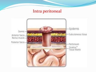
IPOM (Intraperitoneal Onlay Mesh) vs Sublay
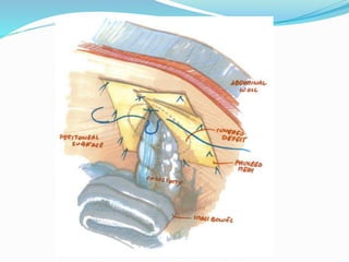
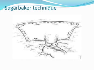
Keyhole technique vs Sugarbaker technique
(bowel entering lateral to the mesh)
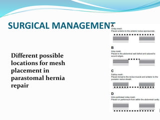
36.
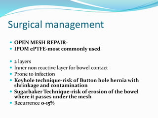
Surgical management
OPENMESH REPAIR-
IPOM ePTFE-most commonly used
2 layers
Inner non reactive layer for bowel contact
Prone to infection
Keyhole technique-risk of Button hole hernia with
shrinkage and contamination
Sugarbaker Technique-risk of erosion of the bowel
where it passes under the mesh
Recurrence 0-15%
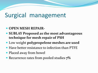
Surgical management
OPENMESH REPAIR-
SUBLAY Proposed as the most advantageous
technique for mesh repair of PSH
Low weight polypropelene meshes are used
Have better resistance to infection than PTFE
Placed away from bowel
Recurrence rates from pooled studies 7%
41.
Surgical management
LAPAROSCOPICAPPROACH
Done in a way similar to open IPOM
Keyhole technique or Sugarbaker
technique
Recurrence rates vary between4-44%
Higher risk of bowel injury 22%
Higher risk of mesh infection (4% in one study)
42.
Surgical management
LAPROSCOPICAPPROACH:
TECHNICAL TIPS
Fashion the mesh before insertion in the abdomen
with a circular defect and a slit
If the mesh is cut in a linear fashion the slit
will enlarge with intraabdominal pressure
A good way to reduce recurrence may be to
place 2 pieces of mesh one on top of the
other
43.
Laparoscopic techniques
Lapvs Open
McLemore – 49 pt with PSH
Laparoscopic vs Open suture repair
No significant difference in morbidity or short term outcomes
Pastor – 25 pts
4/12 laparoscopic had recurrence
7/13 open had recurrence
44.
Laparoscopic Keyhole vs
Sugarbaker
Muysoms,et. al.
Keyhole – recurrence 72.7%
Sugarbaker – recurrence 14.2%
Mancini, et al
Retrospective review of 25 pts with Sugarbaker
technique
1 recurrence at 30 months. (4%)
Surgical management
BIOPROSTHETICS
Studies reporting the use of bioprosthetics for treatment of
parastomal hernias are scant, low powered and have a short
F/U
Most advantages are extrapolated from the use of bioprosthetics
in incisional hernias
Most studies seem to show a low incidence of complications and
an equivalent incidence of recurrence as synthetics
BIOPROSTHETICS Recurrence rates vary between 9-27%
depending on the studies and the type of mesh used
(human dermis vs porcine small bowel submucosa)
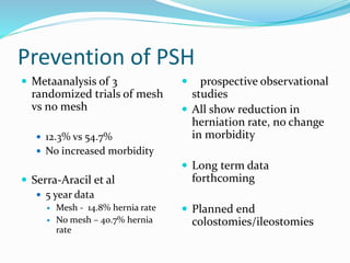
Prevention of PSH
Metaanalysis of 3
randomized trials of mesh
vs no mesh
12.3% vs 54.7%
No increased morbidity
Serra-Aracil et al
5 year data
Mesh - 14.8% hernia rate
No mesh – 40.7% hernia
rate
5 prospective observational
studies
All show reduction in
herniation rate, no change
in morbidity
Long term data
forthcoming
Planned end
colostomies/ileostomies
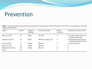
Prevention
Janeset al.randomized 54 patients to stoma
creation with sublay mesh vs no mesh with a
mean F/U of 24 months
1 hernia occurred in the mesh group vs13 in the
non mesh group
There was no complications
Retrospective studies were also in favor of prophylactic
mesh placement
55.
Prevention
CONCLUSION
Placementof mesh at the primary operation is
safe
Reduces the occurrence of parastomal hernia
Prophylactic meshes were also placed in contaminated
cases without infection
More randomized studies needed
56.
Conclusions
Very commoncondition
Only a small proportion will require surgical
therapy
The high recurrence rates underline the fact that
there is no perfect operation for this condition
Promising results with laparoscopy and
bioprosthetics
Prophylactic mesh placement seems to be the
way to go
A Difficult Situation
65 year old man
350 lbs
Diabetic with CHF
Perforated diverticulitis
5 laparotomies
Septic with peritonitis
Get the idea?
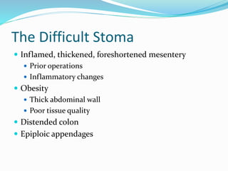
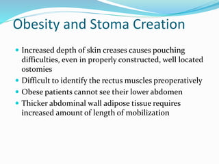
Obesity and StomaCreation
Increased depth of skin creases causes pouching
difficulties, even in properly constructed, well located
ostomies
Difficult to identify the rectus muscles preoperatively
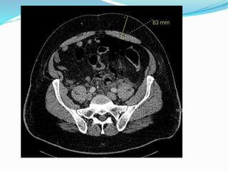
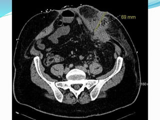
Obese patients cannot see their lower abdomen
Thicker abdominal wall adipose tissue requires
increased amount of length of mobilization
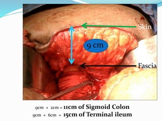
62.
Skin
Fascia
9 cm
9cm +2cm = 11cm of Sigmoid Colon
9cm + 6cm = 15cm of Terminal ileum
BMI
48.7
63.
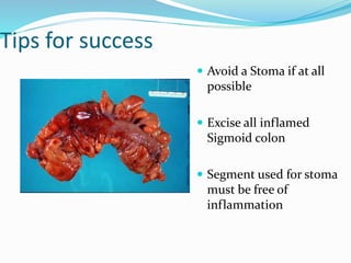
Tips for success
Avoid a Stoma if at all
possible
Excise all inflamed
Sigmoid colon
Segment used for stoma
must be free of
inflammation
64.
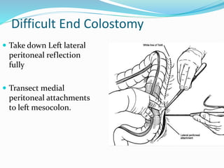
Difficult End Colostomy
Take down Left lateral
peritoneal reflection
fully
Transect medial
peritoneal attachments
to left mesocolon.
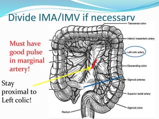
Divide IMA/IMV ifnecessary
Must have
good pulse
in marginal
artery!
Stay
proximal to
Left colic!
67.
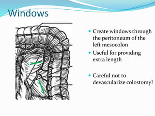
Windows
Create windowsthrough
the peritoneum of the
left mesocolon
Useful for providing
extra length
Careful not to
devascularize colostomy!
68.
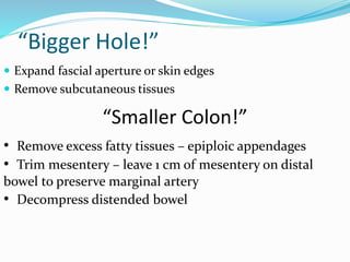
“Bigger Hole!”
Expandfascial aperture or skin edges
Remove subcutaneous tissues
“Smaller Colon!”
• Remove excess fatty tissues – epiploic appendages
• Trim mesentery – leave 1 cm of mesentery on distal
bowel to preserve marginal artery
• Decompress distended bowel
69.
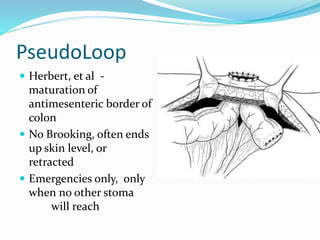
PseudoLoop
Herbert, etal -
maturation of
antimesenteric border of
colon
No Brooking, often ends
up skin level, or
retracted
Emergencies only, only
when no other stoma
will reach
70.
“Better to createan ugly stoma
in a good location than a pretty
stoma in an ugly location.”
--Peter Cataldo
71.
Thinner wall?
Abdominalwall modification
Lipectomy
Meguid (1997) described technique of excision of
subcutaneous fat to reduce abdominal wall thickness
Leave convex contour to abdominal wall – can lead to
pouching issues
Liposuction
Margulies elucidated technique of peristomal suction
lipectomy for removal of excess fat during stomal revision
Thinner Wall?
Flaps
Good for Retraction and pyoderma/skin ulcerations in
Obese people.
Functionally Better than Lipectomy because of
restoration of flat abdominal wall, but have risk of
potential flap necrosis
Not described for initial placement of ostomy
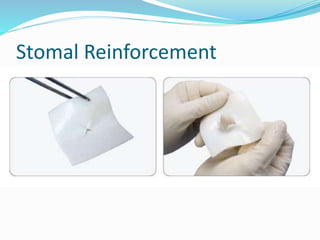
77.
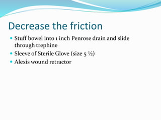
Decrease the friction
Stuff bowel into 1 inch Penrose drain and slide
through trephine
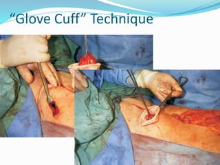
Sleeve of Sterile Glove (size 5 ½)
Alexis wound retractor
78.
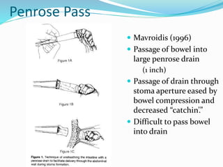
Penrose Pass
Mavroidis(1996)
Passage of bowel into
large penrose drain
(1 inch)
Passage of drain through
stoma aperture eased by
bowel compression and
decreased “catchin’.”
Difficult to pass bowel
into drain
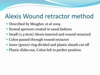
Alexis Wound retractormethod
Described by Meagher, et al 2009
Stomal aperture created in usual fashion
Small (2.5-6cm) Alexis inserted and wound retracted
Colon passed through wound retractor
Inner (green) ring divided and plastic sheath cut off
Plastic slides out, Colon left in perfect position
(Anecdotal) Benefits
“Noticeably”Smaller size of aperture
Less tissue damage/bruising
F/U < 14 months, but no retractions nor parastomal
hernia
Abdominal wall 7-8 cm
83.
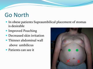
Go North
Inobese patients Supraumbilical placement of stomas
is desirable
Improved Pouching
Decreased skin irritation
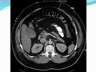
Thinner abdominal wall
above umbilicus
Patients can see it
89.
Stoma Formation
Islife altering for
patients
Is not a benign
procedure
Is associated with a high
rate of early and late
technical complications
May require Operative
imagination
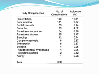
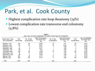
Park, et al.Cook County
Retrospective analysis of 1616 pts (20 years)
Data compiled by EST
553/1616 complications (448 early/105 late)
Early complications (28%)
Skin irritation 12%
Pain/poor location 7%
Partial necrosis 5%
94.
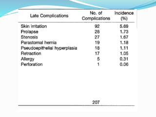
Park, et al.Cook County
Late complications (6%)
Skin irritation 6%
Prolapse 2%
Stenosis 2%
Parastomal hernia not mentioned
Trauma/colorectal had lowest complication rate
No difference in emergent vs elective
96.
Park, et al.Cook County
Highest complication rate loop ileostomy (74%)
Lowest complication rate transverse end colostomy
(5.8%)
98.
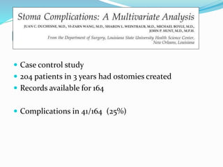
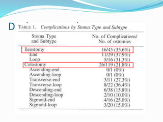
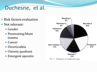
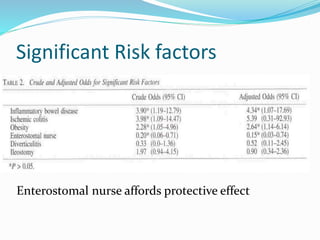
Duchesne, et al-- LSU
Case control study
204 patients in 3 years had ostomies created
Records available for 164
Complications in 41/164 (25%)
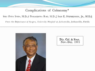
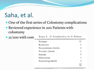
Saha, et al.
One of the first series of Colostomy complications
Reviewed experience in 200 Patients with
colostomy
21/200 with complications (11%)