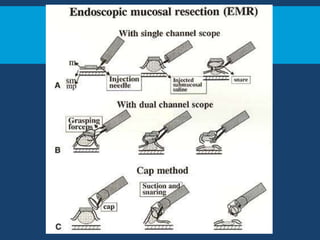
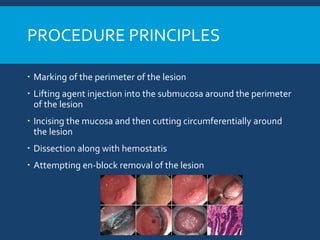
ENDOSCOPIC
SUBMUCOSAL DISSECTION
ShankarZanwar
BACKGROUND
 Endoscopic removal of tumors was first described in the era of
rigid sigmoidoscope in 1955
 Saline injection in submucosal space as safety cushion for
elevation of lesion was described in 1973
 EMR- endoscopic mucosal resection is the precursor of
endoscopic submucosal dissection(ESD)
 Endoscopic submucosal dissection was described first in 1988 by
Hirao et al from Japan
PROCEDURE PRINCIPLES
 Marking of the perimeter of the lesion
 Lifting agent injection into the submucosa around the perimeter
of the lesion
 Incising the mucosa and then cutting circumferentially around
the lesion
 Dissection along with hemostatis
 Attempting en-block removal of the lesion
DEVICES FORTHE ESD
 Dyes
 Knives
 Injection agents and delivery devices
 Hemostatic forceps and other devices
 Tissue retractors
 Endoscopes
 Electrosurgical unit
DYES
 Colorants such as indigo carmine, methylene blue and Lugols
iodine are used
 Used for initial evaluation of marking lesion of interest.
 NBI can be used instead but no comparative studies available.
KNIVES
 The earliest dedicated ESD device simply added a ceramic tip to
an existing needle-knife to prevent in advertent deep dissection
and there by perforation
 Various varieties of knives are available with no randomized
studies to prove superiority of one type over another
 Most of these are compatible with the 2.8mm channel in the
endoscope
IT KNIFE (INSULATEDTIP)
 IT - It has 2.2mm ceramic ball at end of 4mm
cutting knife
 IT2 triangular electrode
 IT nano - 1.7 ball, 3.5mm cutting knife
 Circumferential incision and submucosal
dissection
2. HOOK KNIFE
 Hook –Tip bent at 90 degree, L shaped
 Length 4.5mm, hook 1.3mm
 Knife length and direction both adjustable
 Allow hooking and retraction of tissue
 Can be used for pre incision marking
 This can used any where, particularly when fibrotic
tissue is present.
DUALKNIFE AND FLEXKNIFE
 DualKnife –Very small dome shaped non insulated tip, 2mm in
length
 For marking full retraction in the sheath –leaves only 0.3mm tip out
of the catheter.
 Full extension for cutting and dissection
 FlexKnife – Braided 0.8mm diameter cutting knife with looped tip,
variably extensible.
HYBRIDKNIFE
 Developed by ERBE electrosurgicals.
 Has a central capillary within the cutting knife provides ultrafine 120
micron water jet.
 This single device can perform all the phases of ESD alone.
 Submucosal elevation by water jet, marking, cutting and dissection.
 Has 3 tip configurations, I type,T type(disc) and O type(insulated
dome)
HEMOSTATIC FORCEPS
 Monopolar and bipolar forceps
 Use to treat bleed with coaptive(mutually fitting) thermocoagulation
 Coagrasper is monopolar hemostatic forcep, serrated jaws
 Opening width 5mm
TISSUE RETRACTORS
 Transparent cap applied to the tip of endoscope.
 Servers to keep the resected flap of the mucosa off of the endoscope
lens prevent red out
 Disposable with various size and shapes available.
 Some caps have irrigation port
 Newer caps from Olympus has retractor attached to it - Endolifter,
since cap retraction is not sufficient enough
 For less affording setups
external grasping forceps
can be used.
INJECTION AGENTS
 Inj. agents are first injected around the perimeter of the lesion
to provide a safety margin and later injected below the lesion
during dissection phase
 Goals of an ideal agent
 Safety
 Low cost
 Provision of long lasting cushion
 Normal saline – Safe, low cost not long lasting
 Western endoscopists – Sod. Hyaluronate(0.4%) – safe, long
lasting but expensive
 Another alternative is 0.4% hydroxymethyl cellulose - cheaper
 Injectate is colored with indigo carmine to help differentiate
tissue planes
 Use of adrenaline is debated MI and ischemia reported
 Injectates delivered using 21 to 25G needles
 Viscous injectates require wide bore needle
 ERBE hybridKnife does not need a separate needle.
ENDOSCOPES
 Endoscopes with high definition imaging may allow superior
detection and demarcation of mucosal neoplasia
 Though they help detecting margin extent no added benefit in
the procedure obtained
 A newly developed scope with channel diameter of 3.7mm has
superior optics and suctioning facility.
 Endoscope with double channel may add on the advantage of
using knife and grasping forceps together but add on the cost.
ENDOSURGICAL UNIT (ESU)
 Either a monopolar or bipolar circuit can be used.
 Newer ESUs contain microprocessor that sense voltage change
due to tissue impedance and responsively keep the voltage
constant to attend consistent effect.
 Newer ESUs have wide array of voltage ranges to facilitate
cutting in different tissues.
 ESUs are also capable of APC for marking and hemostasis.
GAS INSUFFLATION
 Standard air insufflation is safe for use in ESD
 CO2 absorbed 160 times more faster than nitrogen and 13 times
more than O2
 CO2 insufflation less prolonged, less pt. discomfort, fewer post
procedure admissions
 Decreased likelihood of tension pneumoperitoneum and
perforation.
SEDATION
 Can be done in moderate or deep sedation using midaz and
propofol respectively
 Preferably by anesthetist
 Need of fine motor movement, longer duration of procedure,
reflux potential, aspiration risk and risk of perforation makes GA
naturally better choice.
INDICATIONS OF ESD
 ESD aiming for curative treatment should be performed in tumors
without LN mets.
 LN mets strongly correlates with
 Depth of invasion
 Histopathologic type
 Lymphatic or vessel involvement
Wehmann Endo. Sur 1994
 Detailed examination, NBI/chromoendoscopy, targeted Bx should be
done preprocedure
 Depth of invasion can be ascertained with mini probe in EUS – accuracy
80-92%.
Okada Surg. Endo -2011
ESOPHAGUS –
 SCCa -
 ESD with complete resection curative only of tumor limited to
epithelium and lamina propria muscle layer
 Those invading the musclaris mucosa and beyond have significant
LN mets risk
 Till date no superficial Barrett esophageal ca. within the mucosa
found to be associated with positive LN mets.
 Therefore, Ca. those limited within the mucosa and high grade
dysplasia can be resected with curative results.
Virchow Arch 2010
GASTRIC ESD
 No limitation of size ( unlike EMR <2cm)
 Japanese gastric cancer asso. – any lesion that can be resected in one
piece and has minimal risk of LN mets
 Absolute indication
 non ulcerated
 Differentiated type
 Mucosal carcinoma < 2cm
 Expanded indication
 Non ulcerated, differentiated >2cm
 Ulcerated, differentiated but <3cm
 Non ulcerated, undifferentiated mucosal type <2cm
 Differentiated minimally invasive submucosal <5oomicron from
muscularis mucosae <3cm.
COLORECTAL ESD
 Considered curative for benign adenoma, non invasive or
minimally invasive ca. without vessel infiltration
 No size limitation
 Additional surgical treatment required if
 Positive vertical margin
 Depth >1000micron
 Vessel infiltration
 Poorly differentiated type
 Considered curative when all above are absent
EFFICACY AND COMPARISONS
 Stomach ESD for EGD
 En bloc resection – 86-97%
 R0 i.e. negative lateral and horizontal margin – 88-93%
 Local recurrence rate 1 %
 5 year survival (overall) 96-100%
 Disease specific survival 99-100%
Tanabe Gastric Cancer 2014
Goto endoscopy 2009
 Two meta-analyses – comparing EMR with ESD
Lian J GIE 2012
 All cause mortality at 3 and 4 years did not differ significantly
 No comparative studies with surgery
 Lap gastrectomy for early ca stomach – 5 year survival 99.8% for
T1a andT1B 98.7
ESD EMR
En block 92% 52%
R0 82-92% 42-43%
Recurrence 0.8% 5.0-6.4%
COLON AND RECTUM
 Generally for laterally spreading tumor >2cm
 R0 resection – 88%
 Incidence of lymphatic metastasis when curative criteria fulfilled – 1.9%
 Comparison with EMR
 Comparing with surgical Rx. (Transanal endoscopic microsurgery) - only
retrospective data
 Similar rates of R0 resection and curative resection between the 2
modalities, but shorted length of stay with ESD.
ESD EMR
Enblock 84-95% 33-57%
Local recurrence 0-2% 12-26%
Park SU Endoscopy 2012
ESOPHAGUS
 For Sq.CC rate of en-block resection 100% with curative
resection rate 68-79%
 Non curative resection patient managed with esophagectomy
 In curative resection group no recurrences or mets for 2 years
follow up
Yamada GIE 2013
 For Barrett’s German study
 90% en-block resection
 R0 38% (any dysplasia precludes R0)
 Follow up for 17 month 96% neoplasia free
 No direct comparison with surgery available, 5 year survival rate,
T1a - adeno ca. 91% and SCC – 62% with surgery
Gertier Surg Endo 2011
COMPLICATIONS, BLEEDING
 Minor oozing treated – coagulation current, significant ooze –
hemostatic forceps
 Severe endoscopically unmanageable – reported prevalence 6 in 1244
early gastric ca. ESDs
 Delayed bleeding rate 4.5% - 15.6%
 Risk factors
 Size of the lesion > 4cm
 Resumption of antithrombotics
 PPI (and H2 blockers) shown to be associated with decreased bleeding
risk
 Mucosal protective agents(sucrafil)  faster healing but no change in
bleeding rates
 Majority (76%) occur in 24 hours
 Non gastric ESD delayed bleed
 Esophagus – 0-5.2%
 Colorectal – 2%
Ishamoto dig Endoscopy - 2013
PERFORATION
 Frequency – Gastric - 4.5%, colorectal - 4.8%, esophageal 0-10%.
 Pooled statistics from Japan , 117 perforations – 115(98%) could be
managed non-operatively
 Treatment – primary clip closure – for defect <1cm, omental patch
for larger lesions using multiple clip after suctioning lesser
omentum.
 Initial management – NG suction – 3days,TPN for 9 days with
antibiotic cover.
 Colorectal ESDs – in a series of 816 ESDs, 16(2%) perforation
occurred.
 Of which 14 treated endoscopically and only 2 needed surgery
 Esophageal perforation can be managed with clips only rare
surgical intervention needed
 Small percentage may present with delayed perforation, in a
series 1159, 6 presented after 10 hours of which 5 needed
surgery.
STRICTURE
 Esophageal stricture(post ESD) defined as – narrowing through
which a standard gastroscope cannot be advanced
 Rate – 12-17% after esophageal ESD
 When circumference >3/4th resected, risk increase
 Serial dilatation, steroids radial electroincision, prophylactic SEMS
can be used.
 After gastric ESD rare - 0.7% of all cases, majority near cardia and
rest near prepyloric antrum
 Post Colorectal ESD strictures not reported
Kim Dig Dis Sci 2014
MISCELLANEOUS
 Learning curve – minimum of 50 human procedures are needed
to be performed for significant improvement across all
outcomes
 Average procedure time in non Japanese institutes – 70-136min
 Cost of knives range between 400-700$(~24000 Rs)
FUTURE RESEARCH
 RCTs comparing EMR and surgery
 Newer agents that would can used for auto-dissection of the
submucosa – Mesna in research, promising reports in animal studies.
 Development of agents that would shield the defects and prevent
bleeding and perforation -- polyglycolic acid sheets and fibrin glue in
development
 Development of endoscopic autologous oral mucosal sheets
transplantation over the defects for stricture prevention.
THANKYOU

Esd

  • 1.
  • 2.
    BACKGROUND  Endoscopic removalof tumors was first described in the era of rigid sigmoidoscope in 1955  Saline injection in submucosal space as safety cushion for elevation of lesion was described in 1973  EMR- endoscopic mucosal resection is the precursor of endoscopic submucosal dissection(ESD)  Endoscopic submucosal dissection was described first in 1988 by Hirao et al from Japan
  • 4.
    PROCEDURE PRINCIPLES  Markingof the perimeter of the lesion  Lifting agent injection into the submucosa around the perimeter of the lesion  Incising the mucosa and then cutting circumferentially around the lesion  Dissection along with hemostatis  Attempting en-block removal of the lesion
  • 5.
    DEVICES FORTHE ESD Dyes  Knives  Injection agents and delivery devices  Hemostatic forceps and other devices  Tissue retractors  Endoscopes  Electrosurgical unit
  • 6.
    DYES  Colorants suchas indigo carmine, methylene blue and Lugols iodine are used  Used for initial evaluation of marking lesion of interest.  NBI can be used instead but no comparative studies available.
  • 7.
    KNIVES  The earliestdedicated ESD device simply added a ceramic tip to an existing needle-knife to prevent in advertent deep dissection and there by perforation  Various varieties of knives are available with no randomized studies to prove superiority of one type over another  Most of these are compatible with the 2.8mm channel in the endoscope
  • 8.
    IT KNIFE (INSULATEDTIP) IT - It has 2.2mm ceramic ball at end of 4mm cutting knife  IT2 triangular electrode  IT nano - 1.7 ball, 3.5mm cutting knife  Circumferential incision and submucosal dissection
  • 9.
    2. HOOK KNIFE Hook –Tip bent at 90 degree, L shaped  Length 4.5mm, hook 1.3mm  Knife length and direction both adjustable  Allow hooking and retraction of tissue  Can be used for pre incision marking  This can used any where, particularly when fibrotic tissue is present.
  • 10.
    DUALKNIFE AND FLEXKNIFE DualKnife –Very small dome shaped non insulated tip, 2mm in length  For marking full retraction in the sheath –leaves only 0.3mm tip out of the catheter.  Full extension for cutting and dissection  FlexKnife – Braided 0.8mm diameter cutting knife with looped tip, variably extensible.
  • 11.
    HYBRIDKNIFE  Developed byERBE electrosurgicals.  Has a central capillary within the cutting knife provides ultrafine 120 micron water jet.  This single device can perform all the phases of ESD alone.  Submucosal elevation by water jet, marking, cutting and dissection.  Has 3 tip configurations, I type,T type(disc) and O type(insulated dome)
  • 12.
    HEMOSTATIC FORCEPS  Monopolarand bipolar forceps  Use to treat bleed with coaptive(mutually fitting) thermocoagulation  Coagrasper is monopolar hemostatic forcep, serrated jaws  Opening width 5mm
  • 13.
    TISSUE RETRACTORS  Transparentcap applied to the tip of endoscope.  Servers to keep the resected flap of the mucosa off of the endoscope lens prevent red out  Disposable with various size and shapes available.  Some caps have irrigation port  Newer caps from Olympus has retractor attached to it - Endolifter, since cap retraction is not sufficient enough  For less affording setups external grasping forceps can be used.
  • 14.
    INJECTION AGENTS  Inj.agents are first injected around the perimeter of the lesion to provide a safety margin and later injected below the lesion during dissection phase  Goals of an ideal agent  Safety  Low cost  Provision of long lasting cushion  Normal saline – Safe, low cost not long lasting  Western endoscopists – Sod. Hyaluronate(0.4%) – safe, long lasting but expensive
  • 15.
     Another alternativeis 0.4% hydroxymethyl cellulose - cheaper  Injectate is colored with indigo carmine to help differentiate tissue planes  Use of adrenaline is debated MI and ischemia reported  Injectates delivered using 21 to 25G needles  Viscous injectates require wide bore needle  ERBE hybridKnife does not need a separate needle.
  • 16.
    ENDOSCOPES  Endoscopes withhigh definition imaging may allow superior detection and demarcation of mucosal neoplasia  Though they help detecting margin extent no added benefit in the procedure obtained  A newly developed scope with channel diameter of 3.7mm has superior optics and suctioning facility.  Endoscope with double channel may add on the advantage of using knife and grasping forceps together but add on the cost.
  • 17.
    ENDOSURGICAL UNIT (ESU) Either a monopolar or bipolar circuit can be used.  Newer ESUs contain microprocessor that sense voltage change due to tissue impedance and responsively keep the voltage constant to attend consistent effect.  Newer ESUs have wide array of voltage ranges to facilitate cutting in different tissues.  ESUs are also capable of APC for marking and hemostasis.
  • 18.
    GAS INSUFFLATION  Standardair insufflation is safe for use in ESD  CO2 absorbed 160 times more faster than nitrogen and 13 times more than O2  CO2 insufflation less prolonged, less pt. discomfort, fewer post procedure admissions  Decreased likelihood of tension pneumoperitoneum and perforation.
  • 19.
    SEDATION  Can bedone in moderate or deep sedation using midaz and propofol respectively  Preferably by anesthetist  Need of fine motor movement, longer duration of procedure, reflux potential, aspiration risk and risk of perforation makes GA naturally better choice.
  • 20.
    INDICATIONS OF ESD ESD aiming for curative treatment should be performed in tumors without LN mets.  LN mets strongly correlates with  Depth of invasion  Histopathologic type  Lymphatic or vessel involvement Wehmann Endo. Sur 1994  Detailed examination, NBI/chromoendoscopy, targeted Bx should be done preprocedure  Depth of invasion can be ascertained with mini probe in EUS – accuracy 80-92%. Okada Surg. Endo -2011
  • 21.
    ESOPHAGUS –  SCCa-  ESD with complete resection curative only of tumor limited to epithelium and lamina propria muscle layer  Those invading the musclaris mucosa and beyond have significant LN mets risk  Till date no superficial Barrett esophageal ca. within the mucosa found to be associated with positive LN mets.  Therefore, Ca. those limited within the mucosa and high grade dysplasia can be resected with curative results. Virchow Arch 2010
  • 22.
    GASTRIC ESD  Nolimitation of size ( unlike EMR <2cm)  Japanese gastric cancer asso. – any lesion that can be resected in one piece and has minimal risk of LN mets  Absolute indication  non ulcerated  Differentiated type  Mucosal carcinoma < 2cm  Expanded indication  Non ulcerated, differentiated >2cm  Ulcerated, differentiated but <3cm  Non ulcerated, undifferentiated mucosal type <2cm  Differentiated minimally invasive submucosal <5oomicron from muscularis mucosae <3cm.
  • 23.
    COLORECTAL ESD  Consideredcurative for benign adenoma, non invasive or minimally invasive ca. without vessel infiltration  No size limitation  Additional surgical treatment required if  Positive vertical margin  Depth >1000micron  Vessel infiltration  Poorly differentiated type  Considered curative when all above are absent
  • 24.
    EFFICACY AND COMPARISONS Stomach ESD for EGD  En bloc resection – 86-97%  R0 i.e. negative lateral and horizontal margin – 88-93%  Local recurrence rate 1 %  5 year survival (overall) 96-100%  Disease specific survival 99-100% Tanabe Gastric Cancer 2014 Goto endoscopy 2009
  • 25.
     Two meta-analyses– comparing EMR with ESD Lian J GIE 2012  All cause mortality at 3 and 4 years did not differ significantly  No comparative studies with surgery  Lap gastrectomy for early ca stomach – 5 year survival 99.8% for T1a andT1B 98.7 ESD EMR En block 92% 52% R0 82-92% 42-43% Recurrence 0.8% 5.0-6.4%
  • 26.
    COLON AND RECTUM Generally for laterally spreading tumor >2cm  R0 resection – 88%  Incidence of lymphatic metastasis when curative criteria fulfilled – 1.9%  Comparison with EMR  Comparing with surgical Rx. (Transanal endoscopic microsurgery) - only retrospective data  Similar rates of R0 resection and curative resection between the 2 modalities, but shorted length of stay with ESD. ESD EMR Enblock 84-95% 33-57% Local recurrence 0-2% 12-26% Park SU Endoscopy 2012
  • 27.
    ESOPHAGUS  For Sq.CCrate of en-block resection 100% with curative resection rate 68-79%  Non curative resection patient managed with esophagectomy  In curative resection group no recurrences or mets for 2 years follow up Yamada GIE 2013
  • 28.
     For Barrett’sGerman study  90% en-block resection  R0 38% (any dysplasia precludes R0)  Follow up for 17 month 96% neoplasia free  No direct comparison with surgery available, 5 year survival rate, T1a - adeno ca. 91% and SCC – 62% with surgery Gertier Surg Endo 2011
  • 29.
    COMPLICATIONS, BLEEDING  Minoroozing treated – coagulation current, significant ooze – hemostatic forceps  Severe endoscopically unmanageable – reported prevalence 6 in 1244 early gastric ca. ESDs  Delayed bleeding rate 4.5% - 15.6%  Risk factors  Size of the lesion > 4cm  Resumption of antithrombotics
  • 30.
     PPI (andH2 blockers) shown to be associated with decreased bleeding risk  Mucosal protective agents(sucrafil)  faster healing but no change in bleeding rates  Majority (76%) occur in 24 hours  Non gastric ESD delayed bleed  Esophagus – 0-5.2%  Colorectal – 2% Ishamoto dig Endoscopy - 2013
  • 31.
    PERFORATION  Frequency –Gastric - 4.5%, colorectal - 4.8%, esophageal 0-10%.  Pooled statistics from Japan , 117 perforations – 115(98%) could be managed non-operatively  Treatment – primary clip closure – for defect <1cm, omental patch for larger lesions using multiple clip after suctioning lesser omentum.  Initial management – NG suction – 3days,TPN for 9 days with antibiotic cover.
  • 32.
     Colorectal ESDs– in a series of 816 ESDs, 16(2%) perforation occurred.  Of which 14 treated endoscopically and only 2 needed surgery  Esophageal perforation can be managed with clips only rare surgical intervention needed  Small percentage may present with delayed perforation, in a series 1159, 6 presented after 10 hours of which 5 needed surgery.
  • 33.
    STRICTURE  Esophageal stricture(postESD) defined as – narrowing through which a standard gastroscope cannot be advanced  Rate – 12-17% after esophageal ESD  When circumference >3/4th resected, risk increase  Serial dilatation, steroids radial electroincision, prophylactic SEMS can be used.  After gastric ESD rare - 0.7% of all cases, majority near cardia and rest near prepyloric antrum  Post Colorectal ESD strictures not reported Kim Dig Dis Sci 2014
  • 34.
    MISCELLANEOUS  Learning curve– minimum of 50 human procedures are needed to be performed for significant improvement across all outcomes  Average procedure time in non Japanese institutes – 70-136min  Cost of knives range between 400-700$(~24000 Rs)
  • 35.
    FUTURE RESEARCH  RCTscomparing EMR and surgery  Newer agents that would can used for auto-dissection of the submucosa – Mesna in research, promising reports in animal studies.  Development of agents that would shield the defects and prevent bleeding and perforation -- polyglycolic acid sheets and fibrin glue in development  Development of endoscopic autologous oral mucosal sheets transplantation over the defects for stricture prevention.
  • 36.