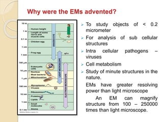
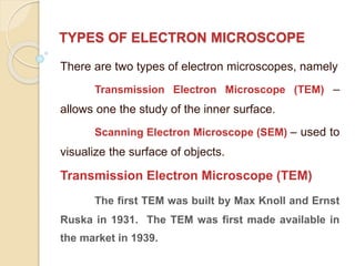
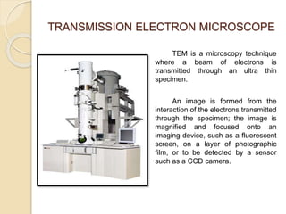
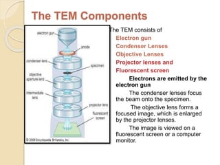
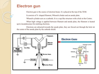
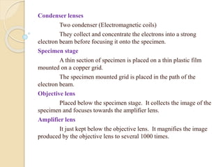
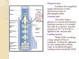
ELECTRON MICROSCOPE
MRS. R. NITHYA M. A., M. Sc., M. Phil., PGDCA.,
ASSISTANT PROFESSOR
SRI ADI CHUNCHANAGIRI WOMEN’S COLLEGE
CUMBUM - 625516
Electron microscope
What are electron microscopes?
Electron microscopes are scientific instruments that use
highly energetic electron to examine objects on a very fine scale
which yield the following information:
1. Topography: The surface features of an object (hardness,
reflectivity… etc.)
2. Morphology: The shape and size of the particles
3. Composition: The elements and compounds that the object
is composed of and the relative amount of them.
4. Crystallographic information: How the atoms are arranged in
the object.
Why were the EMs advented?
 To study objects of < 0.2
micrometer
 For analysis of sub cellular
structures
 Intra cellular pathogens –
viruses
 Cell metabolism
 Study of minute structures in the
nature.
EMs have greater resolving
power than light microscope
An EM can magnify
structure from 100 – 250000
times than light microscope.
TYPES OF ELECTRON MICROSCOPE
There are two types of electron microscopes, namely
Transmission Electron Microscope (TEM) –
allows one the study of the inner surface.
Scanning Electron Microscope (SEM) – used to
visualize the surface of objects.
Transmission Electron Microscope (TEM)
The first TEM was built by Max Knoll and Ernst
Ruska in 1931. The TEM was first made available in
the market in 1939.
TRANSMISSION ELECTRON MICROSCOPE
TEM is a microscopy technique
where a beam of electrons is
transmitted through an ultra thin
specimen.
An image is formed from the
interaction of the electrons transmitted
through the specimen; the image is
magnified and focused onto an
imaging device, such as a fluorescent
screen, on a layer of photographic
film, or to be detected by a sensor
such as a CCD camera.
The TEM Components
The TEM consists of
Electron gun
Condenser Lenses
Objective Lenses
Projector lenses and
Fluorescent screen
Electrons are emitted by the
electron gun
The condenser lenses focus
the beam onto the specimen.
The objective lens forms a
focused image, which is enlarged
by the projector lenses.
The image is viewed on a
fluorescent screen or a computer
monitor.
Electron gun
Electron gun is the source of electron beam. It is placed at the top of the TEM.
It consists of V- shaped filament, Whenelt clinder and an anode plate.
Whenelt cylinder acts as a cathode. It is a cup-like structure with a hole at the Centre.
When high voltage is applied between filament and anode plate, the filament is heated
up to incandescence for emitting electrons.
Electrons are attracted towards the anode plate, they are forced out through the hole int
the centre of the anode plate by the cathode shield.
Condenser lenses
Two condenser (Electromagnetic coils)
They collect and concentrate the electrons into a strong
electron beam before focusing it onto the specimen.
Specimen stage
A thin section of specimen is placed on a thin plastic film
mounted on a copper grid.
The specimen mounted grid is placed in the path of the
electron beam.
Objective lens
Placed below the specimen stage. It collects the image of the
specimen and focuses towards the amplifier lens.
Amplifier lens
It just kept below the objective lens. It magnifies the image
produced by the objective lens to several 1000 times.
Projector lens
It collects the magnified
image and focuses it onto
fluorescent screen or
photographic plate.
Vacuum tube
The entire setup is
placed in a vacuum tube because
electrons can move in a straight
line only in a vacuum. Vacuum
pressure of 10-7 to 10-9 Pa is
applied in the vacuum tube.
Cooling System
While TEM is working,
a large amount of heat is
produced. To keep the apparatus
at a low temperature cooling
water is circulated through a
cooling system around the TEM.
SAMPLE PREPARATION
DEHYDRATION
The wet sample is dehydrated by keeping in
increasing concentration of ethanol or acetone
FIXATION
Fixation is done by immersing the specimen in
chemical preservatives called fixative.
Osmium tetroxide, glutaraldehyde, potassium
permanganate, formalin, etc. are common fixatives.
These fixatives form covalent bond with biological
molecules like proteins and lipids.
They stabilize the structural organization in the
specimen.
EMBEDDING
The specimen is embedded in a hard embedding medium like
araldite
vestopal-W or
Epson-812 or
Plastic medium
The embedded specimen is cut into thin section of 50-100nm
thickness using a glass or diamond knife fixed in an ultramicrotome.
The thin section is mounted on a copper grid of 3mm
diameter and covered with parlodoan.
Ultramicrotome
STAINING
The specimen is kept dipped in a solution containing heavy metal ions for
metallic staining.
Solutions of phosphotungstate, Lead acetate, Lead hydroxide
Osmium tetroxide are useful for this staining.
The specimen mounted grid is then placed on the specimen stage.
The image of the selected section is then viewed on the fluorescent screen.
Image of the specimen is viewed through optical system.
Gamma phage
APPLICATIONS OF TEM
Ideal tool for the study of
ultra structure of cells Chloroplast
Plant cell
Employed in the localization of nucleic acid,
enzymes and protein in cells
and cell organelles.
Baculo virus release DNA Uncoiled DNA
Used in cancer research for the cytological
observation of cancer cells.
Colon cancer cells
Used in identification of plant and animal viruses
Plant virus – tobacco mosaic virus
Animal virus – Polio virus
ADAVANTAGES
 TEM offer the most powerful magnification, potentially
over one million times or more
 TEMs provide information on element and compound
structure
 Images are high- quality and detailed
 They are easy to operate with proper training
SCANNING ELECTRON MICROSCOPE
 SEM produces the
image by scanning it
with focus beam of
electron.
 Electrons interact with
electrons in sample and
convey information in
form of signals to
detectors.
 SEM can achieve
resolution better than
1nanometer.
SEM COMPONENTS
SEM consists of an
Electron gun – produces an e-beam
Condenser lens – collect and
concentrate the electrons into a
strong electron beam before
focusing it onto the specimen
Deflection coil – change the direction
of electron path helps for focussing
the e- beam on the specimen
Specimen stage – Specimen
mounted grid is p[laced on the
specimen stage in a 450 slanting
position.
When e- beam is focused on
the specimen , it produces
secondary electrons(SE), back-
scattered electrons (BSE) and
characteristic x-rays.
Secondary e- detector
The emitted SE is collected by
SED and converted into signal that is
sent to a screen which produces the final
image.
Additional sensors – detect
backscattered electrons and x-rays.
Amplifiers
Different kind of
amplifiers(grid, scintillator and PMT) are
used to measure the electrical signals.
The electrical signals are
converted into bright spots of varying
density by scanning circuit to give the
image of the specimen.
Photographic plate
The image may be captured on a
photographic plate or computer monitor
or hard disk.
Vacuum tube
The entire setup is placed in a vacuum tube because
electrons can move in a straight line only in a vacuum.
Vacuum pressure of 10-7 to 10-9 Pa is applied in the vacuum
tube.
Cooling System
While TEM is working, a large amount of heat is
produced. To keep the apparatus at a low temperature cooling
water is circulated through a cooling system around the TEM.
Blood cells Head of bee
Specimen preparation - Dry sample
Metallic Coating
Dry materials such as wood, bone, feathers,
insect’s wings and shells are coated with electronically
conducting materials.
They are metallic gold, platinum, tungsten,
iridium, osmium, chromium and graphite.
Metallic coating prevents the accumulation of
electrostatic charges on the specimen.
Penguin feather Peacock feather
Wet sample preparation
Fixation
Fixation is done by immersing the specimen in
chemical preservatives. It stabilize the molecular
organization.
Common fixatives are Osmium tetraoxide,
glutaraldehyde, potassium permanganate, formalin etc.
Dehydration
After fixation the specimen is dehydrated by
keeping it in concentrated ethanol or acetone.
Staining
The specimen is then coated with an ultrathin layer
of electroconductive alloy. Then it is placed in slanting
position on the specimen holder.
APPLICATIONS OF SEM
 SEM is very useful to view the surface architecture of microscopic
creatures like bacteria, diatoms, pollen grains, nematodes and others.
 SEM gives 3-D structure of objects.
Pollen grains of sunflower
Hairs and scales on plant and animal surfaces are
characterized with SEM.
Plant trichome
Hairs on the legs of
palnt bug
SEM is employed in the analysis of structural
features of compound eyes of insects.
Eye of black garden ant
Mosquito eyes
Eyes of yellow dung
fly
Electron microscope ppt

Electron microscope ppt

  • 1.
    ELECTRON MICROSCOPE MRS. R.NITHYA M. A., M. Sc., M. Phil., PGDCA., ASSISTANT PROFESSOR SRI ADI CHUNCHANAGIRI WOMEN’S COLLEGE CUMBUM - 625516
  • 2.
    Electron microscope What areelectron microscopes? Electron microscopes are scientific instruments that use highly energetic electron to examine objects on a very fine scale which yield the following information: 1. Topography: The surface features of an object (hardness, reflectivity… etc.) 2. Morphology: The shape and size of the particles 3. Composition: The elements and compounds that the object is composed of and the relative amount of them. 4. Crystallographic information: How the atoms are arranged in the object.
  • 3.
    Why were theEMs advented?  To study objects of < 0.2 micrometer  For analysis of sub cellular structures  Intra cellular pathogens – viruses  Cell metabolism  Study of minute structures in the nature. EMs have greater resolving power than light microscope An EM can magnify structure from 100 – 250000 times than light microscope.
  • 4.
    TYPES OF ELECTRONMICROSCOPE There are two types of electron microscopes, namely Transmission Electron Microscope (TEM) – allows one the study of the inner surface. Scanning Electron Microscope (SEM) – used to visualize the surface of objects. Transmission Electron Microscope (TEM) The first TEM was built by Max Knoll and Ernst Ruska in 1931. The TEM was first made available in the market in 1939.
  • 5.
    TRANSMISSION ELECTRON MICROSCOPE TEMis a microscopy technique where a beam of electrons is transmitted through an ultra thin specimen. An image is formed from the interaction of the electrons transmitted through the specimen; the image is magnified and focused onto an imaging device, such as a fluorescent screen, on a layer of photographic film, or to be detected by a sensor such as a CCD camera.
  • 6.
    The TEM Components TheTEM consists of Electron gun Condenser Lenses Objective Lenses Projector lenses and Fluorescent screen Electrons are emitted by the electron gun The condenser lenses focus the beam onto the specimen. The objective lens forms a focused image, which is enlarged by the projector lenses. The image is viewed on a fluorescent screen or a computer monitor.
  • 7.
    Electron gun Electron gunis the source of electron beam. It is placed at the top of the TEM. It consists of V- shaped filament, Whenelt clinder and an anode plate. Whenelt cylinder acts as a cathode. It is a cup-like structure with a hole at the Centre. When high voltage is applied between filament and anode plate, the filament is heated up to incandescence for emitting electrons. Electrons are attracted towards the anode plate, they are forced out through the hole int the centre of the anode plate by the cathode shield.
  • 8.
    Condenser lenses Two condenser(Electromagnetic coils) They collect and concentrate the electrons into a strong electron beam before focusing it onto the specimen. Specimen stage A thin section of specimen is placed on a thin plastic film mounted on a copper grid. The specimen mounted grid is placed in the path of the electron beam. Objective lens Placed below the specimen stage. It collects the image of the specimen and focuses towards the amplifier lens. Amplifier lens It just kept below the objective lens. It magnifies the image produced by the objective lens to several 1000 times.
  • 9.
    Projector lens It collectsthe magnified image and focuses it onto fluorescent screen or photographic plate. Vacuum tube The entire setup is placed in a vacuum tube because electrons can move in a straight line only in a vacuum. Vacuum pressure of 10-7 to 10-9 Pa is applied in the vacuum tube. Cooling System While TEM is working, a large amount of heat is produced. To keep the apparatus at a low temperature cooling water is circulated through a cooling system around the TEM.
  • 10.
    SAMPLE PREPARATION DEHYDRATION The wetsample is dehydrated by keeping in increasing concentration of ethanol or acetone FIXATION Fixation is done by immersing the specimen in chemical preservatives called fixative. Osmium tetroxide, glutaraldehyde, potassium permanganate, formalin, etc. are common fixatives. These fixatives form covalent bond with biological molecules like proteins and lipids. They stabilize the structural organization in the specimen.
  • 11.
    EMBEDDING The specimen isembedded in a hard embedding medium like araldite vestopal-W or Epson-812 or Plastic medium The embedded specimen is cut into thin section of 50-100nm thickness using a glass or diamond knife fixed in an ultramicrotome. The thin section is mounted on a copper grid of 3mm diameter and covered with parlodoan. Ultramicrotome
  • 12.
    STAINING The specimen iskept dipped in a solution containing heavy metal ions for metallic staining. Solutions of phosphotungstate, Lead acetate, Lead hydroxide Osmium tetroxide are useful for this staining. The specimen mounted grid is then placed on the specimen stage. The image of the selected section is then viewed on the fluorescent screen. Image of the specimen is viewed through optical system. Gamma phage
  • 13.
    APPLICATIONS OF TEM Idealtool for the study of ultra structure of cells Chloroplast Plant cell
  • 14.
    Employed in thelocalization of nucleic acid, enzymes and protein in cells and cell organelles. Baculo virus release DNA Uncoiled DNA
  • 15.
    Used in cancerresearch for the cytological observation of cancer cells. Colon cancer cells
  • 16.
    Used in identificationof plant and animal viruses Plant virus – tobacco mosaic virus Animal virus – Polio virus
  • 17.
    ADAVANTAGES  TEM offerthe most powerful magnification, potentially over one million times or more  TEMs provide information on element and compound structure  Images are high- quality and detailed  They are easy to operate with proper training
  • 18.
    SCANNING ELECTRON MICROSCOPE SEM produces the image by scanning it with focus beam of electron.  Electrons interact with electrons in sample and convey information in form of signals to detectors.  SEM can achieve resolution better than 1nanometer.
  • 19.
    SEM COMPONENTS SEM consistsof an Electron gun – produces an e-beam Condenser lens – collect and concentrate the electrons into a strong electron beam before focusing it onto the specimen Deflection coil – change the direction of electron path helps for focussing the e- beam on the specimen Specimen stage – Specimen mounted grid is p[laced on the specimen stage in a 450 slanting position. When e- beam is focused on the specimen , it produces secondary electrons(SE), back- scattered electrons (BSE) and characteristic x-rays.
  • 20.
    Secondary e- detector Theemitted SE is collected by SED and converted into signal that is sent to a screen which produces the final image. Additional sensors – detect backscattered electrons and x-rays. Amplifiers Different kind of amplifiers(grid, scintillator and PMT) are used to measure the electrical signals. The electrical signals are converted into bright spots of varying density by scanning circuit to give the image of the specimen. Photographic plate The image may be captured on a photographic plate or computer monitor or hard disk.
  • 21.
    Vacuum tube The entiresetup is placed in a vacuum tube because electrons can move in a straight line only in a vacuum. Vacuum pressure of 10-7 to 10-9 Pa is applied in the vacuum tube. Cooling System While TEM is working, a large amount of heat is produced. To keep the apparatus at a low temperature cooling water is circulated through a cooling system around the TEM. Blood cells Head of bee
  • 22.
    Specimen preparation -Dry sample Metallic Coating Dry materials such as wood, bone, feathers, insect’s wings and shells are coated with electronically conducting materials. They are metallic gold, platinum, tungsten, iridium, osmium, chromium and graphite. Metallic coating prevents the accumulation of electrostatic charges on the specimen. Penguin feather Peacock feather
  • 23.
    Wet sample preparation Fixation Fixationis done by immersing the specimen in chemical preservatives. It stabilize the molecular organization. Common fixatives are Osmium tetraoxide, glutaraldehyde, potassium permanganate, formalin etc. Dehydration After fixation the specimen is dehydrated by keeping it in concentrated ethanol or acetone. Staining The specimen is then coated with an ultrathin layer of electroconductive alloy. Then it is placed in slanting position on the specimen holder.
  • 24.
    APPLICATIONS OF SEM SEM is very useful to view the surface architecture of microscopic creatures like bacteria, diatoms, pollen grains, nematodes and others.  SEM gives 3-D structure of objects. Pollen grains of sunflower
  • 25.
    Hairs and scaleson plant and animal surfaces are characterized with SEM. Plant trichome Hairs on the legs of palnt bug
  • 26.
    SEM is employedin the analysis of structural features of compound eyes of insects. Eye of black garden ant
  • 27.
    Mosquito eyes Eyes ofyellow dung fly