The document summarizes the bones that make up the human skeletal system, including those of the axial skeleton, appendicular skeleton, pectoral girdle, upper extremities, pelvic girdle, and lower extremities. It lists the specific bones in each region, such as the humerus, radius, ulna, carpals, metacarpals and phalanges that comprise the upper extremities, and the femur, tibia, fibula, tarsals, metatarsals and phalanges that make up the lower extremities.
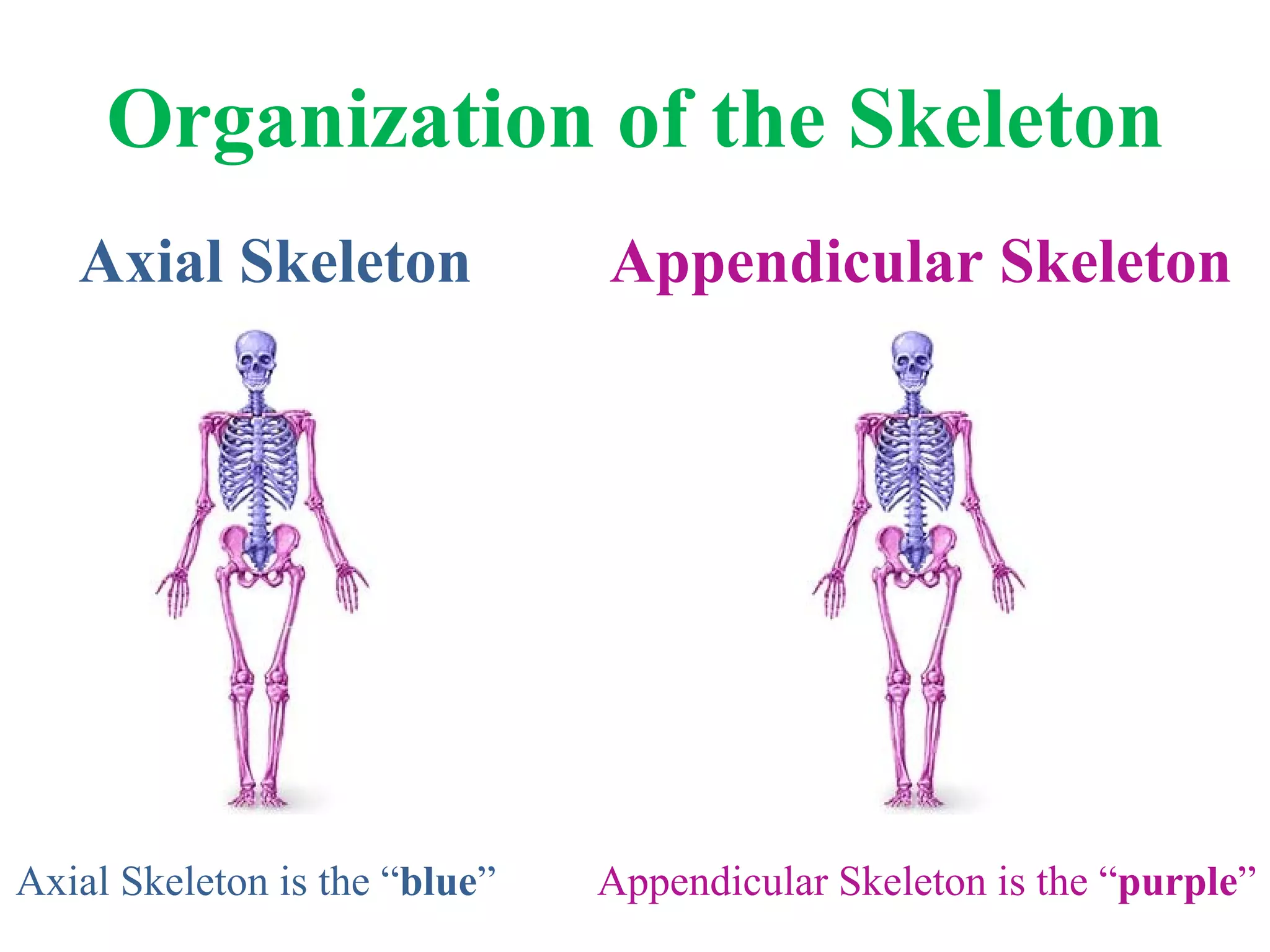
Organization of theSkeleton Axial Skeleton Appendicular Skeleton Axial Skeleton is the “ blue ” Appendicular Skeleton is the “ purple ”
2.
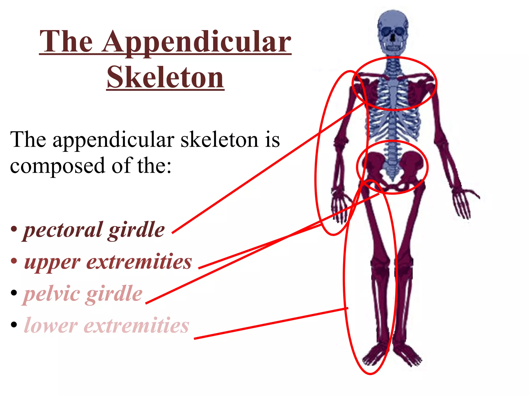
The Appendicular SkeletonThe appendicular skeleton is composed of the: pectoral girdle upper extremities pelvic girdle lower extremities
3.
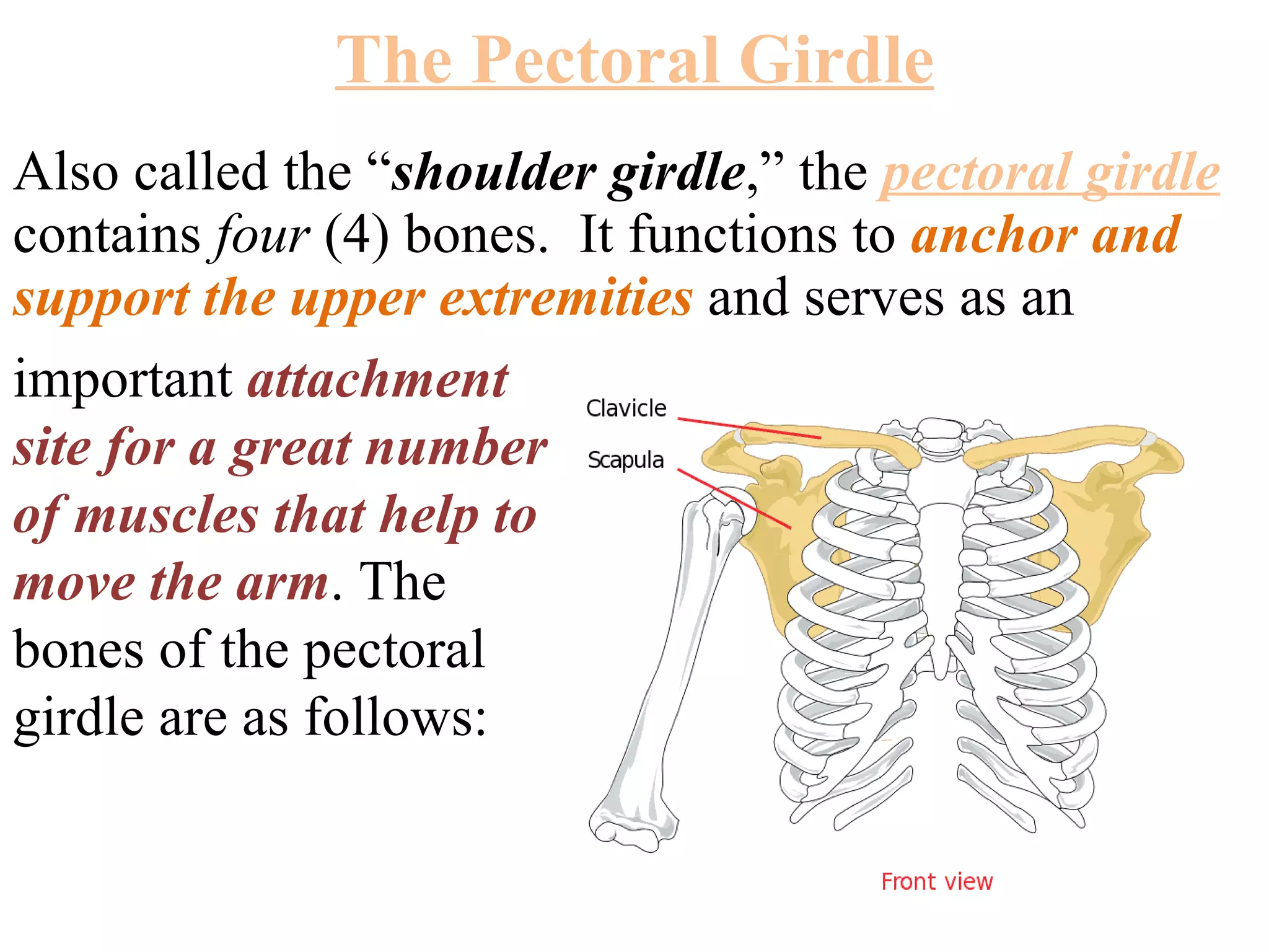
The Pectoral GirdleAlso called the “ shoulder girdle ,” the pectoral girdle contains four (4) bones. It functions to anchor and support the upper extremities and serves as an important attachment site for a great number of muscles that help to move the arm . The bones of the pectoral girdle are as follows:
4.
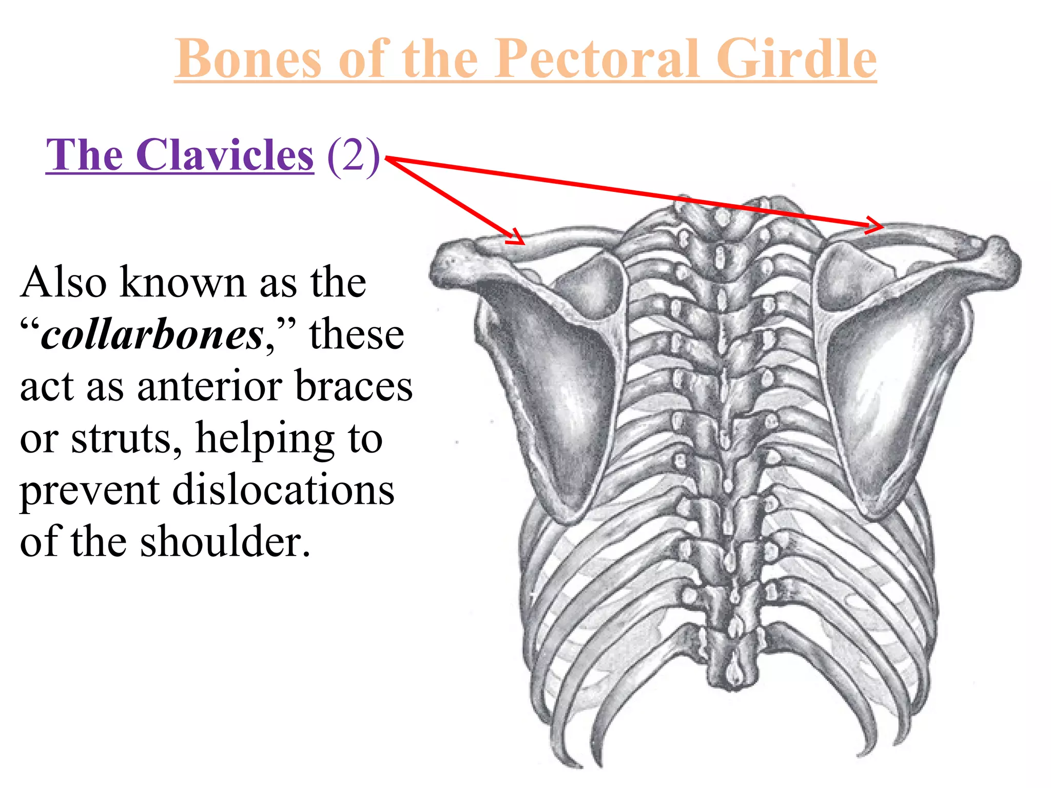
The Clavicles (2) Also known as the “ collarbones ,” these act as anterior braces or struts, helping to prevent dislocations of the shoulder. Bones of the Pectoral Girdle
5.
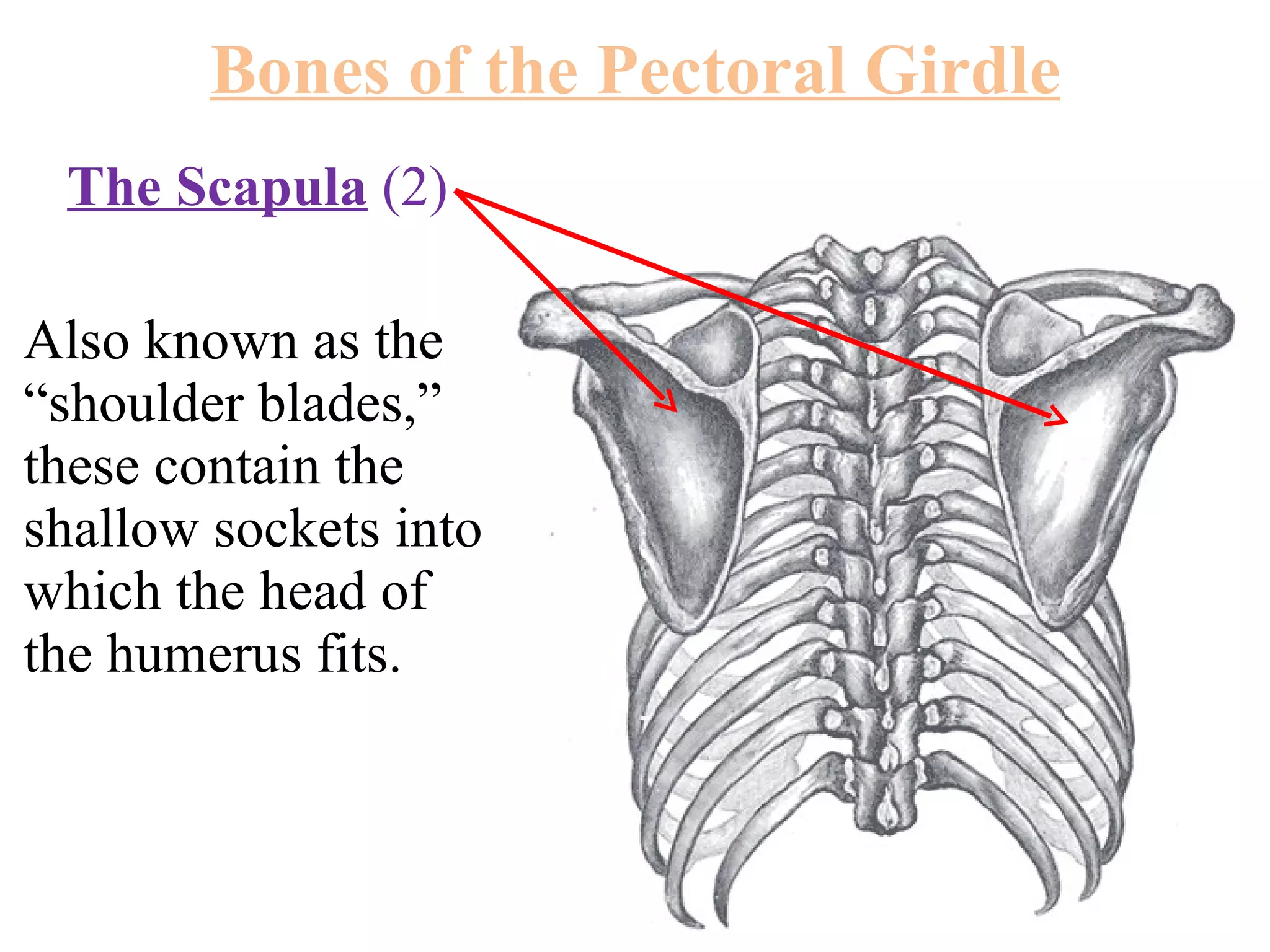
The Scapula (2) Also known as the “shoulder blades,” these contain the shallow sockets into which the head of the humerus fits. Bones of the Pectoral Girdle
6.
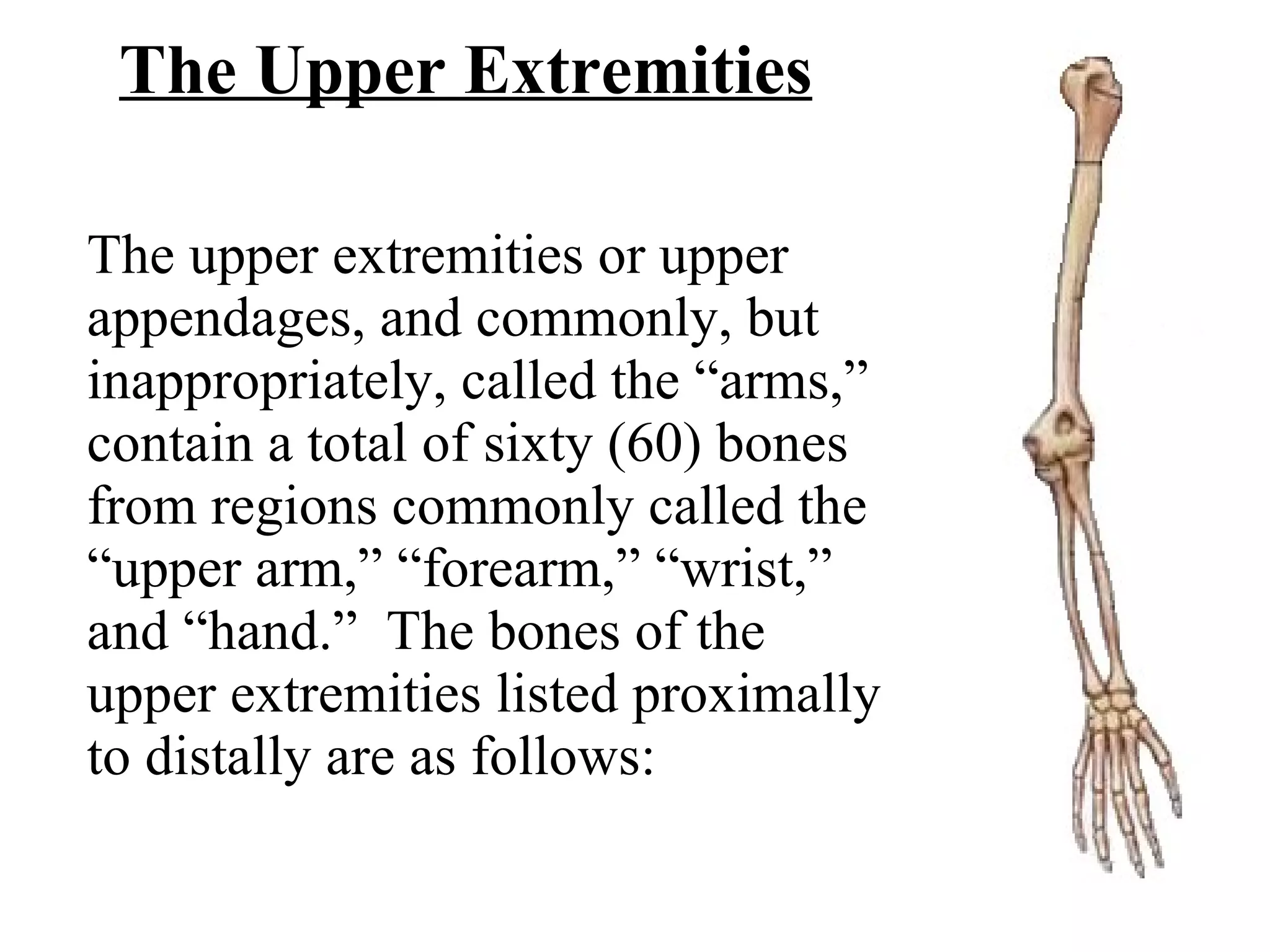
The Upper ExtremitiesThe upper extremities or upper appendages, and commonly, but inappropriately, called the “arms,” contain a total of sixty (60) bones from regions commonly called the “upper arm,” “forearm,” “wrist,” and “hand.” The bones of the upper extremities listed proximally to distally are as follows:
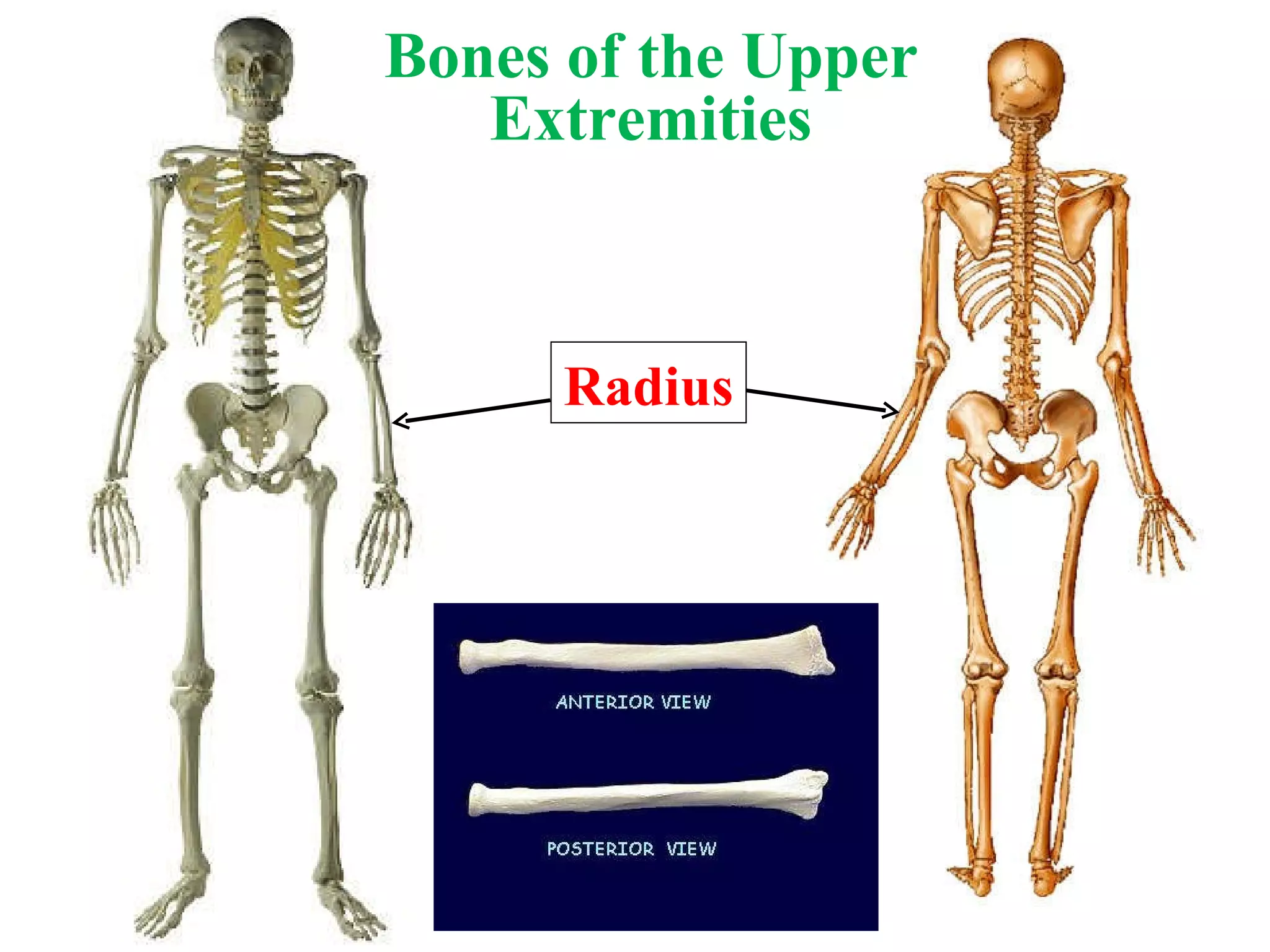
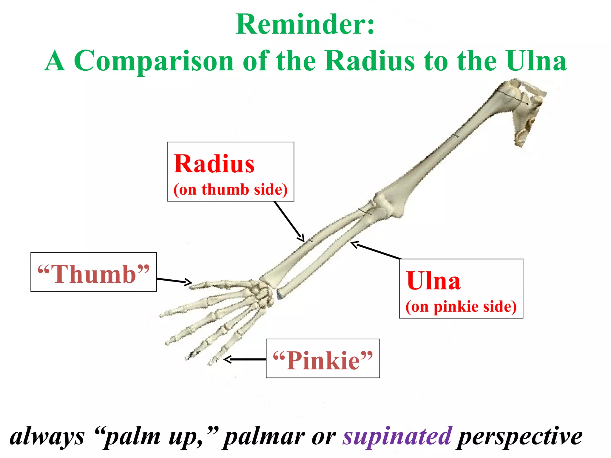
Reminder: A Comparisonof the Radius to the Ulna always “palm up,” palmar or supinated perspective “ Thumb” “ Pinkie” Radius (on thumb side) Ulna (on pinkie side)
11.
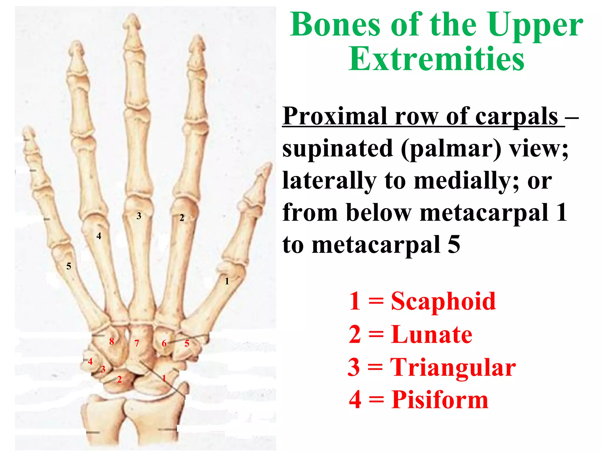
5 1 23 4 4 3 2 1 5 6 7 8 Proximal row of carpals – supinated (palmar) view; laterally to medially; or from below metacarpal 1 to metacarpal 5 Bones of the Upper Extremities 1 = Scaphoid 3 = Triangular 2 = Lunate 4 = Pisiform
12.
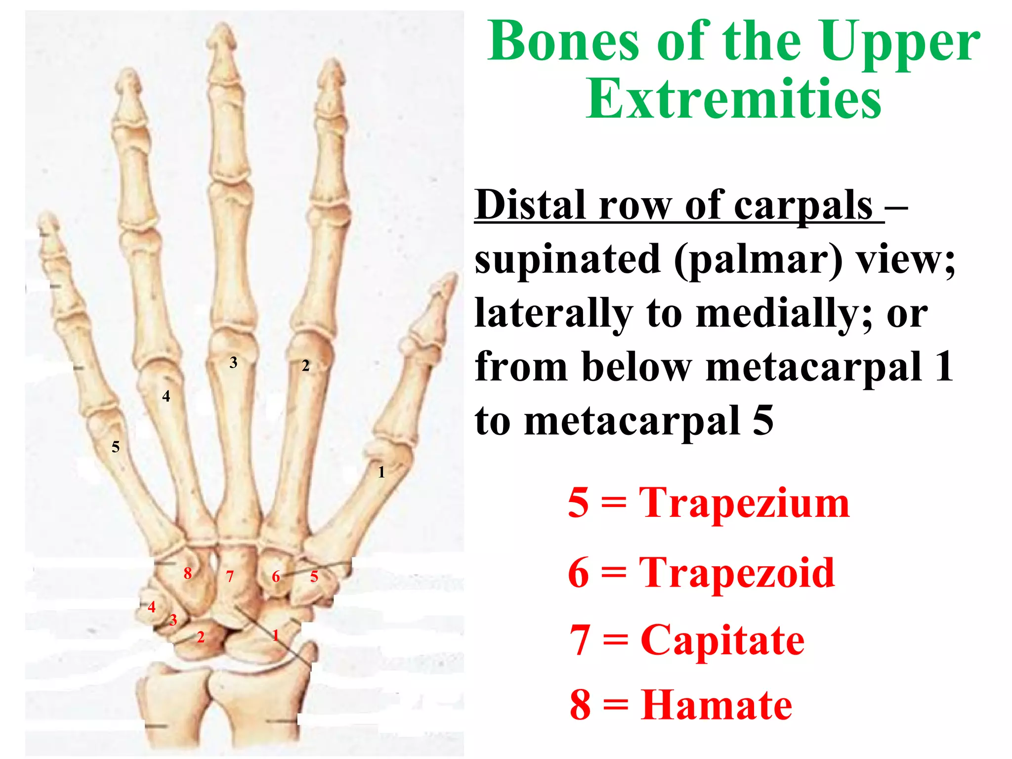
5 = Trapezium6 = Trapezoid 7 = Capitate 8 = Hamate Distal row of carpals – supinated (palmar) view; laterally to medially; or from below metacarpal 1 to metacarpal 5 5 1 2 3 4 4 3 2 1 5 6 7 8 Bones of the Upper Extremities
13.
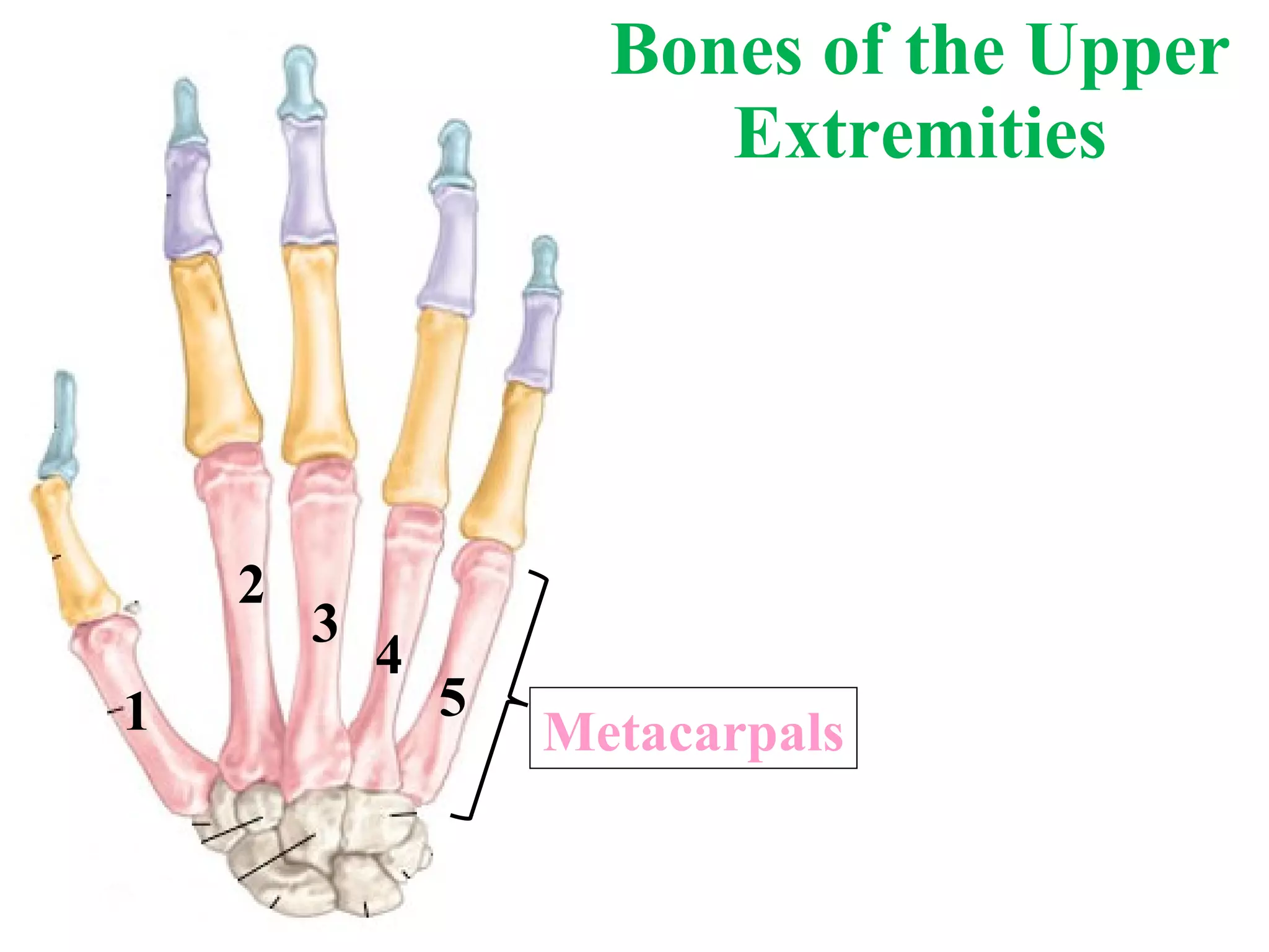
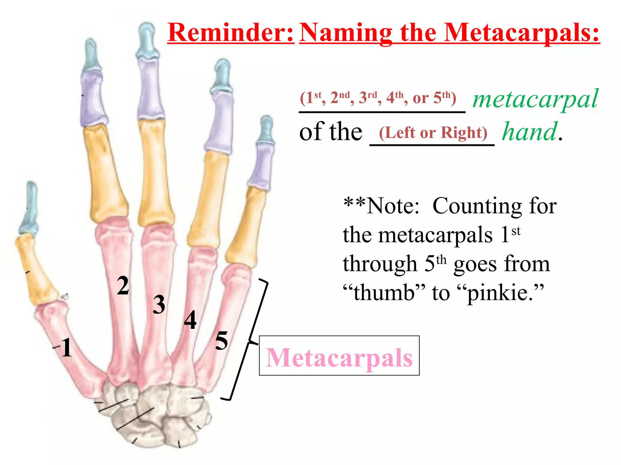
Bones of theUpper Extremities 1 2 3 4 5 Metacarpals
14.
Naming the Metacarpals:____________ metacarpal of the _________ hand . (1 st , 2 nd , 3 rd , 4 th , or 5 th ) (Left or Right) **Note: Counting for the metacarpals 1 st through 5 th goes from “thumb” to “pinkie.” Reminder: 1 2 3 4 5 Metacarpals
15.
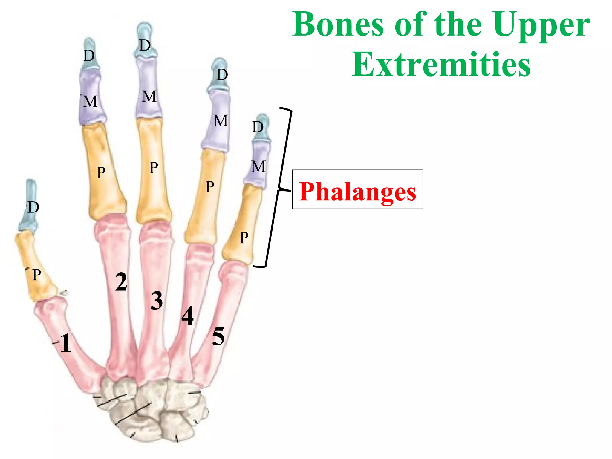
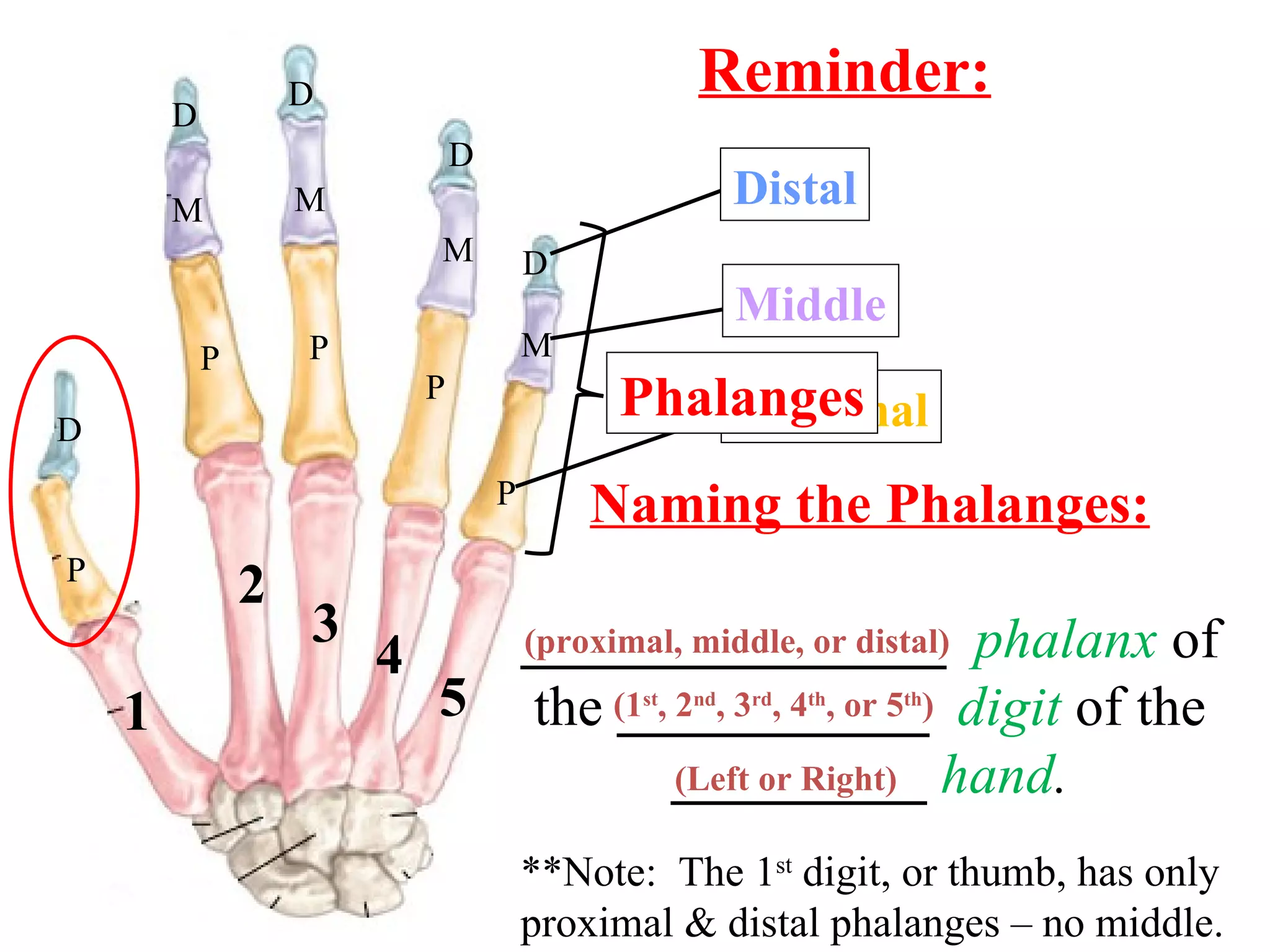
Bones of theUpper Extremities P P M D D P M D P M D P M D 1 2 3 4 5 Phalanges
16.
Naming the Phalanges:_______________ phalanx of the ___________ digit of the _________ hand . (1 st , 2 nd , 3 rd , 4 th , or 5 th ) (Left or Right) (proximal, middle, or distal) **Note: The 1 st digit, or thumb, has only proximal & distal phalanges – no middle. P P M D D P M D P M D P M D Reminder: 1 2 3 4 5 Proximal Middle Distal Phalanges
17.
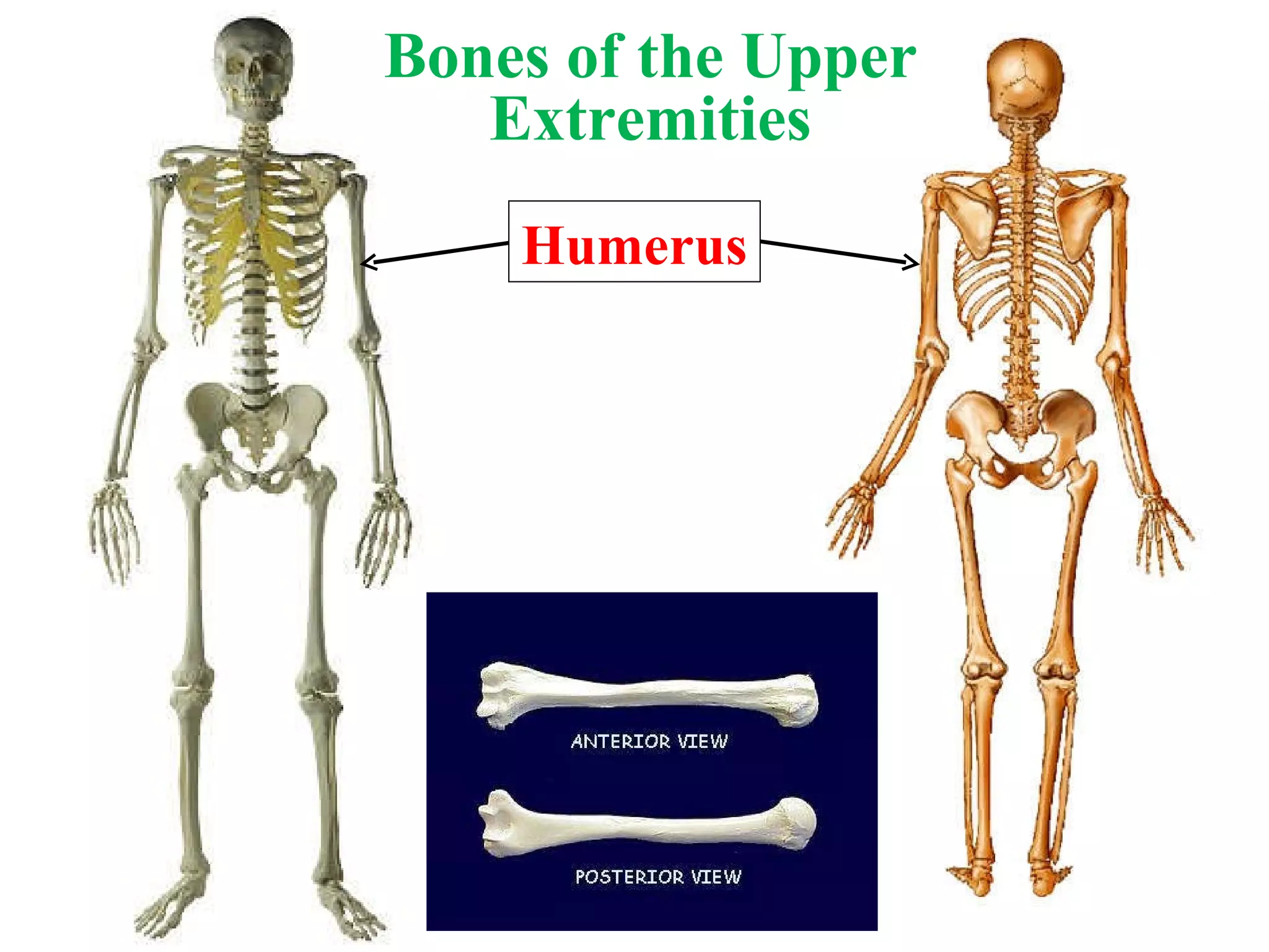
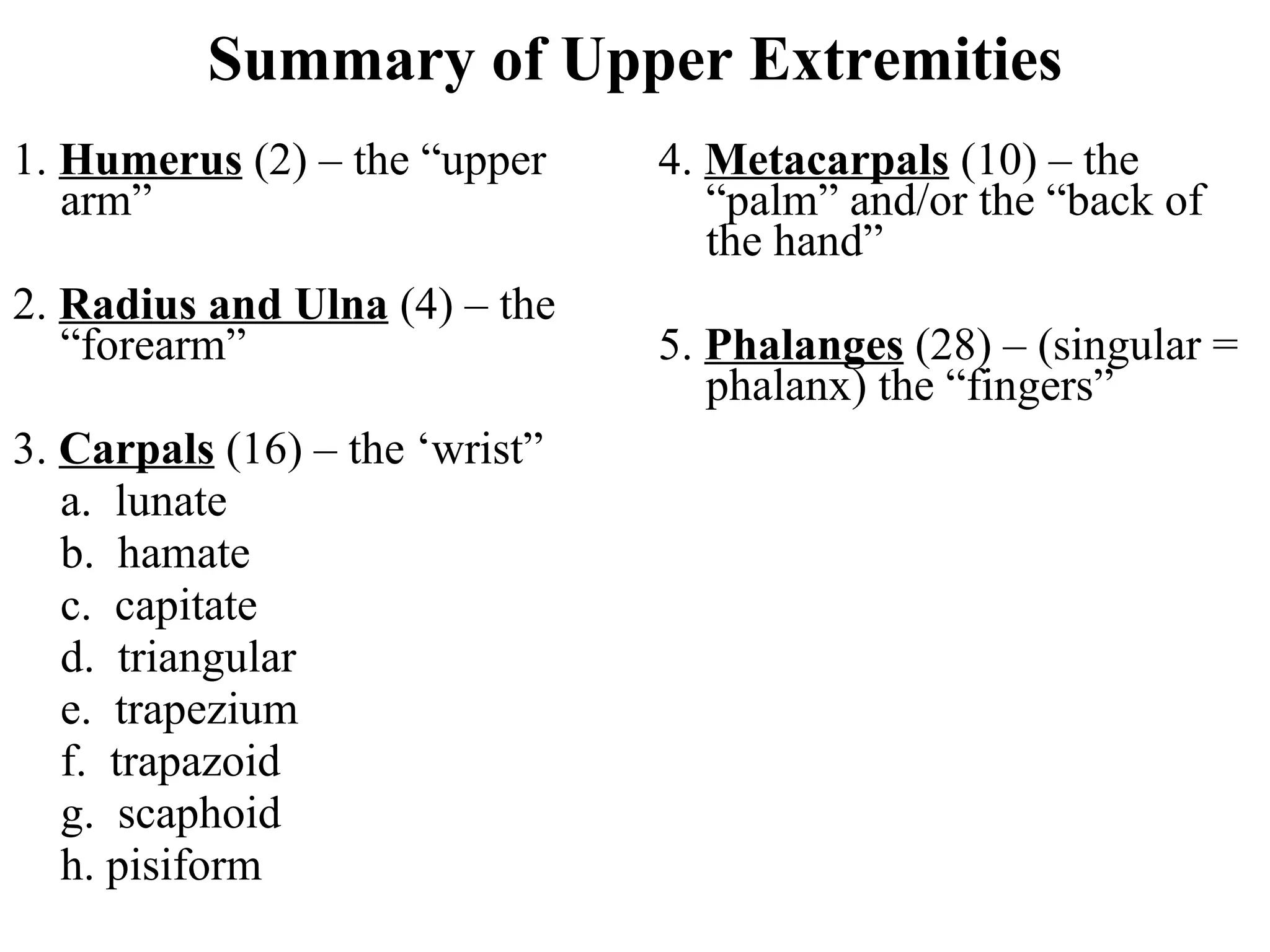
Summary of UpperExtremities 4. Metacarpals (10) – the “palm” and/or the “back of the hand” 5. Phalanges (28) – (singular = phalanx) the “fingers” 1. Humerus (2) – the “upper arm” 2. Radius and Ulna (4) – the “forearm” 3. Carpals (16) – the ‘wrist” a. lunate b. hamate c. capitate d. triangular e. trapezium f. trapazoid g. scaphoid h. pisiform
18.
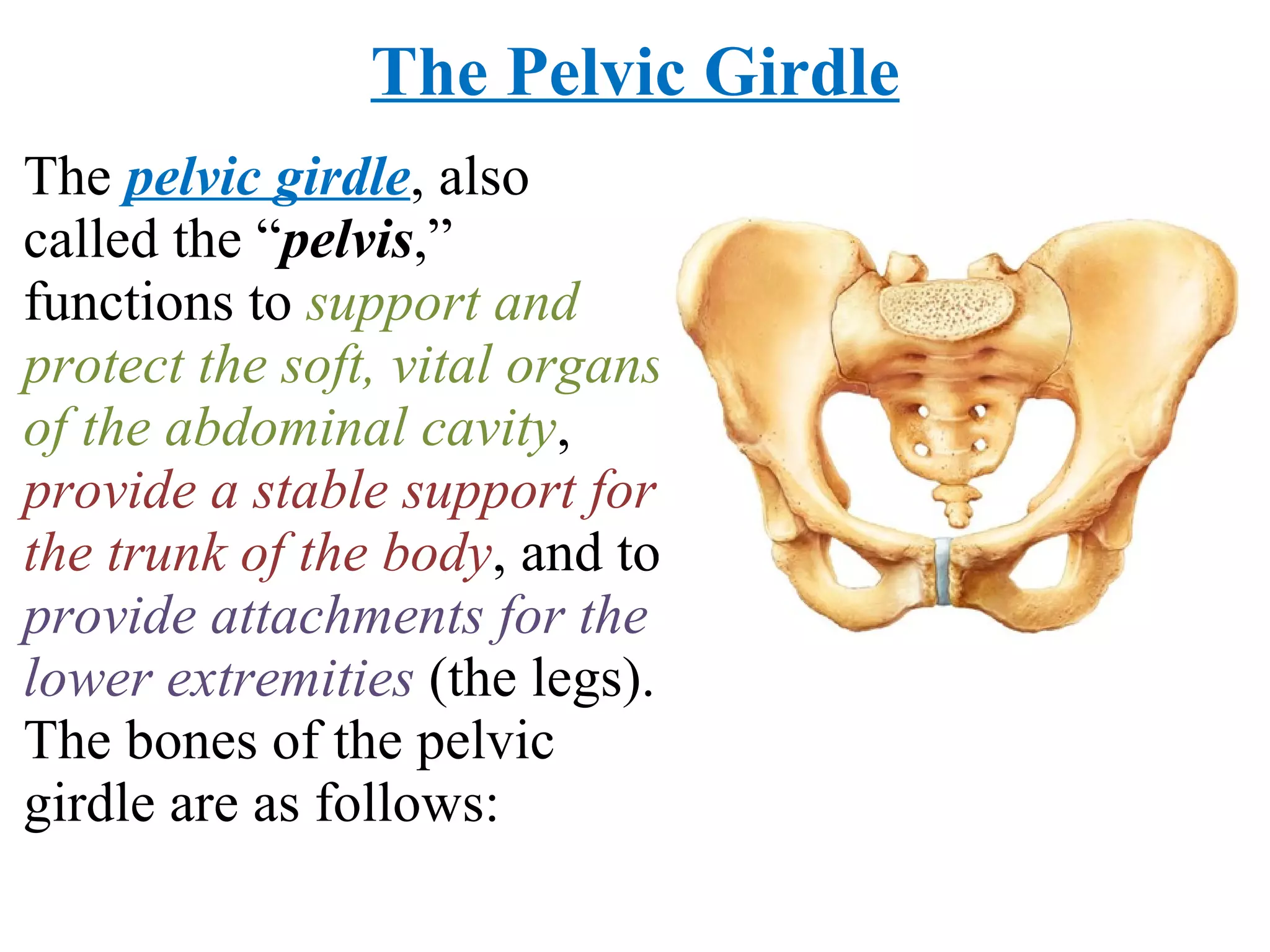
The Pelvic GirdleThe pelvic girdle , also called the “ pelvis ,” functions to support and protect the soft, vital organs of the abdominal cavity , provide a stable support for the trunk of the body , and to provide attachments for the lower extremities (the legs). The bones of the pelvic girdle are as follows:
19.
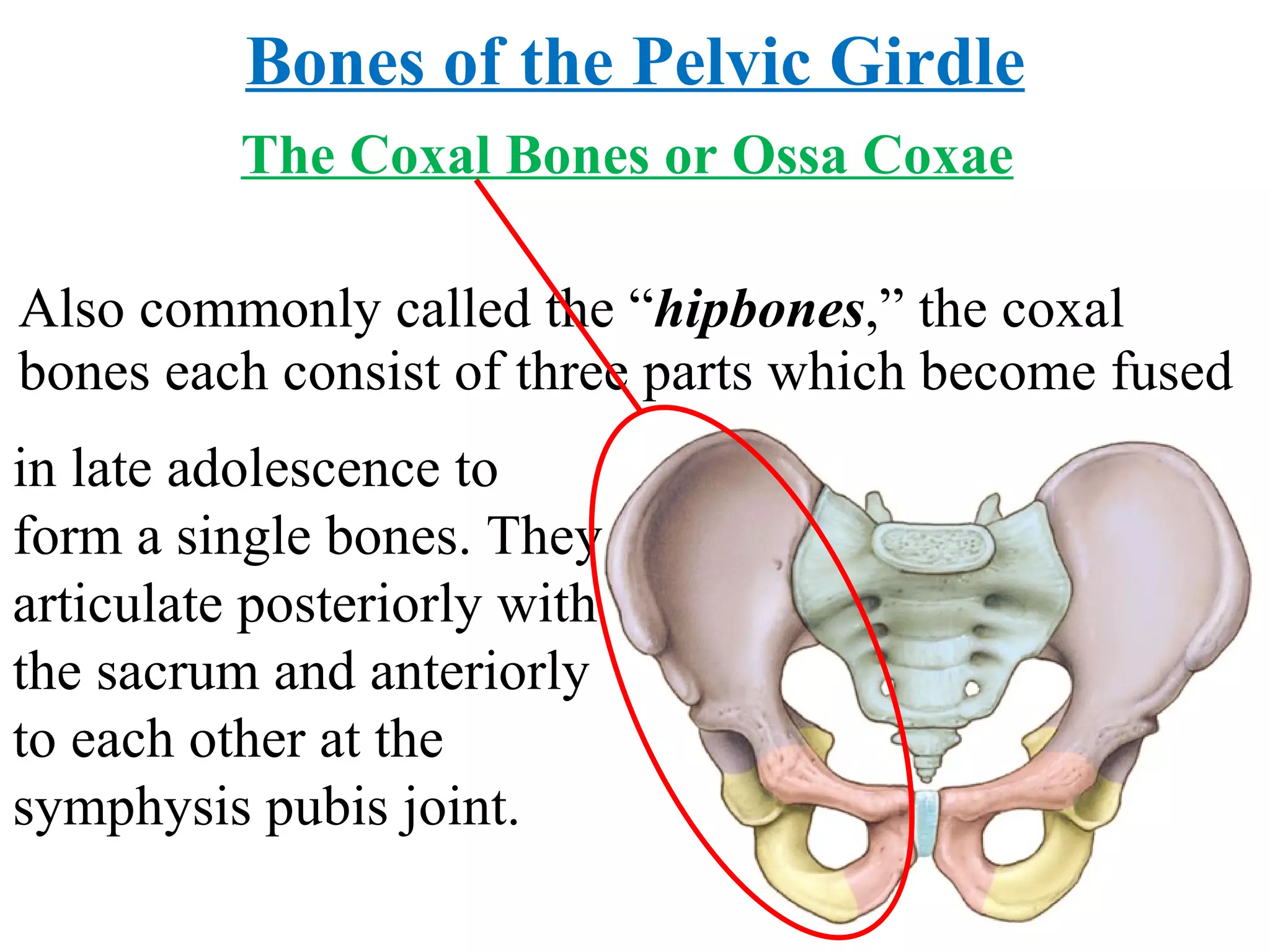
The Coxal Bonesor Ossa Coxae Also commonly called the “ hipbones ,” the coxal bones each consist of three parts which become fused Bones of the Pelvic Girdle in late adolescence to form a single bones. They articulate posteriorly with the sacrum and anteriorly to each other at the symphysis pubis joint.
20.
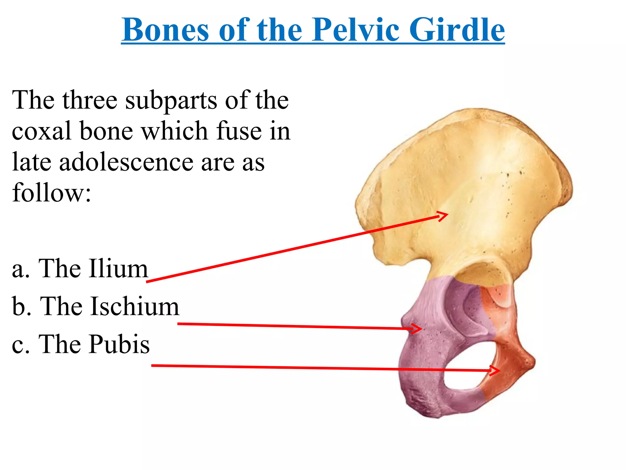
The three subpartsof the coxal bone which fuse in late adolescence are as follow: a. The Ilium b. The Ischium c. The Pubis Bones of the Pelvic Girdle
21.
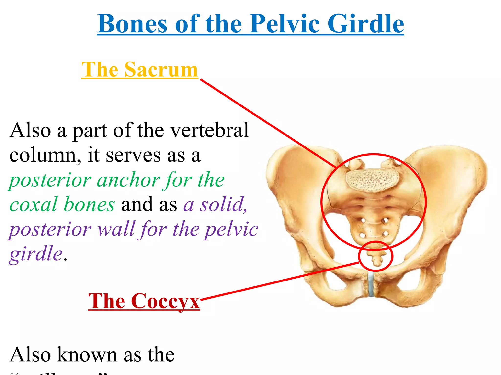
The Sacrum Alsoa part of the vertebral column, it serves as a posterior anchor for the coxal bones and as a solid, posterior wall for the pelvic girdle . Bones of the Pelvic Girdle The Coccyx Also known as the “ tailbone .”
22.
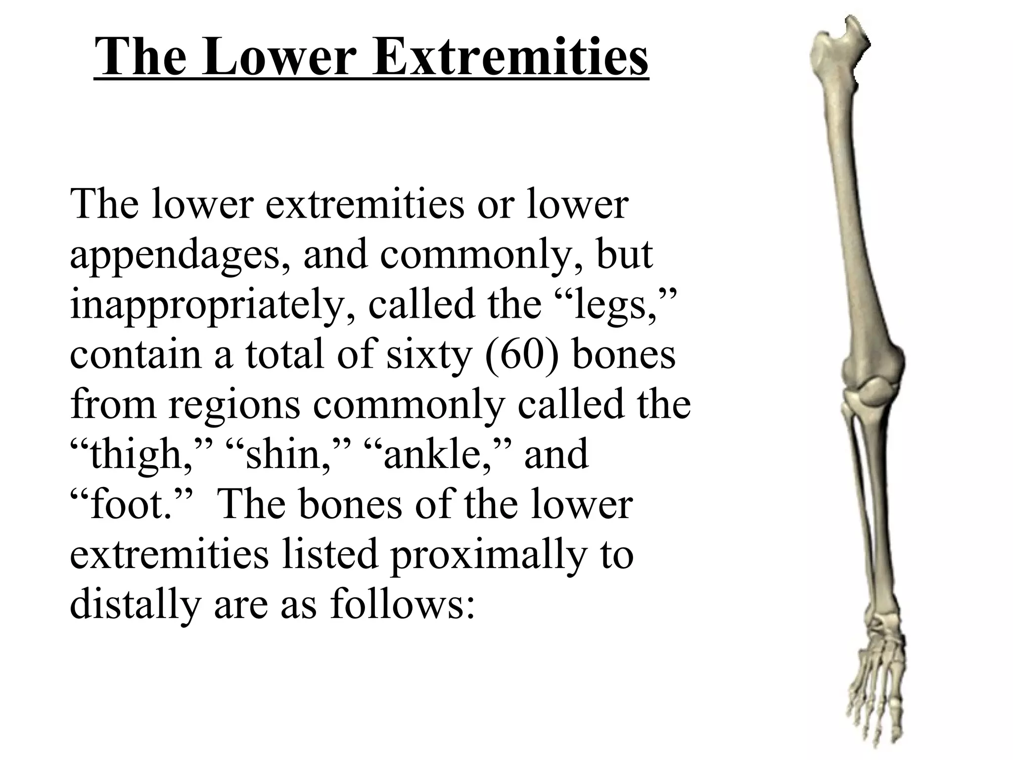
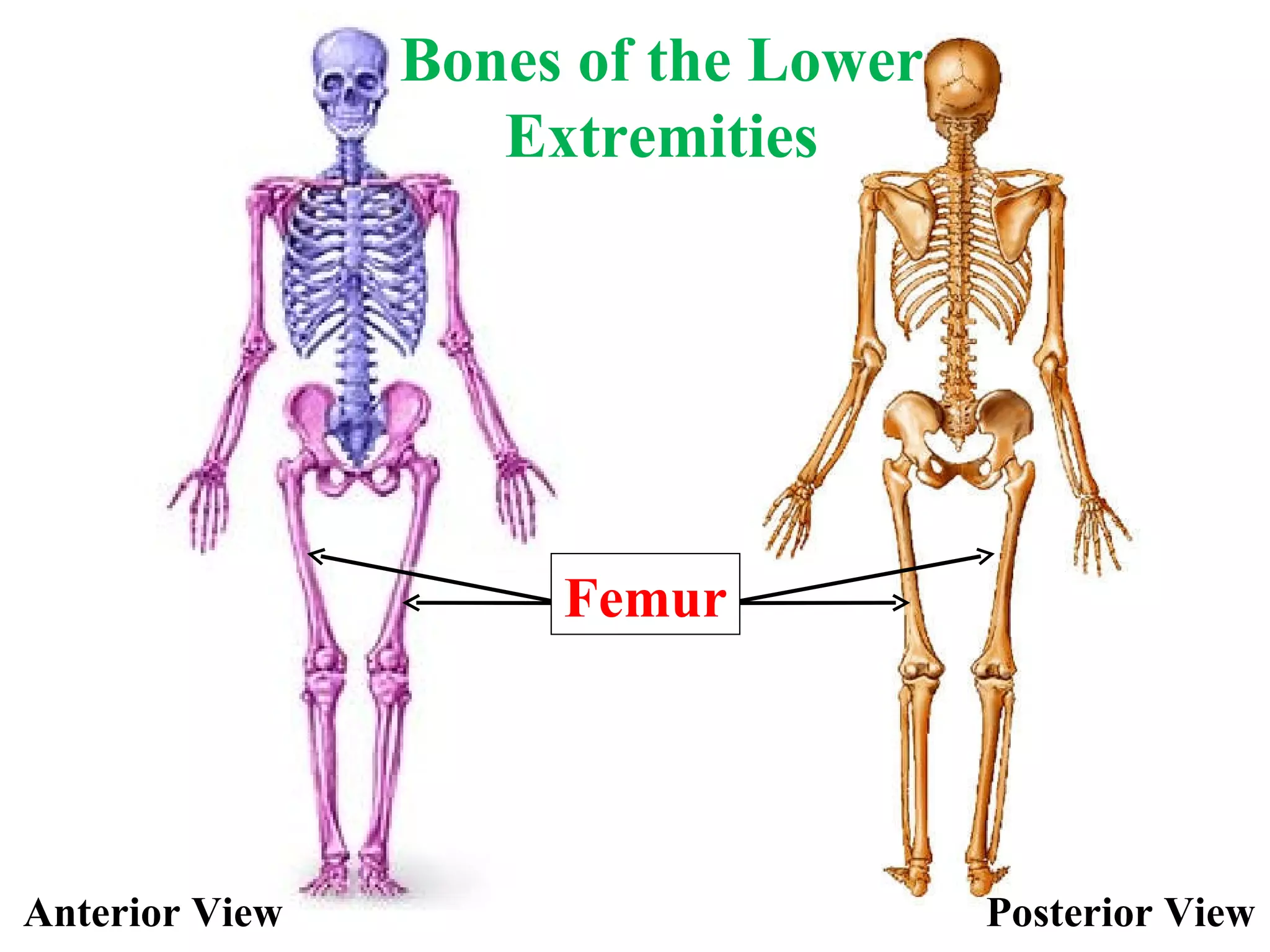
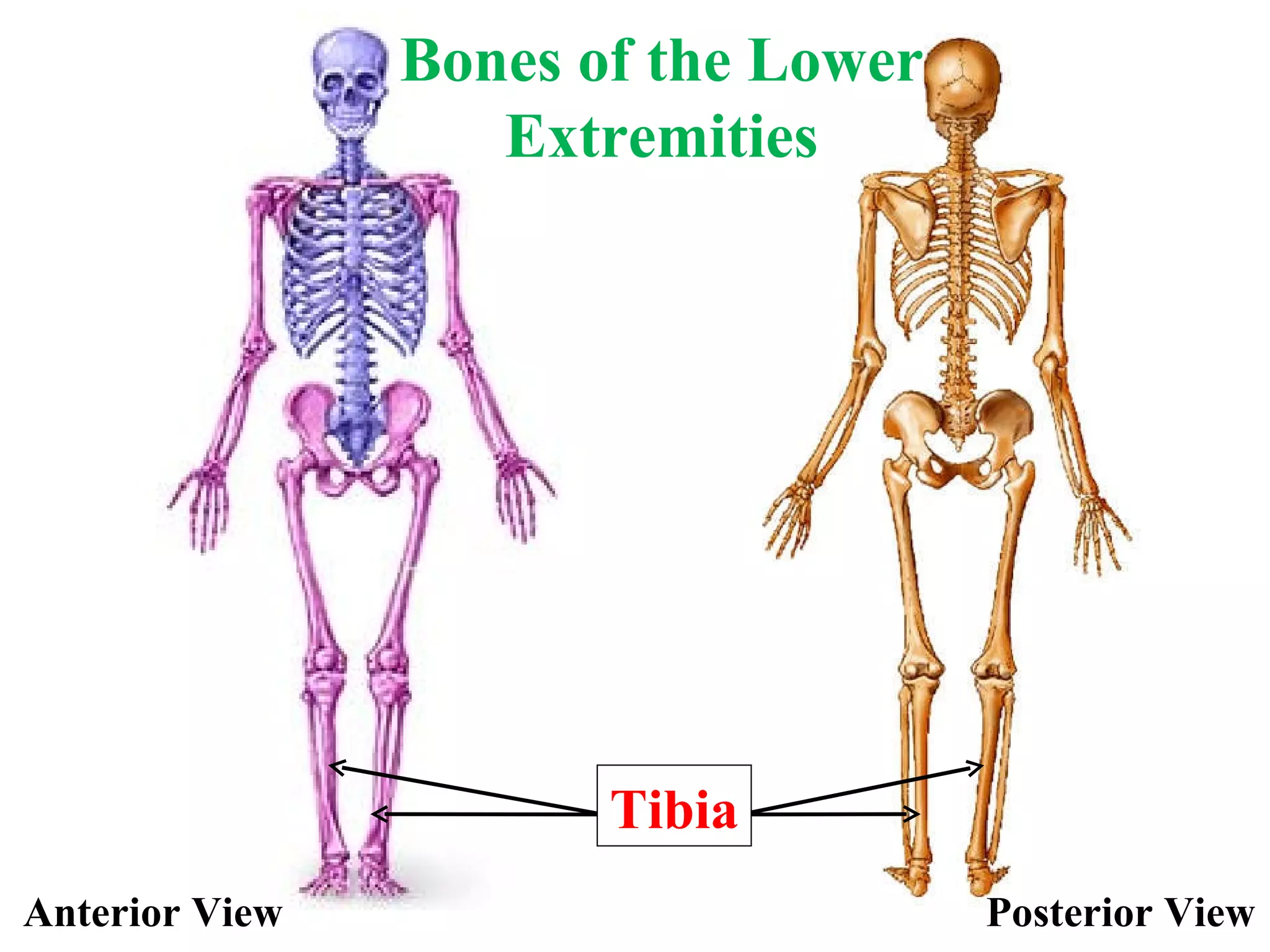
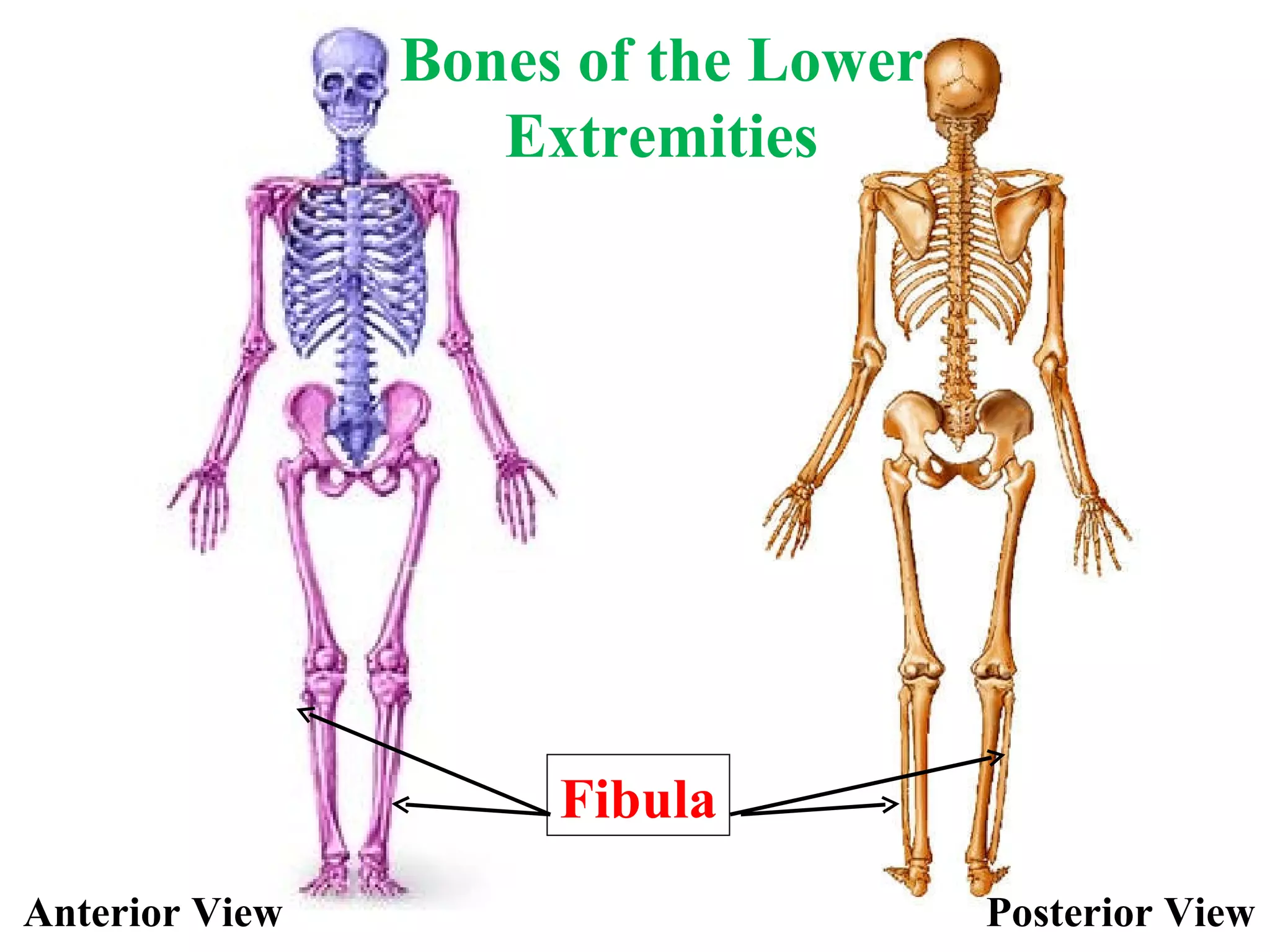
The Lower ExtremitiesThe lower extremities or lower appendages, and commonly, but inappropriately, called the “legs,” contain a total of sixty (60) bones from regions commonly called the “thigh,” “shin,” “ankle,” and “foot.” The bones of the lower extremities listed proximally to distally are as follows:
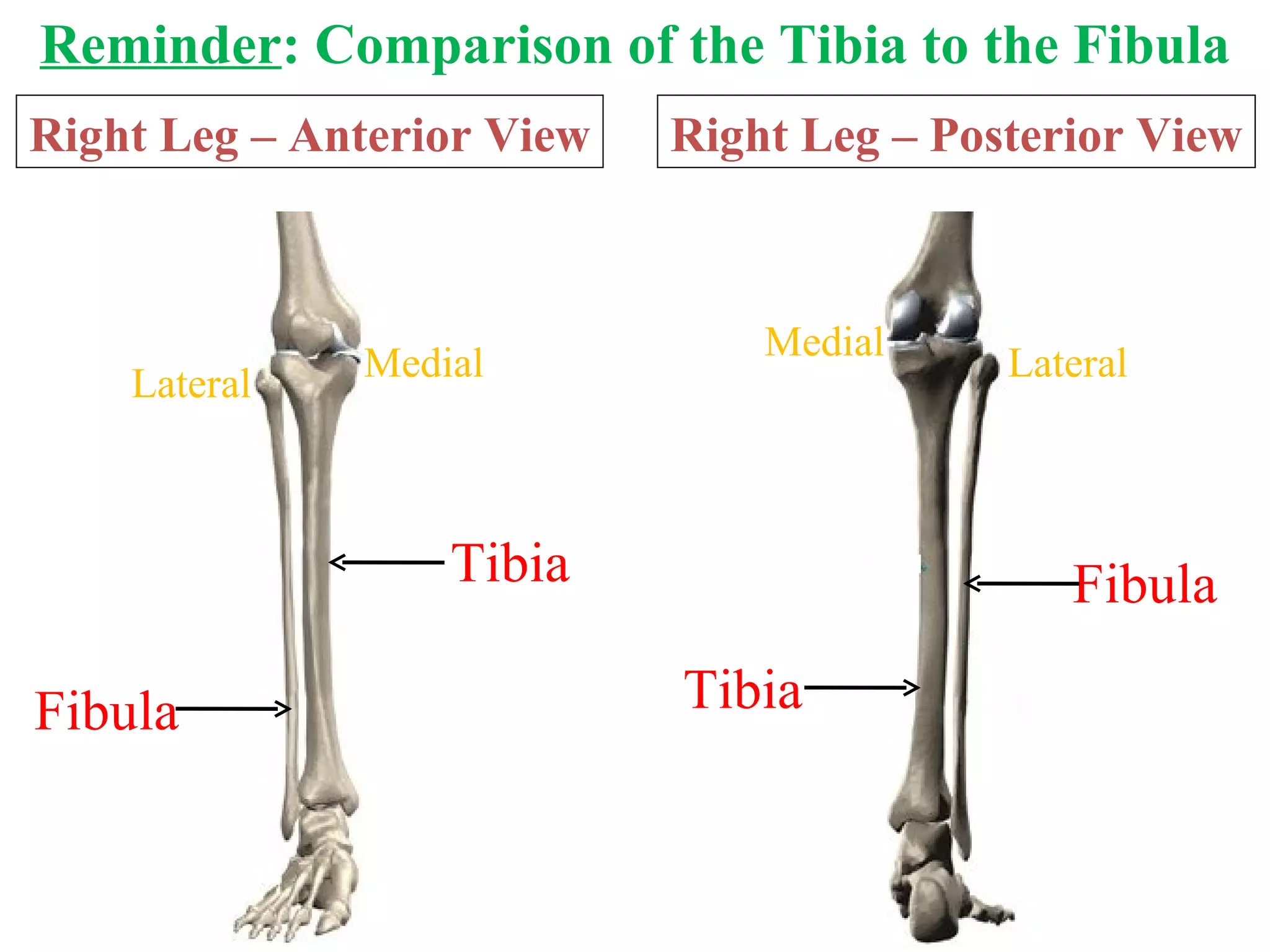
Reminder : Comparisonof the Tibia to the Fibula Right Leg – Anterior View Tibia Fibula Lateral Medial Right Leg – Posterior View Fibula Tibia Lateral Medial
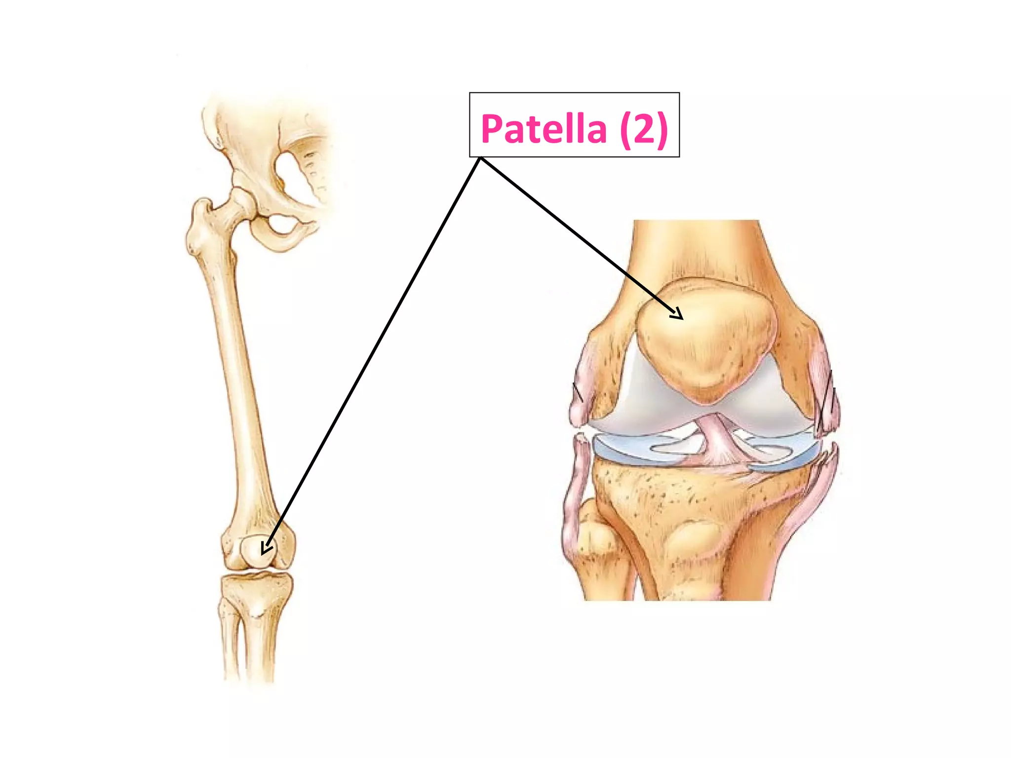
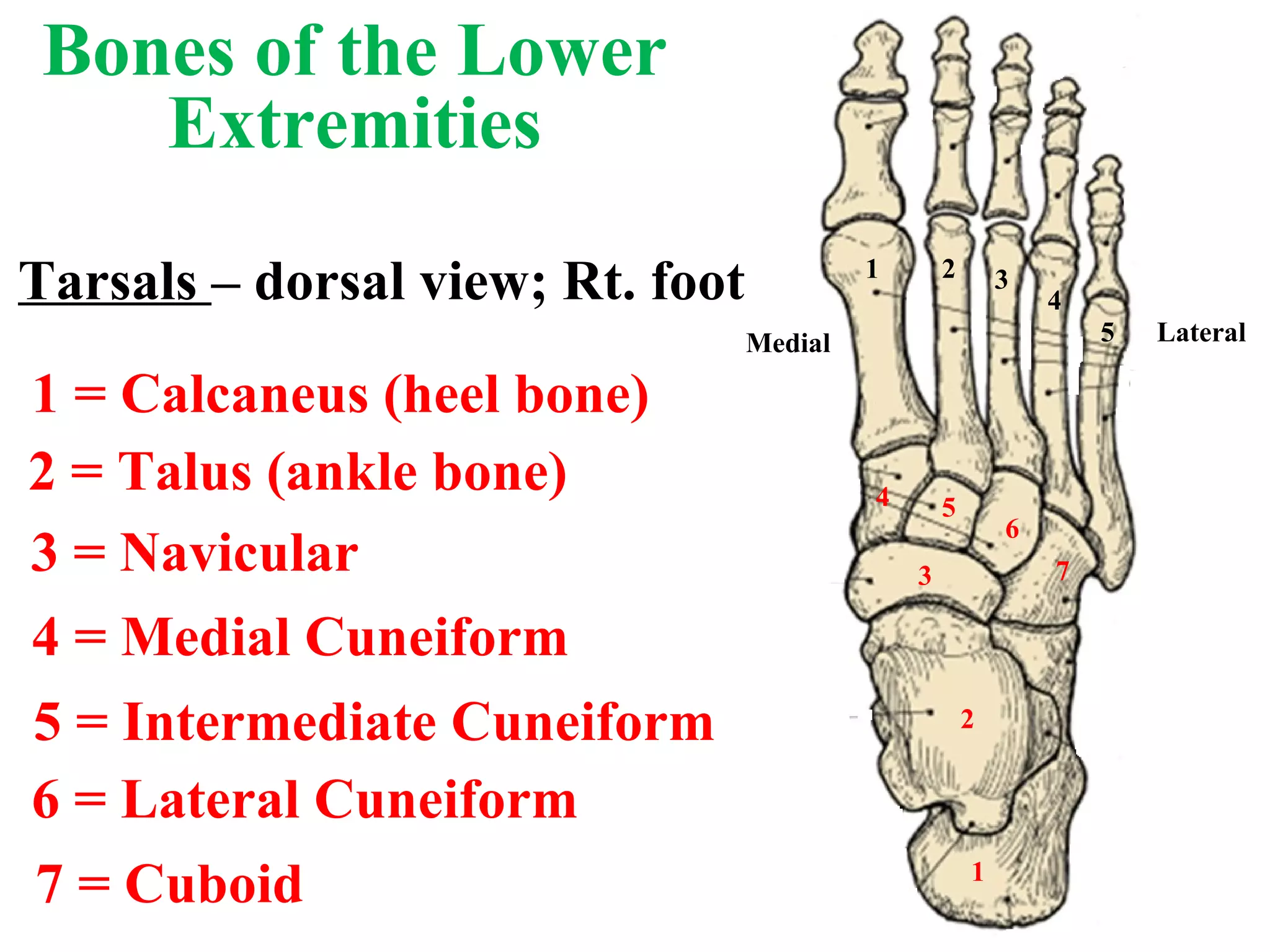
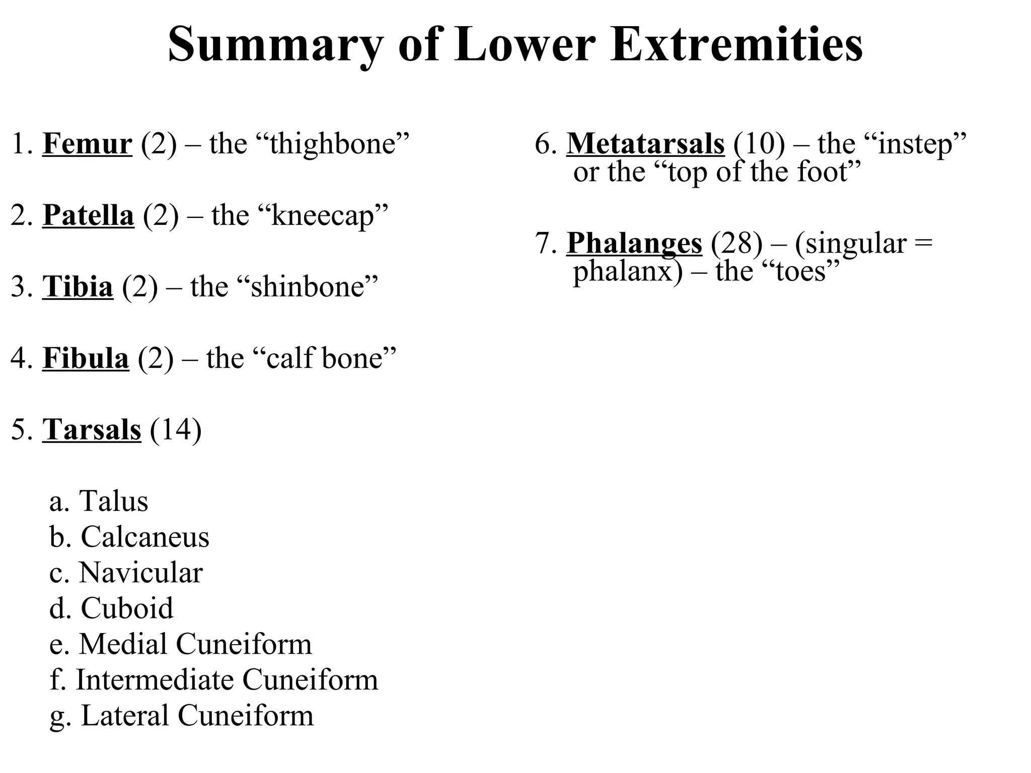
Summary of LowerExtremities 1. Femur (2) – the “thighbone” 2. Patella (2) – the “kneecap” 3. Tibia (2) – the “shinbone” 4. Fibula (2) – the “calf bone” 5. Tarsals (14) a. Talus b. Calcaneus c. Navicular d. Cuboid e. Medial Cuneiform f. Intermediate Cuneiform g. Lateral Cuneiform 6. Metatarsals (10) – the “instep” or the “top of the foot” 7. Phalanges (28) – (singular = phalanx) – the “toes”


![08 [chapter 8 the skeletal system appendicular skeleton]](https://cdn.slidesharecdn.com/ss_thumbnails/08chapter8theskeletalsystem-appendicularskeleton-170828041008-thumbnail.jpg?width=640&height=640&fit=bounds)


