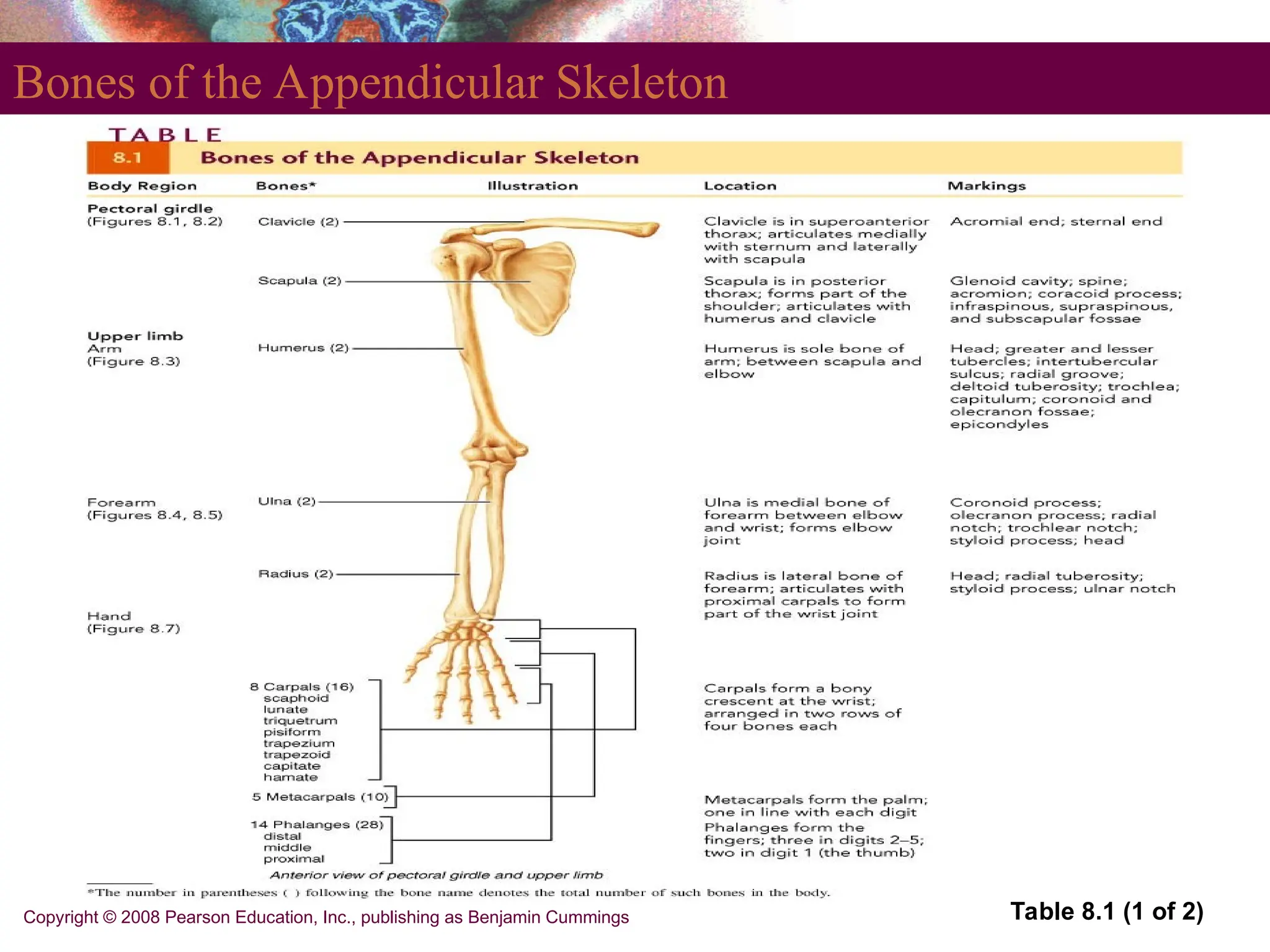
PowerPoint®
Lecture Slides
prepared by Leslie Hendon,
University of Alabama,
Birmingham
HUMAN
ANATOMY
fifth edition
MARIEB | MALLATT | WILHELM
8
Copyright © 2008 Pearson Education, Inc.,
publishing as Benjamin Cummings
Bones,
Part 1: The
Appendicular
Skeleton
PART 1
Copyright © 2008 Pearson Education, Inc., publishing as Benjamin Cummings
The Appendicular Skeleton
 Pectoral girdle
 Attaches the upper limbs to the trunk
 Pelvic girdle
 Attaches the lower limbs to the trunk
 Upper and lower limbs differ in function
 Share the same structural plan
Copyright © 2008 Pearson Education, Inc., publishing as Benjamin Cummings
The Pectoral Girdle
 Consists of the clavicle and the scapula
 Pectoral girdles do not quite encircle the body
completely
 Medial end of each clavicle articulates with the
manubrium and first rib
 Laterally – the ends of the clavicles join the
scapulae
 Scapulae do not join each other or the axial
skeleton
Copyright © 2008 Pearson Education, Inc., publishing as Benjamin Cummings
The Pectoral Girdle
 Provides attachment for many muscles that move
the upper limb
 Girdle is very light and upper limbs are mobile
 Only clavicle articulates with the axial skeleton
 Socket of the shoulder joint (glenoid cavity) is
shallow
Good for flexibility – bad for stability
Copyright © 2008 Pearson Education, Inc., publishing as Benjamin Cummings
Articulated Pectoral Girdle
Figure 8.1a
PLAY Shoulder
Copyright © 2008 Pearson Education, Inc., publishing as Benjamin Cummings
Clavicles
 Extend horizontally across the superior thorax
 Sternal end articulates with the manubrium
 Acromial end articulates with scapula
Copyright © 2008 Pearson Education, Inc., publishing as Benjamin Cummings
Clavicles
Figure 8.1b, c
Copyright © 2008 Pearson Education, Inc., publishing as Benjamin Cummings
PLAY Shoulder
Clavicles
 Provide attachment for muscles
 Hold the scapulae and arms laterally
 Transmit compression forces from the upper limbs
to the axial skeleton
Copyright © 2008 Pearson Education, Inc., publishing as Benjamin Cummings
Scapulae
 Lie on the dorsal surface of the rib cage
 Located between ribs 2 – 7
 Have three borders
 Superior
 Medial (vertebral)
 Lateral (axillary)
 Have three angles
 Lateral, superior, and inferior
Copyright © 2008 Pearson Education, Inc., publishing as Benjamin Cummings
Structures of the Scapula
Figure 8.2a
Copyright © 2008 Pearson Education, Inc., publishing as Benjamin Cummings
Structures of the Scapula
Figure 8.2b
Copyright © 2008 Pearson Education, Inc., publishing as Benjamin Cummings
Structures of the Scapula
Figure 8.2c
Copyright © 2008 Pearson Education, Inc., publishing as Benjamin Cummings
The Upper Limb
 30 bones form each upper limb
 Grouped into bones of the
 Arm
 Forearm
 Hand
Copyright © 2008 Pearson Education, Inc., publishing as Benjamin Cummings
Arm
 Region of the upper limb between the shoulder
and elbow
 Humerus
 The only bone of the arm
 Longest and strongest bone of the upper limb
 Articulates with the scapula at the shoulder
 Articulates with the radius and ulna at the elbow
Copyright © 2008 Pearson Education, Inc., publishing as Benjamin Cummings
Arm
 Humerus
 Many structures of the humerus provide sites for
muscle attachment
 Other structures of the humerus provide
articulation sites for other bones
Copyright © 2008 Pearson Education, Inc., publishing as Benjamin Cummings
Structures of the Humerus of the Right Arm
Figure 8.3a, b
Copyright © 2008 Pearson Education, Inc., publishing as Benjamin Cummings
Forearm
 Formed from the radius and ulna
 Proximal ends articulate with the humerus
 Distal ends articulate with carpals
PLAY Elbow
Copyright © 2008 Pearson Education, Inc., publishing as Benjamin Cummings
Forearm
 Radius and ulna articulate with each other
 At the proximal and distal radioulnar joints
 The interosseous membrane
 Interconnects radius and ulna
 In anatomical position
 The radius is lateral and the ulna is medial
Copyright © 2008 Pearson Education, Inc., publishing as Benjamin Cummings
Details of Arm and Forearm
Figure 8.5a
Copyright © 2008 Pearson Education, Inc., publishing as Benjamin Cummings
Ulna
 Main bone responsible for forming the elbow joint
with the humerus
 Hinge joint allows forearm to bend on arm
 Distal end is separated from carpals by
fibrocartilage
 Plays little to no role in hand movement
Copyright © 2008 Pearson Education, Inc., publishing as Benjamin Cummings
Proximal Part of the Ulna
Figure 8.5b
Copyright © 2008 Pearson Education, Inc., publishing as Benjamin Cummings
Radius and Ulna
Figure 8.4a, b
Copyright © 2008 Pearson Education, Inc., publishing as Benjamin Cummings
Radius
 Superior surface of the head of the radius
articulates with the capitulum
 Medially – the head of the radius articulates with
the radial notch of the ulna
 Contributes heavily to the wrist joint
 Distal radius articulates with carpal bones
 When radius moves, the hand moves with it
Copyright © 2008 Pearson Education, Inc., publishing as Benjamin Cummings
Distal Ends of the Radius and Ulna
Figure 8.5c
PowerPoint®
Lecture Slides
prepared by Leslie Hendon,
University of Alabama,
Birmingham
HUMAN
ANATOMY
fifth edition
MARIEB | MALLATT | WILHELM
8
Copyright © 2008 Pearson Education, Inc.,
publishing as Benjamin Cummings
Bones,
Part 1: The
Appendicular
Skeleton
PART 2
Copyright © 2008 Pearson Education, Inc., publishing as Benjamin Cummings
Hand
 Includes the following bones
 Carpus – wrist
 Metacarpals – palm
 Phalanges – fingers
Copyright © 2008 Pearson Education, Inc., publishing as Benjamin Cummings
Carpus
 Forms the true wrist – the proximal region of the
hand
 Gliding movements occur between carpals
 Composed of eight marble-sized bones
Copyright © 2008 Pearson Education, Inc., publishing as Benjamin Cummings
Carpus
 Carpal bones
 Are arranged in two irregular rows
 Proximal row from lateral to medial
Scaphoid, lunate, triquetral, and pisiform
 Distal row from lateral to medial
Trapezium, trapezoid, capitate, and hamate
 A mnemonic to help remember carpals
Sally left the party to take Carmen home
Copyright © 2008 Pearson Education, Inc., publishing as Benjamin Cummings
Bones of the Hand
Figure 8.7a, b
Copyright © 2008 Pearson Education, Inc., publishing as Benjamin Cummings
Metacarpus
 Five metacarpals radiate distally from the wrist
 Metacarpals form the palm
 Numbered 1–5, beginning with the pollex (thumb)
 Articulate proximally with the distal row of carpals
 Articulate distally with the proximal phalanges
Copyright © 2008 Pearson Education, Inc., publishing as Benjamin Cummings
Phalanges
 Numbered 1–5, beginning with the pollex (thumb)
 Except for the thumb, each finger has three
phalanges
 Proximal, middle, and distal
Copyright © 2008 Pearson Education, Inc., publishing as Benjamin Cummings
Bones of the Appendicular Skeleton
Table 8.1 (1 of 2)
Copyright © 2008 Pearson Education, Inc., publishing as Benjamin Cummings
Pelvic Girdle
 Attaches lower limbs to the spine
 Supports visceral organs
 Attaches to the axial skeleton by strong ligaments
 Acetabulum is a deep cup that holds the head of
the femur
 Lower limbs have less freedom of movement
Are more stable than the arm
Copyright © 2008 Pearson Education, Inc., publishing as Benjamin Cummings
Pelvic Girdle
 Consists of paired hip bones (coxal bones)
 Hip bones unite anteriorly with each other
 Articulates posteriorly with the sacrum
PLAY Hip
Copyright © 2008 Pearson Education, Inc., publishing as Benjamin Cummings
Bony Pelvis
 A deep, basin-like structure
 Formed by
 Coxal bones, sacrum, and coccyx
Copyright © 2008 Pearson Education, Inc., publishing as Benjamin Cummings
Bony Pelvis
Figure 8.8a
PLAY Pelvis
Copyright © 2008 Pearson Education, Inc., publishing as Benjamin Cummings
Coxal Bones
 Consist of three separate bones in childhood
 Ilium, ischium, and pubis
 Bones fuse – retain separate names to regions of
the coxal bones
 Acetabulum
 A deep hemispherical socket on lateral pelvic
surface
Copyright © 2008 Pearson Education, Inc., publishing as Benjamin Cummings
Ilium
 Large, flaring bone
 Forms the superior region of the coxal bone
 Site of attachment for many muscles
 Articulation with the sacrum forms sacroiliac joint
Copyright © 2008 Pearson Education, Inc., publishing as Benjamin Cummings
Ischium
 Forms posteroinferior region of the coxal bone
 Anteriorly – joins the pubis
 Ischial tuberosities
 Are the strongest part of the hip bone
Copyright © 2008 Pearson Education, Inc., publishing as Benjamin Cummings
Pubis
 Forms the anterior region of the coxal bone
 Lies horizontally in anatomical position
 Pubic symphysis
 The two pubic bones are joined by fibrocartilage at
the midline
Copyright © 2008 Pearson Education, Inc., publishing as Benjamin Cummings
Lateral and Medial Views of the Hip Bone
Figure 8.8b, c
Copyright © 2008 Pearson Education, Inc., publishing as Benjamin Cummings
True and False Pelves
 Bony pelvis is divided into two regions
 False (greater) pelvis – bounded by alae of the iliac
bones
 True (lesser) pelvis – inferior to pelvic brim
Forms a bowl containing the pelvic organs
Copyright © 2008 Pearson Education, Inc., publishing as Benjamin Cummings
True and False Pelves
Figure 8.9b
PowerPoint®
Lecture Slides
prepared by Leslie Hendon,
University of Alabama,
Birmingham
HUMAN
ANATOMY
fifth edition
MARIEB | MALLATT | WILHELM
8
Copyright © 2008 Pearson Education, Inc.,
publishing as Benjamin Cummings
Bones,
Part 1: The
Appendicular
Skeleton
PART 3
Copyright © 2008 Pearson Education, Inc., publishing as Benjamin Cummings
Pelvic Structures and Childbearing
 Major differences between male and female pelves
 Female pelvis is adapted for childbearing
Pelvis is lighter, wider, and shallower than in the
male
Provides more room in the true pelvis
Copyright © 2008 Pearson Education, Inc., publishing as Benjamin Cummings
Female and Male Pelves
Table 8.2 (1 of 2)
Copyright © 2008 Pearson Education, Inc., publishing as Benjamin Cummings
Female and Male Pelves
Table 8.2 (2 of 2)
Copyright © 2008 Pearson Education, Inc., publishing as Benjamin Cummings
The Lower Limb
 Carries the entire weight of the erect body
 Bones of lower limb are thicker and stronger than
those of upper limb
 Divided into three segments
 Thigh, leg, and foot
Copyright © 2008 Pearson Education, Inc., publishing as Benjamin Cummings
Thigh
 The region of the lower limb between the hip and
the knee
 Femur – the single bone of the thigh
 Longest and strongest bone of the body
 Ball-shaped head articulates with the acetabulum
Copyright © 2008 Pearson Education, Inc., publishing as Benjamin Cummings
Structures of the Femur
Figure 8.10b
Copyright © 2008 Pearson Education, Inc., publishing as Benjamin Cummings
Patella
 Triangular sesamoid bone
 Imbedded in the tendon that secures the
quadriceps muscles
 Protects the knee anteriorly
 Improves leverage of the thigh muscles across the
knee
Copyright © 2008 Pearson Education, Inc., publishing as Benjamin Cummings
Leg
 Refers to the region of the lower limb between the
knee and the ankle
 Composed of the tibia and fibula
 Tibia – more massive medial bone of the leg
Receives weight of the body from the femur
 Fibula – stick-like lateral bone of the leg
 Interosseous membrane
 Connects the tibia and fibula
PLAY Knee
Copyright © 2008 Pearson Education, Inc., publishing as Benjamin Cummings
Leg
 Tibia articulates with femur at superior end
 Forms the knee joint
 Tibia articulates with talus at the inferior end
 Forms the ankle joint
 Fibula does not contribute to the knee joint
 Stabilizes the ankle joint
Copyright © 2008 Pearson Education, Inc., publishing as Benjamin Cummings
Structures of the Tibia and Fibula
Figure 8.11a, b
Copyright © 2008 Pearson Education, Inc., publishing as Benjamin Cummings
The Foot
 Foot is composed of
 Tarsus, metatarsus, and the phalanges
 Important functions
 Supports body weight
 Acts as a lever to propel body forward when
walking
 Segmentation makes foot pliable and adapted to
uneven ground
Copyright © 2008 Pearson Education, Inc., publishing as Benjamin Cummings
Tarsus
 Makes up the posterior half of the foot
 Contains seven bones called tarsals
 Body weight is primarily borne by the talus and
calcaneus
Copyright © 2008 Pearson Education, Inc., publishing as Benjamin Cummings
Metatarsus
 Consists of five small long bones called
metatarsals
 Numbered 1–5 beginning with the hallux
(great toe)
 First metatarsal supports body weight
Copyright © 2008 Pearson Education, Inc., publishing as Benjamin Cummings
Phalanges of the Toes
 14 phalanges of the toes
 Smaller and less nimble than those of the fingers
 Structure and arrangement are similar to phalanges
of fingers
 Except for the great toe, each toe has three
phalanges
Proximal, middle, and distal
Copyright © 2008 Pearson Education, Inc., publishing as Benjamin Cummings
Bones of the Foot
Figure 8.12a
Copyright © 2008 Pearson Education, Inc., publishing as Benjamin Cummings
Bones of the Foot
Figure 8.12b
Copyright © 2008 Pearson Education, Inc., publishing as Benjamin Cummings
Bones of the Foot
Figure 8.12c
PLAY Bones of the Foot
Copyright © 2008 Pearson Education, Inc., publishing as Benjamin Cummings
Arches of the Foot
 Foot has three important arches
 Medial and lateral longitudinal arch
 Transverse arch
 Arches are maintained by
 Interlocking shapes of tarsals
 Ligaments and tendons
Copyright © 2008 Pearson Education, Inc., publishing as Benjamin Cummings
Arches of the Foot
Figure 8.13
Copyright © 2008 Pearson Education, Inc., publishing as Benjamin Cummings
Lower Limb and Pelvis
Table 8.1 (2 of 2)
Copyright © 2008 Pearson Education, Inc., publishing as Benjamin Cummings
Disorders of the Appendicular Skeleton
 Bone fractures
 Hip dysplasia
 Head of the femur slips out of acetabulum
 Clubfoot
 Soles of the feet turn medially
Copyright © 2008 Pearson Education, Inc., publishing as Benjamin Cummings
The Appendicular Skeleton Throughout Life
 Growth of the appendicular skeleton
 Increases height
 Changes body proportions
 Upper-lower body ratio changes with age
 At birth head and trunk are 1.5 times as long as
lower limbs
 Lower limbs grow faster than the trunk
 Upper-lower body ratio of 1 to 1 by age 10
Copyright © 2008 Pearson Education, Inc., publishing as Benjamin Cummings
Changes in Body Proportions
Figure 8.14
Copyright © 2008 Pearson Education, Inc., publishing as Benjamin Cummings
The Appendicular Skeleton Throughout Life
 Few changes occur in adult skeleton until middle
age, when
 Skeleton loses mass
 Osteoporosis and limb fractures become more
common

Bones of appendicular skeleton lecture.ppt

  • 1.
    PowerPoint® Lecture Slides prepared byLeslie Hendon, University of Alabama, Birmingham HUMAN ANATOMY fifth edition MARIEB | MALLATT | WILHELM 8 Copyright © 2008 Pearson Education, Inc., publishing as Benjamin Cummings Bones, Part 1: The Appendicular Skeleton PART 1
  • 2.
    Copyright © 2008Pearson Education, Inc., publishing as Benjamin Cummings The Appendicular Skeleton  Pectoral girdle  Attaches the upper limbs to the trunk  Pelvic girdle  Attaches the lower limbs to the trunk  Upper and lower limbs differ in function  Share the same structural plan
  • 3.
    Copyright © 2008Pearson Education, Inc., publishing as Benjamin Cummings The Pectoral Girdle  Consists of the clavicle and the scapula  Pectoral girdles do not quite encircle the body completely  Medial end of each clavicle articulates with the manubrium and first rib  Laterally – the ends of the clavicles join the scapulae  Scapulae do not join each other or the axial skeleton
  • 4.
    Copyright © 2008Pearson Education, Inc., publishing as Benjamin Cummings The Pectoral Girdle  Provides attachment for many muscles that move the upper limb  Girdle is very light and upper limbs are mobile  Only clavicle articulates with the axial skeleton  Socket of the shoulder joint (glenoid cavity) is shallow Good for flexibility – bad for stability
  • 5.
    Copyright © 2008Pearson Education, Inc., publishing as Benjamin Cummings Articulated Pectoral Girdle Figure 8.1a PLAY Shoulder
  • 6.
    Copyright © 2008Pearson Education, Inc., publishing as Benjamin Cummings Clavicles  Extend horizontally across the superior thorax  Sternal end articulates with the manubrium  Acromial end articulates with scapula
  • 7.
    Copyright © 2008Pearson Education, Inc., publishing as Benjamin Cummings Clavicles Figure 8.1b, c
  • 8.
    Copyright © 2008Pearson Education, Inc., publishing as Benjamin Cummings PLAY Shoulder Clavicles  Provide attachment for muscles  Hold the scapulae and arms laterally  Transmit compression forces from the upper limbs to the axial skeleton
  • 9.
    Copyright © 2008Pearson Education, Inc., publishing as Benjamin Cummings Scapulae  Lie on the dorsal surface of the rib cage  Located between ribs 2 – 7  Have three borders  Superior  Medial (vertebral)  Lateral (axillary)  Have three angles  Lateral, superior, and inferior
  • 10.
    Copyright © 2008Pearson Education, Inc., publishing as Benjamin Cummings Structures of the Scapula Figure 8.2a
  • 11.
    Copyright © 2008Pearson Education, Inc., publishing as Benjamin Cummings Structures of the Scapula Figure 8.2b
  • 12.
    Copyright © 2008Pearson Education, Inc., publishing as Benjamin Cummings Structures of the Scapula Figure 8.2c
  • 13.
    Copyright © 2008Pearson Education, Inc., publishing as Benjamin Cummings The Upper Limb  30 bones form each upper limb  Grouped into bones of the  Arm  Forearm  Hand
  • 14.
    Copyright © 2008Pearson Education, Inc., publishing as Benjamin Cummings Arm  Region of the upper limb between the shoulder and elbow  Humerus  The only bone of the arm  Longest and strongest bone of the upper limb  Articulates with the scapula at the shoulder  Articulates with the radius and ulna at the elbow
  • 15.
    Copyright © 2008Pearson Education, Inc., publishing as Benjamin Cummings Arm  Humerus  Many structures of the humerus provide sites for muscle attachment  Other structures of the humerus provide articulation sites for other bones
  • 16.
    Copyright © 2008Pearson Education, Inc., publishing as Benjamin Cummings Structures of the Humerus of the Right Arm Figure 8.3a, b
  • 17.
    Copyright © 2008Pearson Education, Inc., publishing as Benjamin Cummings Forearm  Formed from the radius and ulna  Proximal ends articulate with the humerus  Distal ends articulate with carpals PLAY Elbow
  • 18.
    Copyright © 2008Pearson Education, Inc., publishing as Benjamin Cummings Forearm  Radius and ulna articulate with each other  At the proximal and distal radioulnar joints  The interosseous membrane  Interconnects radius and ulna  In anatomical position  The radius is lateral and the ulna is medial
  • 19.
    Copyright © 2008Pearson Education, Inc., publishing as Benjamin Cummings Details of Arm and Forearm Figure 8.5a
  • 20.
    Copyright © 2008Pearson Education, Inc., publishing as Benjamin Cummings Ulna  Main bone responsible for forming the elbow joint with the humerus  Hinge joint allows forearm to bend on arm  Distal end is separated from carpals by fibrocartilage  Plays little to no role in hand movement
  • 21.
    Copyright © 2008Pearson Education, Inc., publishing as Benjamin Cummings Proximal Part of the Ulna Figure 8.5b
  • 22.
    Copyright © 2008Pearson Education, Inc., publishing as Benjamin Cummings Radius and Ulna Figure 8.4a, b
  • 23.
    Copyright © 2008Pearson Education, Inc., publishing as Benjamin Cummings Radius  Superior surface of the head of the radius articulates with the capitulum  Medially – the head of the radius articulates with the radial notch of the ulna  Contributes heavily to the wrist joint  Distal radius articulates with carpal bones  When radius moves, the hand moves with it
  • 24.
    Copyright © 2008Pearson Education, Inc., publishing as Benjamin Cummings Distal Ends of the Radius and Ulna Figure 8.5c
  • 25.
    PowerPoint® Lecture Slides prepared byLeslie Hendon, University of Alabama, Birmingham HUMAN ANATOMY fifth edition MARIEB | MALLATT | WILHELM 8 Copyright © 2008 Pearson Education, Inc., publishing as Benjamin Cummings Bones, Part 1: The Appendicular Skeleton PART 2
  • 26.
    Copyright © 2008Pearson Education, Inc., publishing as Benjamin Cummings Hand  Includes the following bones  Carpus – wrist  Metacarpals – palm  Phalanges – fingers
  • 27.
    Copyright © 2008Pearson Education, Inc., publishing as Benjamin Cummings Carpus  Forms the true wrist – the proximal region of the hand  Gliding movements occur between carpals  Composed of eight marble-sized bones
  • 28.
    Copyright © 2008Pearson Education, Inc., publishing as Benjamin Cummings Carpus  Carpal bones  Are arranged in two irregular rows  Proximal row from lateral to medial Scaphoid, lunate, triquetral, and pisiform  Distal row from lateral to medial Trapezium, trapezoid, capitate, and hamate  A mnemonic to help remember carpals Sally left the party to take Carmen home
  • 29.
    Copyright © 2008Pearson Education, Inc., publishing as Benjamin Cummings Bones of the Hand Figure 8.7a, b
  • 30.
    Copyright © 2008Pearson Education, Inc., publishing as Benjamin Cummings Metacarpus  Five metacarpals radiate distally from the wrist  Metacarpals form the palm  Numbered 1–5, beginning with the pollex (thumb)  Articulate proximally with the distal row of carpals  Articulate distally with the proximal phalanges
  • 31.
    Copyright © 2008Pearson Education, Inc., publishing as Benjamin Cummings Phalanges  Numbered 1–5, beginning with the pollex (thumb)  Except for the thumb, each finger has three phalanges  Proximal, middle, and distal
  • 32.
    Copyright © 2008Pearson Education, Inc., publishing as Benjamin Cummings Bones of the Appendicular Skeleton Table 8.1 (1 of 2)
  • 33.
    Copyright © 2008Pearson Education, Inc., publishing as Benjamin Cummings Pelvic Girdle  Attaches lower limbs to the spine  Supports visceral organs  Attaches to the axial skeleton by strong ligaments  Acetabulum is a deep cup that holds the head of the femur  Lower limbs have less freedom of movement Are more stable than the arm
  • 34.
    Copyright © 2008Pearson Education, Inc., publishing as Benjamin Cummings Pelvic Girdle  Consists of paired hip bones (coxal bones)  Hip bones unite anteriorly with each other  Articulates posteriorly with the sacrum PLAY Hip
  • 35.
    Copyright © 2008Pearson Education, Inc., publishing as Benjamin Cummings Bony Pelvis  A deep, basin-like structure  Formed by  Coxal bones, sacrum, and coccyx
  • 36.
    Copyright © 2008Pearson Education, Inc., publishing as Benjamin Cummings Bony Pelvis Figure 8.8a PLAY Pelvis
  • 37.
    Copyright © 2008Pearson Education, Inc., publishing as Benjamin Cummings Coxal Bones  Consist of three separate bones in childhood  Ilium, ischium, and pubis  Bones fuse – retain separate names to regions of the coxal bones  Acetabulum  A deep hemispherical socket on lateral pelvic surface
  • 38.
    Copyright © 2008Pearson Education, Inc., publishing as Benjamin Cummings Ilium  Large, flaring bone  Forms the superior region of the coxal bone  Site of attachment for many muscles  Articulation with the sacrum forms sacroiliac joint
  • 39.
    Copyright © 2008Pearson Education, Inc., publishing as Benjamin Cummings Ischium  Forms posteroinferior region of the coxal bone  Anteriorly – joins the pubis  Ischial tuberosities  Are the strongest part of the hip bone
  • 40.
    Copyright © 2008Pearson Education, Inc., publishing as Benjamin Cummings Pubis  Forms the anterior region of the coxal bone  Lies horizontally in anatomical position  Pubic symphysis  The two pubic bones are joined by fibrocartilage at the midline
  • 41.
    Copyright © 2008Pearson Education, Inc., publishing as Benjamin Cummings Lateral and Medial Views of the Hip Bone Figure 8.8b, c
  • 42.
    Copyright © 2008Pearson Education, Inc., publishing as Benjamin Cummings True and False Pelves  Bony pelvis is divided into two regions  False (greater) pelvis – bounded by alae of the iliac bones  True (lesser) pelvis – inferior to pelvic brim Forms a bowl containing the pelvic organs
  • 43.
    Copyright © 2008Pearson Education, Inc., publishing as Benjamin Cummings True and False Pelves Figure 8.9b
  • 44.
    PowerPoint® Lecture Slides prepared byLeslie Hendon, University of Alabama, Birmingham HUMAN ANATOMY fifth edition MARIEB | MALLATT | WILHELM 8 Copyright © 2008 Pearson Education, Inc., publishing as Benjamin Cummings Bones, Part 1: The Appendicular Skeleton PART 3
  • 45.
    Copyright © 2008Pearson Education, Inc., publishing as Benjamin Cummings Pelvic Structures and Childbearing  Major differences between male and female pelves  Female pelvis is adapted for childbearing Pelvis is lighter, wider, and shallower than in the male Provides more room in the true pelvis
  • 46.
    Copyright © 2008Pearson Education, Inc., publishing as Benjamin Cummings Female and Male Pelves Table 8.2 (1 of 2)
  • 47.
    Copyright © 2008Pearson Education, Inc., publishing as Benjamin Cummings Female and Male Pelves Table 8.2 (2 of 2)
  • 48.
    Copyright © 2008Pearson Education, Inc., publishing as Benjamin Cummings The Lower Limb  Carries the entire weight of the erect body  Bones of lower limb are thicker and stronger than those of upper limb  Divided into three segments  Thigh, leg, and foot
  • 49.
    Copyright © 2008Pearson Education, Inc., publishing as Benjamin Cummings Thigh  The region of the lower limb between the hip and the knee  Femur – the single bone of the thigh  Longest and strongest bone of the body  Ball-shaped head articulates with the acetabulum
  • 50.
    Copyright © 2008Pearson Education, Inc., publishing as Benjamin Cummings Structures of the Femur Figure 8.10b
  • 51.
    Copyright © 2008Pearson Education, Inc., publishing as Benjamin Cummings Patella  Triangular sesamoid bone  Imbedded in the tendon that secures the quadriceps muscles  Protects the knee anteriorly  Improves leverage of the thigh muscles across the knee
  • 52.
    Copyright © 2008Pearson Education, Inc., publishing as Benjamin Cummings Leg  Refers to the region of the lower limb between the knee and the ankle  Composed of the tibia and fibula  Tibia – more massive medial bone of the leg Receives weight of the body from the femur  Fibula – stick-like lateral bone of the leg  Interosseous membrane  Connects the tibia and fibula PLAY Knee
  • 53.
    Copyright © 2008Pearson Education, Inc., publishing as Benjamin Cummings Leg  Tibia articulates with femur at superior end  Forms the knee joint  Tibia articulates with talus at the inferior end  Forms the ankle joint  Fibula does not contribute to the knee joint  Stabilizes the ankle joint
  • 54.
    Copyright © 2008Pearson Education, Inc., publishing as Benjamin Cummings Structures of the Tibia and Fibula Figure 8.11a, b
  • 55.
    Copyright © 2008Pearson Education, Inc., publishing as Benjamin Cummings The Foot  Foot is composed of  Tarsus, metatarsus, and the phalanges  Important functions  Supports body weight  Acts as a lever to propel body forward when walking  Segmentation makes foot pliable and adapted to uneven ground
  • 56.
    Copyright © 2008Pearson Education, Inc., publishing as Benjamin Cummings Tarsus  Makes up the posterior half of the foot  Contains seven bones called tarsals  Body weight is primarily borne by the talus and calcaneus
  • 57.
    Copyright © 2008Pearson Education, Inc., publishing as Benjamin Cummings Metatarsus  Consists of five small long bones called metatarsals  Numbered 1–5 beginning with the hallux (great toe)  First metatarsal supports body weight
  • 58.
    Copyright © 2008Pearson Education, Inc., publishing as Benjamin Cummings Phalanges of the Toes  14 phalanges of the toes  Smaller and less nimble than those of the fingers  Structure and arrangement are similar to phalanges of fingers  Except for the great toe, each toe has three phalanges Proximal, middle, and distal
  • 59.
    Copyright © 2008Pearson Education, Inc., publishing as Benjamin Cummings Bones of the Foot Figure 8.12a
  • 60.
    Copyright © 2008Pearson Education, Inc., publishing as Benjamin Cummings Bones of the Foot Figure 8.12b
  • 61.
    Copyright © 2008Pearson Education, Inc., publishing as Benjamin Cummings Bones of the Foot Figure 8.12c PLAY Bones of the Foot
  • 62.
    Copyright © 2008Pearson Education, Inc., publishing as Benjamin Cummings Arches of the Foot  Foot has three important arches  Medial and lateral longitudinal arch  Transverse arch  Arches are maintained by  Interlocking shapes of tarsals  Ligaments and tendons
  • 63.
    Copyright © 2008Pearson Education, Inc., publishing as Benjamin Cummings Arches of the Foot Figure 8.13
  • 64.
    Copyright © 2008Pearson Education, Inc., publishing as Benjamin Cummings Lower Limb and Pelvis Table 8.1 (2 of 2)
  • 65.
    Copyright © 2008Pearson Education, Inc., publishing as Benjamin Cummings Disorders of the Appendicular Skeleton  Bone fractures  Hip dysplasia  Head of the femur slips out of acetabulum  Clubfoot  Soles of the feet turn medially
  • 66.
    Copyright © 2008Pearson Education, Inc., publishing as Benjamin Cummings The Appendicular Skeleton Throughout Life  Growth of the appendicular skeleton  Increases height  Changes body proportions  Upper-lower body ratio changes with age  At birth head and trunk are 1.5 times as long as lower limbs  Lower limbs grow faster than the trunk  Upper-lower body ratio of 1 to 1 by age 10
  • 67.
    Copyright © 2008Pearson Education, Inc., publishing as Benjamin Cummings Changes in Body Proportions Figure 8.14
  • 68.
    Copyright © 2008Pearson Education, Inc., publishing as Benjamin Cummings The Appendicular Skeleton Throughout Life  Few changes occur in adult skeleton until middle age, when  Skeleton loses mass  Osteoporosis and limb fractures become more common