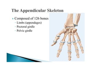
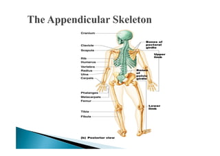
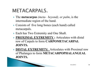
Appendicular Skeleton.
                  BY:
      Shahzad Bashir.
Identify the bones of the pectoral (shoulder) girdle and
their principal markings.
Identify the bones of the upper limb and their principal
markings.
Describe the joints between the upper limb bones.


                                           4/6/2013        2
Composed of 126 bones
◦ Limbs (appendages)
◦ Pectoral girdle
◦ Pelvic girdle
Bones of the upper limbs.

 S. No   REGIONS     BONES NAME      NUMBER.
   1.    Shoulder    Scapula.        1
         Girdle      Clavicle.       1
    2.   Upper Arm   Humerus         1
    3.   Fore Arm    Ulna.           1
                     Radius          1
    4.   Wrist       Carpels         8
    5.   Hands       Meta- Carpels   5
    6.   Fingers     Phalanges       14


                                               4/6/2013   6
Bone of the upper limbs are attached to the skeleton of
the trunk by the shoulder girdle.
It is made up of two bones:
◦ Clavicle — Collarbone
◦ Scapula — Shoulder blade


                                           4/6/2013       7
One in number.
Long bone lying at root of neck.
Has an S- shaped Shaft.
It has two Extremities
◦ Sternal Extremity
◦ Acromial Extremity.
Sternal Extremity articulates with STERNUM MANUBRIUM.
Acromial Extremity articulates with ACROMION PROCESS
OF SCUPULA.
It is also called COLLAR BONE.
                                         4/6/2013       9
Sternoclavicular
Acromioclavicular    joint
joint                                   Sternum
                       CLAVICLE

                                                                                  CLAVICLE
Glenohumeral
joint

SCAPULA
                                                                                  SCAPULA

  Rib

                                                                                      Rib

Humerus                                                                                Humerus


                                                                                      Vertebrae


         Anterior view of pectoral girdle         Posterior view of pectoral girdle          10
STERNO – CLAVICULAR JOINT. (With Sternum)
ACROMIO – CLAVICULAR JOINT. (With
Acromion Process of Scapula).


                                4/6/2013    12
One in number.
Flat bone lying over the ribs on the upper back between
the levels of the second and seventh ribs.
Also know as SHOULDER BLADE.
Triangular Shaped has 2 Surfaces, 3 Angles and 3
Borders.


                                             4/6/2013     13
Subscapular fossa


Supraspinous
fossa


  Infraspinous
  fossa
SURFACES: Has two Surfaces.
ANTERIOR SURFACE: Consists of Sub scapular Fossa
lying against ribs.
POSTERIOR SURFACE: Divided by Spine into SUPRA
SPINOUS FOSSA above and INFRA SPINOUS
FOSSA below.
Spine ends in ACROMION PROCESS, which articulates
with Clavicle to form the ACROMIOCLAVICULAR
JOINT.

                                      4/6/2013      15
ANGLES: Scapula has three angles.

Superior Angle.
Inferior Angle.
Lateral Angle.

◦ LATERAL ANGLE contains GLENOID CAVITY into
  which head of Humerus fits to form the
  GLENOHUMERAL (shoulder) JOINT.


                                    4/6/2013   16
BORDERS: Scapula has three borders.

Medical Border.
Lateral Border.
Superior Border.


◦ SUPERIOR BORDER ends in CORICOID PROCESS to
 which the tendons and ligaments attach.


                                           4/6/2013   17
ACROMIO – CLAVICULAR Joint. (With Clavicle)
HUMERO – SCAPIULAR/ GLENOHUMERAL
(shoulder) JOINT ( With Humerus).


                                    4/6/2013   18
Each upper limb includes a Humerus, ulna,          Clavicle
radius, carpals, metacarpals, and phalanges.        Scapula

                                               HUMERUS


                                               RADIUS

                                                                      ULNA


                                                                     CARPALS
                                                                      METACARPALS

                                                                      PHALANGES


                                                          4/6/2013             19
One in number.
The humerus, or arm bone, is the longest and largest
bone of the upper limb.
It articulates proximally with the scapula and distally at
the elbow with two bones, the ulna and the radius.
Long bone of upper limbs extending from shoulder to
elbow.
It has two Extremities and One Shaft.

                                           4/6/2013          20
UPPER EXTREMITY:
Head is 1/3rd sphere and articulates with GLENOID
CAVITY OF SCAPPULA to form the
GLENOHUMERAL JOINT.
Anatomical neck.
Greater Tubercle: (Laterally Below neck).
Less Tubercle:( Anteriorly)
Intertubercular sulcus : ( B/W the 2 Tubercle for
Tendons of Biceps Muscles).
                                         4/6/2013   22
SHAFT:
Long and thin.
Rounded (Superiorly)
Flattened (Inferiorly)
Deltoid Tuberosity: (Laterally V-shaped area for the
tendons of Deltoid Muscle)
Radial Groove: (Posteriorly for Radial Nerve)


                                          4/6/2013     23
LOWER EXTREMITY:
 Broad and flat.
Has TROCHLEA (Pulley) (Medially) for articulation with
ULNA.
Has CAPITULLAM (Capit = head) (Laterally) for
articulation with RADIUS.
Medial and lateral Epicondyles.
CORONOID FOSSA: (Anteriorly) receives Coronoid
Process of ulna when arm is flexed.
OLECRANON FOSSA: (Posteriorly) receives Olecraon
process of ulna when arm is Extended.


                                            4/6/2013     24
4/6/2013   25
HEMERO –SCAPULAR JOINT ( Shoulder Joint)
HEMERO – ULNAR JOINT
HEMERO – RADIAL JOINT.    (Elbow Joint)


                                   4/6/2013   26
One in each arm.
Long Medial bone of Fore-arm.
Long than Radius.
It has Two Extremities and One Shaft.

UPPER EXTREMITY:
Strong and Thick.
Articulates with Humerus to form part of Elbow Joint.
CORONOID PROCESS: Projects Anteriorly and fits into
CORONOID FOSSA OF HUMERUS when elbow is bent.
OLERANON PROESS: The prominence of ulna anteriorly
fits into the OLECRANON FOSSA OF HUMERUS when
elbow is straight.

                                          4/6/2013      27
OLECRANON

TROCHLEAR NOTCH


   CORONOID PROCESS

 RADIAL NOTCH
TROCHLEAR NOTCH: Is a large curved area
between Olecranon and Coronoid Process.
RADIAL NOTCH: Is an articulating surface for head
of radius to form PROXIMAL/ SUPERIOR RADIO-
ULNAR JOINT.

SHAFT:
Thinner at lower end.


                                      4/6/2013      29
LOWER EXTREMITY:
Small rounded head articulates with Radius to form
INFERIOR/DISTAL RADIO – ULNAR JOINT.
STYLOID PROCESS: Projects down wards
posteriorly. It provides attachment for the ulnar
collateral ligament to the wrist.

JOINTS OF ULNA:
HUMERO – ULNAR (Elbow Joint)
SUPERIOR/PROXIMAL – RADIO- ULNAR JOINT.
INFERIOR/DISTAL – RTADIO – ULNAR JOINT.

                                         4/6/2013    30
One bone in each fore-arm.
Long bone LATERAL BONE OF FORE ARM.
Smaller bone of the forearm and is located on the lateral aspect (thumb
side) of the forearm.
Nat its proximal end and widens at its distal end.
It has two Extremity and One Shaft.

UPPER EXTREMITY:
Button shaped/disc-shaped Head with shallow upper surface for
articulation with Capitulum of HUMERUS.
Sides of the head articulates with RADIAL NOTCH OF ULNA.
Neck lies below the head.
RADIAL TUBEROSITY: Lies medially below neck for insertion of
BICEPS- MUSCLES.

                                                      4/6/2013            31
SHAFT.
Narrow above and wide below.

LOWER EXTREMITY:
Square in shape.
ULNAR NOTCH: Is an articulating surface on medial side
for articulation with head of Ulna to form
INFERIOR/DISTAL RADIO- ULNAR JOINT.
STYLOD PROCESS: Laterally projecting downwards to
provides attachment for muscle.
Distal end of the radius articulates with three bones of the
wrist — the Lunate, the Scaphoid, and the Triquetrum —to
form the RADIOCARPAL (WRIST) JOINT.
                                              4/6/2013         33
HUMERO- RADIAL (Elbow Joint)
SUPERIOR/PROXIMAL – RADIO- ULNAR JOINT.
INFERIOR/DISTAL – RADIO – ULNAR JOINT.
RADIO – CARPAL JOINT (Wrist Joint)


                                     4/6/2013   34
The Carpus (wrist) is the proximal region of the hand.
Consists of eight (each wrist)small bones called carpals.
The Capitate is the largest carpal bone.
Carpals joined to one another by ligaments.
Short bones arranged in two transverse Rows of four
bones from medial to lateral.
UPPER ROW:
Scaphoid or Navicular (Boat – Shaped)
Lunate: ( Cresent, Moon – Shaped)
Triquetrum: (Three Sided)
Pisiform: (Pea – Shaped)

                                           4/6/2013         35
MNEMONIC for carpal bones*:
Stop Letting Those People Touch The
Cadaver’s Hand.
LOWER ROWS:
Trapezium: ( Four – Sided)
Trapezoid: (Four – Sided)
Capitate: (Head – Shaped)
Hamate: (Hammer, Hooked – Shaped)
              JOINTS OF CARPALS.

INTERCARPAL JOINTS. (Between Each other).
RADIO – CARPALS JOINT. (Wrist Joint)
CARPO- METACARPAL JOINT.
                                    4/6/2013   37
The metacarpus (meta- beyond), or palm, is the
intermediate region of the hand.
Consists of five long bones (each hand) called
metacarpals.
Each has Two Extremity and One Shaft.
PROXIMAL EXTREMITY : Articulates with distal
row of Carpals to form CARPOMETACARPAL
JOINTS.
DISTAL EXTREMITY: Articulates with Proximal row
of Phalanges to form METACARPOPHALANGEAL
JOINTS.

                                   4/6/2013       38
JOINTS OF METACARPALS.

CARPO- METACARPAL JOINT.(With Carpals)
METACARPO – PHALANGEAL JOINT.(With
Phalanges)


                                 4/6/2013   39
The phalanges (Phalan = a battle line), or bones of the
digits, make up the distal part of the hand.
Fourteen in each hand.
Long bones forming fingers.
Three in each finger.
Two in Thumb(pollex).

         JOINTS OF PHALANGES.
METACARPO- PHALANGES JOINT.
INTER-PHALANGEAL JOINT.


                                          4/6/2013        40
Acromio-Clavicular Joint.
Sterno-Clavicular Joint.
Humero -Scapular Joint. (Shoulder Joint)
Humero- Ulnar Joint.         (Elbow Joint)
Humero- Radial Joint.
Superior -Radio Ulnar Joint.
Inferior- Radio Ulnar Joint.
Radio –Carpal Joint. (Wrist Joint)
Carpo –Meta-Carpal Joint.
Meta Carpo –Phalangeal Joint.
Inter- Phalangeal Joint.

                                             4/6/2013   41
Tortora, G. J. & Grabowski, S.R. (2000). Principles
of anatomy and physiology (12th ed.). New York:
John Wiley & sons Inc.
Eline, N.M, Essential of human anatomy &
physiology (9th ed.). New York: John Wiley & sons
Inc.


                                         4/6/2013     42

Final appendicular skeleton(upper limbs).

  • 1.
    Appendicular Skeleton. BY: Shahzad Bashir.
  • 2.
    Identify the bonesof the pectoral (shoulder) girdle and their principal markings. Identify the bones of the upper limb and their principal markings. Describe the joints between the upper limb bones. 4/6/2013 2
  • 3.
    Composed of 126bones ◦ Limbs (appendages) ◦ Pectoral girdle ◦ Pelvic girdle
  • 6.
    Bones of theupper limbs. S. No REGIONS BONES NAME NUMBER. 1. Shoulder Scapula. 1 Girdle Clavicle. 1 2. Upper Arm Humerus 1 3. Fore Arm Ulna. 1 Radius 1 4. Wrist Carpels 8 5. Hands Meta- Carpels 5 6. Fingers Phalanges 14 4/6/2013 6
  • 7.
    Bone of theupper limbs are attached to the skeleton of the trunk by the shoulder girdle. It is made up of two bones: ◦ Clavicle — Collarbone ◦ Scapula — Shoulder blade 4/6/2013 7
  • 9.
    One in number. Longbone lying at root of neck. Has an S- shaped Shaft. It has two Extremities ◦ Sternal Extremity ◦ Acromial Extremity. Sternal Extremity articulates with STERNUM MANUBRIUM. Acromial Extremity articulates with ACROMION PROCESS OF SCUPULA. It is also called COLLAR BONE. 4/6/2013 9
  • 10.
    Sternoclavicular Acromioclavicular joint joint Sternum CLAVICLE CLAVICLE Glenohumeral joint SCAPULA SCAPULA Rib Rib Humerus Humerus Vertebrae Anterior view of pectoral girdle Posterior view of pectoral girdle 10
  • 12.
    STERNO – CLAVICULARJOINT. (With Sternum) ACROMIO – CLAVICULAR JOINT. (With Acromion Process of Scapula). 4/6/2013 12
  • 13.
    One in number. Flatbone lying over the ribs on the upper back between the levels of the second and seventh ribs. Also know as SHOULDER BLADE. Triangular Shaped has 2 Surfaces, 3 Angles and 3 Borders. 4/6/2013 13
  • 14.
  • 15.
    SURFACES: Has twoSurfaces. ANTERIOR SURFACE: Consists of Sub scapular Fossa lying against ribs. POSTERIOR SURFACE: Divided by Spine into SUPRA SPINOUS FOSSA above and INFRA SPINOUS FOSSA below. Spine ends in ACROMION PROCESS, which articulates with Clavicle to form the ACROMIOCLAVICULAR JOINT. 4/6/2013 15
  • 16.
    ANGLES: Scapula hasthree angles. Superior Angle. Inferior Angle. Lateral Angle. ◦ LATERAL ANGLE contains GLENOID CAVITY into which head of Humerus fits to form the GLENOHUMERAL (shoulder) JOINT. 4/6/2013 16
  • 17.
    BORDERS: Scapula hasthree borders. Medical Border. Lateral Border. Superior Border. ◦ SUPERIOR BORDER ends in CORICOID PROCESS to which the tendons and ligaments attach. 4/6/2013 17
  • 18.
    ACROMIO – CLAVICULARJoint. (With Clavicle) HUMERO – SCAPIULAR/ GLENOHUMERAL (shoulder) JOINT ( With Humerus). 4/6/2013 18
  • 19.
    Each upper limbincludes a Humerus, ulna, Clavicle radius, carpals, metacarpals, and phalanges. Scapula HUMERUS RADIUS ULNA CARPALS METACARPALS PHALANGES 4/6/2013 19
  • 20.
    One in number. Thehumerus, or arm bone, is the longest and largest bone of the upper limb. It articulates proximally with the scapula and distally at the elbow with two bones, the ulna and the radius. Long bone of upper limbs extending from shoulder to elbow. It has two Extremities and One Shaft. 4/6/2013 20
  • 22.
    UPPER EXTREMITY: Head is1/3rd sphere and articulates with GLENOID CAVITY OF SCAPPULA to form the GLENOHUMERAL JOINT. Anatomical neck. Greater Tubercle: (Laterally Below neck). Less Tubercle:( Anteriorly) Intertubercular sulcus : ( B/W the 2 Tubercle for Tendons of Biceps Muscles). 4/6/2013 22
  • 23.
    SHAFT: Long and thin. Rounded(Superiorly) Flattened (Inferiorly) Deltoid Tuberosity: (Laterally V-shaped area for the tendons of Deltoid Muscle) Radial Groove: (Posteriorly for Radial Nerve) 4/6/2013 23
  • 24.
    LOWER EXTREMITY: Broadand flat. Has TROCHLEA (Pulley) (Medially) for articulation with ULNA. Has CAPITULLAM (Capit = head) (Laterally) for articulation with RADIUS. Medial and lateral Epicondyles. CORONOID FOSSA: (Anteriorly) receives Coronoid Process of ulna when arm is flexed. OLECRANON FOSSA: (Posteriorly) receives Olecraon process of ulna when arm is Extended. 4/6/2013 24
  • 25.
  • 26.
    HEMERO –SCAPULAR JOINT( Shoulder Joint) HEMERO – ULNAR JOINT HEMERO – RADIAL JOINT. (Elbow Joint) 4/6/2013 26
  • 27.
    One in eacharm. Long Medial bone of Fore-arm. Long than Radius. It has Two Extremities and One Shaft. UPPER EXTREMITY: Strong and Thick. Articulates with Humerus to form part of Elbow Joint. CORONOID PROCESS: Projects Anteriorly and fits into CORONOID FOSSA OF HUMERUS when elbow is bent. OLERANON PROESS: The prominence of ulna anteriorly fits into the OLECRANON FOSSA OF HUMERUS when elbow is straight. 4/6/2013 27
  • 28.
    OLECRANON TROCHLEAR NOTCH CORONOID PROCESS RADIAL NOTCH
  • 29.
    TROCHLEAR NOTCH: Isa large curved area between Olecranon and Coronoid Process. RADIAL NOTCH: Is an articulating surface for head of radius to form PROXIMAL/ SUPERIOR RADIO- ULNAR JOINT. SHAFT: Thinner at lower end. 4/6/2013 29
  • 30.
    LOWER EXTREMITY: Small roundedhead articulates with Radius to form INFERIOR/DISTAL RADIO – ULNAR JOINT. STYLOID PROCESS: Projects down wards posteriorly. It provides attachment for the ulnar collateral ligament to the wrist. JOINTS OF ULNA: HUMERO – ULNAR (Elbow Joint) SUPERIOR/PROXIMAL – RADIO- ULNAR JOINT. INFERIOR/DISTAL – RTADIO – ULNAR JOINT. 4/6/2013 30
  • 31.
    One bone ineach fore-arm. Long bone LATERAL BONE OF FORE ARM. Smaller bone of the forearm and is located on the lateral aspect (thumb side) of the forearm. Nat its proximal end and widens at its distal end. It has two Extremity and One Shaft. UPPER EXTREMITY: Button shaped/disc-shaped Head with shallow upper surface for articulation with Capitulum of HUMERUS. Sides of the head articulates with RADIAL NOTCH OF ULNA. Neck lies below the head. RADIAL TUBEROSITY: Lies medially below neck for insertion of BICEPS- MUSCLES. 4/6/2013 31
  • 33.
    SHAFT. Narrow above andwide below. LOWER EXTREMITY: Square in shape. ULNAR NOTCH: Is an articulating surface on medial side for articulation with head of Ulna to form INFERIOR/DISTAL RADIO- ULNAR JOINT. STYLOD PROCESS: Laterally projecting downwards to provides attachment for muscle. Distal end of the radius articulates with three bones of the wrist — the Lunate, the Scaphoid, and the Triquetrum —to form the RADIOCARPAL (WRIST) JOINT. 4/6/2013 33
  • 34.
    HUMERO- RADIAL (ElbowJoint) SUPERIOR/PROXIMAL – RADIO- ULNAR JOINT. INFERIOR/DISTAL – RADIO – ULNAR JOINT. RADIO – CARPAL JOINT (Wrist Joint) 4/6/2013 34
  • 35.
    The Carpus (wrist)is the proximal region of the hand. Consists of eight (each wrist)small bones called carpals. The Capitate is the largest carpal bone. Carpals joined to one another by ligaments. Short bones arranged in two transverse Rows of four bones from medial to lateral. UPPER ROW: Scaphoid or Navicular (Boat – Shaped) Lunate: ( Cresent, Moon – Shaped) Triquetrum: (Three Sided) Pisiform: (Pea – Shaped) 4/6/2013 35
  • 36.
    MNEMONIC for carpalbones*: Stop Letting Those People Touch The Cadaver’s Hand.
  • 37.
    LOWER ROWS: Trapezium: (Four – Sided) Trapezoid: (Four – Sided) Capitate: (Head – Shaped) Hamate: (Hammer, Hooked – Shaped) JOINTS OF CARPALS. INTERCARPAL JOINTS. (Between Each other). RADIO – CARPALS JOINT. (Wrist Joint) CARPO- METACARPAL JOINT. 4/6/2013 37
  • 38.
    The metacarpus (meta-beyond), or palm, is the intermediate region of the hand. Consists of five long bones (each hand) called metacarpals. Each has Two Extremity and One Shaft. PROXIMAL EXTREMITY : Articulates with distal row of Carpals to form CARPOMETACARPAL JOINTS. DISTAL EXTREMITY: Articulates with Proximal row of Phalanges to form METACARPOPHALANGEAL JOINTS. 4/6/2013 38
  • 39.
    JOINTS OF METACARPALS. CARPO-METACARPAL JOINT.(With Carpals) METACARPO – PHALANGEAL JOINT.(With Phalanges) 4/6/2013 39
  • 40.
    The phalanges (Phalan= a battle line), or bones of the digits, make up the distal part of the hand. Fourteen in each hand. Long bones forming fingers. Three in each finger. Two in Thumb(pollex). JOINTS OF PHALANGES. METACARPO- PHALANGES JOINT. INTER-PHALANGEAL JOINT. 4/6/2013 40
  • 41.
    Acromio-Clavicular Joint. Sterno-Clavicular Joint. Humero-Scapular Joint. (Shoulder Joint) Humero- Ulnar Joint. (Elbow Joint) Humero- Radial Joint. Superior -Radio Ulnar Joint. Inferior- Radio Ulnar Joint. Radio –Carpal Joint. (Wrist Joint) Carpo –Meta-Carpal Joint. Meta Carpo –Phalangeal Joint. Inter- Phalangeal Joint. 4/6/2013 41
  • 42.
    Tortora, G. J.& Grabowski, S.R. (2000). Principles of anatomy and physiology (12th ed.). New York: John Wiley & sons Inc. Eline, N.M, Essential of human anatomy & physiology (9th ed.). New York: John Wiley & sons Inc. 4/6/2013 42