LYMPH NODES
•Download as PPTX, PDF•
1 like•3,182 views

The lymph node is a small bean-shaped object which factors in the body's immune system. Lymph nodes clean out substances that travel through the lymphatic fluid, and they contain white blood cells that are the body's defense when fighting off ailments. There are many lymph nodes found throughout the body.

Recommended
More Related Content
What's hot
What's hot (20)
Similar to LYMPH NODES
Similar to LYMPH NODES (20)
More from Jai Narain Vyas University Jodhpur Rajasthan India 342003
More from Jai Narain Vyas University Jodhpur Rajasthan India 342003 (20)
Recently uploaded
Recently uploaded (20)
LYMPH NODES
- 2. LYMPH NODES JAI NARAIN VYAS UNIVERSITY, JODHPUR ASSISTANT PROFESSOR:- ASHWIN SINGH CHOUHAN DEPARTMENT:- PHARMACOLOGY E-mail:- anshukavya1993@gmail.com
- 3. LYMPH A clear fluid composed mainly of water, electrolytes, and some small plasma proteins. Transported in the lymphatic pathway form the lymphatic capillaries to the collecting ducts, and at the end disposed into the venous blood. Reabsorbed by lymphatic capillaries in all body tissues (especially in extremities) where excessive tissue fluids occur. Because of the lack of a pumping organ in the lymphatic pathway, lymph movement is largely dependent on skeletal muscle activity (similar to blood flow in the veins). Lymph movement is normally constant and smooth, except when obstruction (small blood clot, tumor) occurs which tend to back up the lymph, and results in edema (fluid accumulation in tissues). JNVU PHARMACY, JODHPUR
- 4. Lymph nodes are small solid structures placed at varying points along the lymphatic system such as the groin, armpit and mesentery. They contain both T and B lymphocytes as well as accessory cells and are primarily responsible for mounting immune responses against foreign antigens entering the tissues. Lymph nodes are situated at strategic positions throughout the body and serve to filter the lymph. JNVU PHARMACY, JODHPUR
- 5. JNVU PHARMACY, JODHPUR The lymph nodes are small bean shaped glands or bulbs that tend to occur in clusters much like grapes. Along the lymph channels reside approximately 600 lymph nodes. These act as filters that sieve off the harmful substances brought by the lymphatic channels. The lymphatic channels of the fingers, hand and arm for example comes to be filtered at the lymph nodes that lie at the elbow and the arm pit. Similarly, those of the legs, toes and thighs drain and nodes behind the knees and the groin. Lymph channels from the face, head and scalp drain at the nodes present at the back of the head, behind the ears and sides of the neck.
- 6. JNVU PHARMACY, JODHPUR Some lymph nodes are located deeper within the body at the chest (between the two lobes of the lungs), around the coils of the intestines, in the pelvis etc. The lymph nodes contain 2 regions within them – these include the cortex and the medulla. The cortex contains collections of lymphocytes. These contain predominantly B-lymphocytes and some T- lymphocytes. The B lymphocytes mature completely within the bone marrow while the T lymphocytes exit the bone marrow immature and attain maturity within the thymus. The lymphatic vessels entering the lymph nodes are called afferent lymphatic vessels and those exiting are called efferent lymphatic vessels.
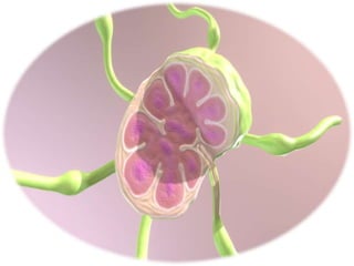
- 8. They range in size from 2 to 10 mm, are spherical in shape and are encapsulated. Lymph node is surrounded by a fibrous capsule which dips down into the node substance forming partition or trabeculae. The node is made by reticular and lymphatic tissues containing mainly lymphocytes and macrophages. Beneath the capsule is the subcapsular sinus, the cortex, a paracortical region and a medulla. The cortex contains many follicles and on antigenic stimulation becomes enlarged with germinal centers. The follicles are comprised mainly of B cells and follicular dendritic cells. The paracortical (thymus-dependent) region contains large numbers of T cells interspersed with interdigitating cells. STRUCTURE OF LYMPH NODES JNVU PHARMACY, JODHPUR
- 9. Each lymph node has 4-5 afferent vessels that bring lymph to the node while only one efferent vessel draining lymph away from the node. It also has a concave surface called the hilum where an artery enters, a vein and the efferent lymph vessel leave. Depending upon the position, the lymph nodes may be superficial or deep lymph nodes. Groups of lymph nodes are present in the neck, collarbone, under the arms (armpit), and groin. JNVU PHARMACY, JODHPUR
- 10. FUNCTION OF LYMPH NODES Filter lymph before returned to blood stream. 99 percent of pathogens (bacteria, toxins etc.) are removed. Located in ideal spots to protect vital organs of body. The primary role of the lymph node is to filter the lymph and then produce an immune response against trapped microbes/antigens. Filtering of the lymph helps in removal of particles not normally found in the serum. The lymphoide tissues in the nodes break down materials which have been filtered off such as microorganisms, tumor cells and cells damaged by inflammation. Lymphocyte develops from the reticular and lymphoid tissue in the nodes. Antibodies and antitoxins are also formed by the cells of lymph nodes. JNVU PHARMACY, JODHPUR
- 11. Lymph arriving from the tissues or from a preceding lymph node in the chain, passes via the afferent lymphatics into the subcapsular sinus and then into the cortex, around the follicles, into the paracortical area and then into the medulla. Lymph in the medullary sinuses then drains into efferent lymphatics and hence through larger lymphatic vessels back into the bloodstream. LYMPH CIRCULATION IN LYMPH NODES JNVU PHARMACY, JODHPUR
- 12. JNVU PHARMACY, JODHPUR THE LYMPHATIC SYSTEM The lymphatic system also consists of other organs like the spleen that lies on the above left sided part of the abdomen. It acts like a large filter to remove worn out and damaged red blood cells from the blood and recycle them. The spleen also contains B lymphocytes and T lymphocytes. When blood passes through the organ these cells pick up the infections. The lymphatic system also contains the thymus that lies behind the chest bone. The thymus is a maturation site for T lymphocytes. Tonsils and adenoids are also part of the lymphatic system. The lie at the back of the throat. These are sentinels that protect the digestive system and the lungs from bacteria and viruses.
- 13. JNVU PHARMACY, JODHPUR FUNCTIONS OF THE LYMPHATIC SYSTEM Drainage of fluid from blood stream into the tissues – The circulating blood through narrow vessels leads to leakage of fluid or plasma into the tissues carrying oxygen and nutrients to the tissues and carrying waste materials from the tissues into the lymph channels.The leaked fluid drains into the lymph vessels. This forms a circulatory system of fluids within the body. Filtration of the lymph at the lymph nodes – The nodes contain white blood cells that can attack any bacteria or viruses they find in the lymph as it flows through the lymph nodes.The cancer cells may also get trapped similarly at the lymph nodes and thus lymph nodes act as indicators of how far the cancer has already spread. Filtering blood – This is done by the spleen. The spleen filters out bacteria, viruses and other foreign particles. Raise an immune reaction and fight infections – The lymphatic system especially the lymph nodes are over active in case of an infection the lymph nodes or glands often swell up in case of a local infection.
- 14. JNVU PHARMACY, JODHPUR Lymphoid tissues are collections of lymphocytes strategically located at potential sites of infection. They can be classified as either primary lymphoid organs (bone marrow and thymus) where de novo synthesis and maturation of lymphocytes occur; or secondary lymphoid organs where activation of lymphocytes occur. Lymph nodes are secondary lymphoid organs widely distributed throughout the body. They are strategically located at areas that are open to foreign microorganisms (e.g. the oral cavity). For the sake of completeness, other secondary lymphoid organs include (but are not limited to) the spleen, mucosa-associated lymphoid tissue (MALT), tonsils, and Peyer’s patches. The average young adult has about 450 lymph nodes throughout the body; most of which are in the abdominopelvic region, then the thorax and the remainder in the head and neck.
- 15. JNVU PHARMACY, JODHPUR LYMPHOID TISSUES Primary: bone marrow, thymus Secondary: lymph nodes, spleen, MALT, Peyer's patches STRUCTURE OF A LYMPH NODE Hilum - passage for the artery, vein, afferent and efferent lymph vessels Capsule - surrounding dense connective tissue Cortex - lymphatic nodules - primary nodule - dormant B-lymphocites - secondary nodule - activated B-lymphocites form germinative center with dark zone (centroblasts), light zone (centrocytes) and mantle zone (small quiescent cells) Paracortex - CD4 and CD8-T cells, migrating dendritic cells Medulla: - medullary cords - plasma cells, small lymphocytes and macrophages - medullary sinuses - drain lymph to the efferent lymph vessel REGIONAL NODES Cervical - superficial (parotid, mastoid, occipital) and deep nodes Waldeyer's ring - pharyngeal tonsils, tubal tonsils of Gerlach, palatine tonsils, lingual tonsils, MALT at the oropharyngeal wall Axillary - apical, central, posterior (subscapular), anterior (pectoral), lateral nodes Supratrochlear - superficial to the deep fascia of the arm and medial to the basillic vein Mediastinal - hilum of the lungs, juxta-oesophageal, superior and inferior tracheobronchial nodes Abdominal - para-aortic, mesenteric, common iliac, external iliac, internal iliac, superior and middle rectal nodes Inguinal - superficial (inferior, superolateral, superomedial), deep nodes VASCULATURE Lymphatic vessels (afferent, efferent), lymphatic capillaries CLINICAL Lymphadenopathy, lymphedema, sentinel lymph nodes
- 16. JNVU PHARMACY, JODHPUR GROSS ANATOMY Lymph nodes are bean-shaped structures about 0.1 – 2.5 cm in length. The node is enclosed in a capsule and has an indentation on one surface (along one of its long axes) known as the hilum. The hilum is the point at which arteries carrying nutrients and lymphocytes enter the lymph node and veins leave it. Afferent lymphatic vessels enter the lymph node through the capsule peripherally and efferent lymphatic vessels leave the node via the hilum. The former takes lymph from peripheral sites to the node, while the latter takes processed lymph from the nodes back to the venous circulation.
- 17. JNVU PHARMACY, JODHPUR HISTOLOGY A horizontal section through the lymph node reveals that the dense connective tissue capsule (composed of elastin, collagen and fibroblasts) projects trabeculae interiorly; giving the lymph node a lobular appearance, while carrying major blood vessels of the lymph node. Additionally, there is a pericapsular adipose tissue layer that surrounds the connective tissue capsule. This layer contains arterioles and venules that supply the lymph node. The lymph node is divided into an outer cortex and an inner medulla. Histological staining of the node reveals that the cortex stains darker than the medulla with hematoxylin and eosin (H&E) due to its higher cell content. The cortex contains lymphoid nodules, which are non- encapsulated, spherical collections of lymphocytes.
- 18. JNVU PHARMACY, JODHPUR Beneath the fibrous capsule is the subcapsular sinus. The subcapsular sinus receives afferent lymphatic ducts at intervals that deposit lymphatic fluid in the space. The subcapsular sinus communicates with the cortical sinuses that travel parallel to the capsular trabeculation. They carry lymph to the medullary sinus.
- 19. JNVU PHARMACY, JODHPUR CORTEX Within the cortex are regions of lymphocyte aggregation (primarily B-cells and some supporting T-cells) and specialized follicular dendritic cells that form the lymphoid follicles or lymphatic nodules. These can either be primary or secondary follicles depending on their cellular population. Primary lymphoid follicles contain small, dormant lymphocytes, while secondary lymphoid follicles contain a lighter staining area of active lymphocyte proliferation known as a germinal centre. The germinal centre supports affinity maturation (high affinity antibody production) of B-cells. It is subdivided into a dark zone, light zone and a mantle zone. The B- cells of the dark zone are known as centroblasts. They rapidly replicate, resulting in hypermutation of their antibody molecules. germinal centre.
- 20. JNVU PHARMACY, JODHPUR Centroblasts migrate to the light zone, where they are referred to as centrocytes. Here they compete for binding with the unprocessed antigens presented on the surface of follicular dendritic cells. Those centrocytes that successfully bind to the follicular dendritic cells will survive, while the others will die. Small, quiescent cells are peripherally marginalized due to the rapid proliferation of the central cells. These cells form the mantle zone of the germinal centre.
- 21. PARACORTEX Deep to the cortical layer and superficial to the medulla is the paracortex. This region contains mostly T-cells of the CD4 (cluster of differentiation) and CD8 subsets. Migrating dendritic cell lines (such as Langerhans cells) found in this area present processed antigen to the T- cells. MEDULLA The lymphocytes of the medulla are less organized and form irregular medullary cords. The cords also contain plasma cells, small lymphocytes and macrophages. The medullary sinuses drain the lymph coming from the cortical sinuses to the efferent lymphatic vessel via the hilum. JNVU PHARMACY, JODHPUR
- 23. JNVU PHARMACY, JODHPUR BLOOD SUPPLY The hilum of the lymph nodes is the primary point of entry for arteries and exit for veins. Once they enter the hilum, they give off straight branches that pass through the medulla. The arteries form bundles of anastomosing arterioles and capillaries in the cortex that return to similarly branched venules and veins. In the paracortical zone, there are large numbers of postcapillary high endothelial veins that act as a point where blood-borne lymphocytes leave the blood vessels and enter the lymph nodes.
- 25. FLOW OF LYMPH IN NODES In the nodes the lymph get enter by afferent lymphatic vessels and out by the efferent lymphatic vessels. The afferent lymphatic vessels consist valve at the opening part and the valve is open in such a direction that once lymph can enter in to the afferent vessels in not go back from that also efferent lymphatic vessels consist valve at the end of their part and the here also the valve is located in same manner of afferent vessels. The efferent lymphatic vessels have wider diameters than the afferent lymphatic vessels. The main function of lymph nodes is filtration of lymph. It filters the foreign substances which are harmful for us because the macrophages, T-lymphocytes and B- lymphocytes of nodes destroy them. JNVU PHARMACY, JODHPUR
- 26. JNVU PHARMACY, JODHPUR LYMPHATIC VESSELS The lymphatic vasculature is comprised of lymphatic capillaries and lymphatic vessels. They are valvular channels responsible for taking lymph to and from the lymph nodes and back to the main systemic circulation. The valves of the lymphatic vessels ensure that lymph flows in a unidirectional manner: from afferent lymphatic vessels to efferent lymphatic vessels. They have very thin walls and their valves can be appreciated histologically when the vessel is cut along its longitudinal axis. Unlike arteries and veins, which are open at both ends, lymphatic vessels begin as blind-ended (closed at one end) channels in the interstitium of specific organs. Their thin endothelial walls permit passive movement of excess interstitial fluid into the lumen of the vessels, which is subsequently returned to the venous circulation.
- 29. JNVU PHARMACY, JODHPUR REGIONAL LYMPH NODES As stated above, lymph nodes are strategically located throughout the body at points susceptible to foreign microorganisms. The following is an overview of these lymph node regions and their subdivisions. Brain It is a long held concept that the brain is the only region of the body devoid of a lymphatic system. The idea was that resident microglia were totally responsible for maintaining immunity within the region. However, the method by which these macrophages entered the central nervous system was still unclear. It has been recently proposed by Louveau et al. (2015) that lymphatic channels line the dural sinuses located between the two layers of dura and drain their contents to the deep group of cervical lymph nodes.
- 30. JNVU PHARMACY, JODHPUR CERVICAL The cervical lymph nodes can be subdivided into two major groups. Those superficial to the sternocleidomastoid muscle are known as the superficial cervical nodes, while those deep to the same muscle are the deep cervical nodes. The superficial cervical nodes are further subdivided into the pre-auricular or parotid nodes (anterior to the external ear), mastoid nodes (posterior to the external ear), and the occipital nodes. The deep cervical nodes are located in relation to the internal jugular vein. The superior deep cervical nodes are adjacent to the upper part of the internal jugular vein, while the inferior deep cervical nodes are adjacent to the lower part of the same vein.
- 32. JNVU PHARMACY, JODHPUR WALDEYER'S RING The nasal and oral passages are two of the major ports of entry that pathogens use to access the human body. The region is guarded by a collection of lymphatic tissue known as Waldeyer’s ring. This circular collection of lymphoid tissue is formed by the pharyngeal tonsils (adenoids) located in the fossa of Rosenmüller, the tubal tonsils of Gerlach in the torus tubaris, the palatine tonsils between the palatoglossal and palatopharyngeal folds, the lingual tonsils at the posterior region of the tongue and mucosa-associated lymphatic tissue lining the oropharyngeal wall.
- 34. JNVU PHARMACY, JODHPUR AXILLARY The axillary lymph nodes were previously divided into three groups by pectoralis minor. Level one nodes are inferior to the muscle, level two nodes are posterior to the muscle and level three nodes are superior to the muscle. Subsequently, axillary nodes have been divided into five groups: apical central (in the fat of the axilla) posterior (subscapular) anterior (pectoral) lateral (medial to the axillary vein) nodes.
- 36. JNVU PHARMACY, JODHPUR EPITROCHLEAR Epitrochlear or supratrochlear nodes are located superficial to the deep fascia of the arm and medial to the basilic vein and proximal to the medial epicondyle. MEDIASTINAL Bronchopulmonary lymph nodes (Nodi lymphoidei bronchopulmonales)Mediastinal lymph nodes are divided into nine stations. They include those found in the hilum of the lungs, the juxta-oesophageal nodes, and superior and inferior tracheobronchial nodes. There are also posterior mediastinal nodes. The thymus, which is a primary lymphatic organ, is also found in the anterior mediastinum.
- 38. JNVU PHARMACY, JODHPUR ABDOMINAL Throughout the abdominopelvic region there are numerous groups of nodes associated with specific viscera and adjacent to the main vascular structures within the region. Those associated with the vascular structures include the para-aortic and mesenteric (superior and inferior) nodes, common, internal and external iliac nodes, and the superior and middle rectal nodes
- 39. JNVU PHARMACY, JODHPUR INGUINAL The inguinal lymph nodes are defined as superficial or deep based on their relationship to the fascia lata of the thigh. The group of nodes superficial to the fascia lata are the superficial inguinal nodes. They are further subdivided into inferior, superolateral and superomedial nodes. Those nodes deep to the fascia are the deep inguinal nodes. The largest and most superior of the deep nodes is the deep inguinal node of Cloquet. It is located in the femoral ring.
- 41. JNVU PHARMACY, JODHPUR DISEASE OF LYMPH NODES
- 42. JNVU PHARMACY, JODHPUR CANCER DIAGNOSIS Although cancerous lesions are the product of unregulated cell proliferation, the metastatic process usually follows a particular pattern. For carcinomas that disseminate by way of the lymphatic system, they most often come in contact with regional nodes (i.e. those nodes that are closest to the origin of the tumor) before moving on to the next tier of nodes in the series. Therefore, these immediate regional nodes are referred to as sentinel lymph nodes.
- 43. JNVU PHARMACY, JODHPUR While classically the word sentinel referred to a soldier who stands guard at a post, from a medical perspective, sentinels are one of the first concrete indicators of disease. This concept contributes to the notion that if cancer is indeed present, then it is more likely to be first found in a sentinel node than other lymph nodes. As a result, clinicians are more inclined to perform a sentinel lymph node biopsy in order to help diagnose and stage individuals suspected to have cancer. If a sentinel node is biopsied and no evidence of cancer is observed, then it is unlikely that the patient has cancer. On the other hand, if there is evidence of cancer cells found on a sentinel lymph node biopsy, then the patient has cancer and it is also likely that the cancer has spread to nearby nodes. This information will assist with staging of the cancer and the mode of therapy that will be employed.
- 44. Lymphedema Lymphedema is where Edema, or fluid collection, leads to swelling of limbs and other regions drained by the affected lymphatic channel. The primary function of the lymph channels are to drain the fluids of the body. When this is impaired there may be swelling. For example, if there is a blockage of the channels draining the legs, there may be excessive swelling of the leg. It may be either primary or secondary. A primary condition is inherited and may be because of impaired or missing lymphatic vessels. This may appear at birth or may develop later in life with no palpable reasons or cause. JNVU PHARMACY, JODHPUR
- 45. Secondary lymphedema usually occurs due to some acquired condition that has led to the lymphatic channel blockage or malfunctioning. This could be after a surgery, injury or an infection. There may be damage to the channels or to the lymph nodes. For therapy compression bandages may be tried to allow for adequate drainage of the accumulated lymph. JNVU PHARMACY, JODHPUR
- 46. Lymphadenopathy Lymphadenopathy occurs when the lymph nodes swell due to infections. For example, an infection of the leg such as an abscess or cellulitis may lead to swelling of lymph nodes at the groin. These lymph nodes may be painful, red, warm and tender to touch. Viral infections like measles, German measles (rubella), glandular fever, HIV AIDS etc. may also cause lymphadenopathy of all the lymph nodes. Some conditions like rheumatoid arthritis affect the immunity and may lead to swollen lymph nodes. JNVU PHARMACY, JODHPUR
- 47. Lymphadenitis Lymphadenitis is inflammation of the lymph nodes usually caused due to infections Lymphoma Lymphoma is cancer of the lymphatic system. This cancer may be of various forms. The major classes are Hodgkin’s and Non-Hodgkin’s lymphomas. These start as the lymphocytes in lymph nodes turn cancerous. The lymphomas can begin in the stomach or intestinal lymph nodes as well. Symptoms of lymphoma include fatigue or tiredness, fever, propensity for infections, unexplained weight loss and excessive sweating at night. Other cancers like breast cancer (affects lymph nodes of the arm pit) may also spread to the nearest lymph nodes. JNVU PHARMACY, JODHPUR
- 48. Filariasis Filariasis is infection of the lymphatic channels by a worm or parasite. Splenomegaly This is where the spleen is swollen due to a viral infection like infectious mononucleosis. Tonsilitis Tonsilitis is where the tonsils become infected, inflamed and swollen. The tonsils normally protect the entrance of the gastrointestinal tract and the respiratory tract. The infection may lead to sore throat, fever, and severe difficulty and pain while swallowing. JNVU PHARMACY, JODHPUR
- 49. THANK YOU