❤️ Call Girls service In Panchkula☎️9815457724☎️ Call Girl service in Panchku...
lymphatic system.pdf
1.
2. •The lymphatic system is part of the circulatory
system and the immune system, comprising a
network of lymphatic vessels that carry a clear
fluid called lymph unidirectionally towards the
heart.
3.
4. Removal of interstitial fluid from tissues
Absorption of Fat: Lymph
vessels called lacteals are present in the lining
of the small intestine. Which absorb fat.
Transportation of WBC
Immune function: Cells in the lymphatic
system react to antigens presented or found
by the cells.
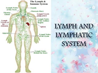
5. The four components of lymphatic system are:
1. LYMPH
2. LYMPH VESSEL
3. LYMPHOID TISSUE/ LYMPHOID ORGAN
4. LYMPHOCYTES and PHAGOCYTES
6. A clear fluid composed mainly of water,
electrolytes and some plasma proteins.
Transported in lymphatic pathway from
lymphatic vessels to collecting ducts and at the
end, disposed into venous blood.
7. When blood circulates in high pressure, the
fluid (plasma) portion seeps through thin
capillary walls into surrounding tissue.
This interstitial fluid is returned to blood
through walls of venules.
The remainder enters a network of thin
walled tubes called lymphatic vessels and
now is called lymph.
8.
9. The flow of lymph in the thoracic duct
in an average resting person usually
approximates 100 ml/ hr.
The total lymph flow in the body is
about 2 to 3 litres per day.
10. Lymphatic capillary run parallel to blood
capillaries in all body tissues and allow
diffusion of fluid from interstitial spaces into
lymphatic pathway.
Structurally identical to veins- vessel wall
composed of 3 thin layers of tissue ( Tunica
intima, Tunica media, Tunica adventia), and
contain valves to prevent backflow.
11.
12. Afferent lymphatic vessels- the vessels that
enters the lymph node
Efferent lymphatic vessels- the vessels that
leaves the lymph node
13. Lymphatic vessels exit lymph nodes and unite to
form lymphatic trunks.
The major trunks are
Lumbar trunks (right and left lumbar trunks)
Intestinal trunk
Bronchomediastinal trunks (right and left)
Subclavian trunks (right and left)
Jugular trunks (right and left)
14.
15. The lumbar trunks drain lymph from the lower
limbs, the pelvis, the kidneys, the adrenal glands,
and the abdominal wall.
The intestinal trunk drains lymph from the
stomach, intestines, pancreas, spleen, and part of
the liver.
The bronchomediastinal trunks drain lymph from
the thoracic wall, lung, and heart.
The subclavian trunks drain the upper limbs.
The jugular trunks drain the head and neck.
17. 1. Thoracic duct / Left lymphatic duct
Main duct for the return of lymph to blood.
38 – 45 cm length.
Begins at cisterna chyli (anterior to 2nd lumbar
vertebrae).
Cisterna chyli is an enlarged lymph sac which
receives lymph from right and left lumbar trunks
and intestinal trunk.
Receives lymph from cisterna chyli, left jugular, left
subclavian, and left bronchomediastinal trunks.
The thoracic duct drains lymph into venous blood
at the junction of the left internal jugular and left
subclavian veins.
18. 2. Right lymphatic duct
About 1.2 cm length.
Receives lymph from the right jugular, right
subclavian, and right bronchomediastinal
trunks.
Drains into venous blood at the junction of
the right internal jugular and right subclavian
veins.
19. The lymphatic vessels
form specialized
lymphatic organs called
lymph nodes which
store macrophages and
lymphocytes to
eliminate foreign
substance in the lymph.
20. Transport lymph away from tissues
Collect and filter lymph (at the nodes) as
it continues to move toward larger vessels
called collecting ducts.
21. Lymphatic organs are divided as primary lymphatic
organs and secondary lymphatic organs:
1. Primary Lymphatic Organs are red bone marrow
and thymus gland. The red bone marrow produce T-
cells in response to an antigen and they get matured
in thymus gland.
2. Secondary Lymphatic Organs are lymph nodes,
spleen, lymphatic nodules. These are the sites of
immune responses.
22. Bone marrow is a semi-solid tissue found
within the spongy portions of bones
It is the primary site of haematopoiesis.
It is composed of hematopoietic cells,
marrow adipose tissue, and
supportive stromal cells
23.
24. The thymus is an organ with two lobes that is
located anterior to the ascending aorta and
posterior to the sternum.
25. Each lobe surrounded by a capsule and
divided into lobules, which are separated
from each other by strans of connective
tissue called trabeculae.
26. Each lobule organized into 2 compartments: the
outer compartment cortex, and the inner
compartment medulla.
27. The maturation of T cells and monocytes
takes place in the medulla which is
supported by epithelial cells and
macrophages present in the thymus.
In addition it also secrete hormone such as
thymosin which stimulate the development
of antibodies.
28. Around 600 bean shaped lymph nodes are
located throughout the body.
Lymph nodes filter substances that travel
through the lymphatic fluid, and contain
lymphocytes (white blood cells) that help
the body fight infection and disease
29. Lymph node can be divided into 3 centric
regions: the Cortex, the Paracortex and the
Medulla.
The outer most cortex contains B
lymphocytes, macrophages and follicular
dendritic cells.
Beneath cortex, paracortex contains T
lymphocytes.
Medulla contains lymphoid cells, and plasma
cells which actively secrets antibodies.
30.
31. Filteration of lymph- lymph node filter and
purify the lymph (remove cell debris, virus,
fungi etc) before return to the venous
circulation
Trapping the antigen- traps the antigen and
presents infront of lymphocytes
Maturation of lymphocytes
Phagocytosis
32. The spleen is a fist-sized
organ in the upper left side of
abdomen, next to the stomach
and behind left ribs.
It is surrounded by a capsule
from which a number of
projections (trabeculae)
extend into interior to form
compartments.
33.
34. Compartments are of two types: the RED pulp and
the WHITE pulp, separated by a diffuse marginal
zone.
The white pulp surrounds the branches of splenic
artery, forming Periarterial Lymphatc Sheath
(PALS) which containsT lymphocytes.
Within the red pulp the following functions such
as removal of defective blood cells and platelets,
storage of platelets for emergency use, and
production of blood cells during fetal life etc.
35. Phagocytosis (destruction of RBC and
Platelets)
Storage of blood (approx 350 ml of blood is
stored which can be used in critical conditions)
Erythropoiesis (in fetus)
Immune response (spleenomegaly in case of any
infection due to activatedT and B lymphocytes)
36. They are egg shaped masses of lymphatic
tissues different from lymph nodes since they
do not have capsules.
They are located at the mucosal lining of GI,
urinary, respiratory and reproductive tracts and
also referred as MALT (mucosa-associated
lymphatic tissue)
The tonsils (in the throat) and the Peyer
patches (within the small intestine) are
examples of MALT.
37.
38.
39. Water and solutes continually filter out from
capillary into interstitial space. To balance this
outflow, fluid continually reenters blood through
lymphatic system.
Fluid (lymph) is filtered through lymph nodes to
remove bacteria, abnormal cells and other foreign
materials.
This fluid is then transported back into the
bloodstream via the lymph vessels.
Lymph only moves in one direction, toward the
heart.
40.
41. T CELLS- A type of white blood cell. T cells are
part of the immune system and develop from stem
cells in the bone marrow. They help protect the
body from infection and may help fight cancer.
Also calledT lymphocyte andThymocyte.
B CELLS- A type of white blood cell that makes
antibodies. B cells are part of the immune system
and develop from stem cells in the bone marrow.
Also called B lymphocyte.
42. MONOCYTES- A monocyte is a type of white
blood cell and a type of phagocyte. These are the
largest of leucocytes.
DENDRITIC CELLS: A dendritic cell is a type of
phagocyte and a type of antigen-presenting
cell (APC). It has long branched projections similar
to neurons.
GRANULOCYTIC CELLS: Neutrophil.
Eosinophil, Basophil
43. Lymphedema: It is a condition of
localized fluid retention and tissue swelling caused
by blocked/ improper lymphatic system.
The main symptom is swelling in an arm or leg
that may be accompanied by pain or discomfort.
44. The condition can be inherited or can be caused
by an injury to lymph vessel or nodes as a result
of treatment of cancer such as radiation, surgery
or a parasitic infection called filariasis.
Lymphedema Filariasis
45. There's no cure for lymphedema.
Therapies for lymphadema:
Exercises. Gentle contraction of the muscles
in the arm or leg can help move the excess
fluid out of the swollen limb.
Manual lymph drainage
Compression bandages
Compression garments