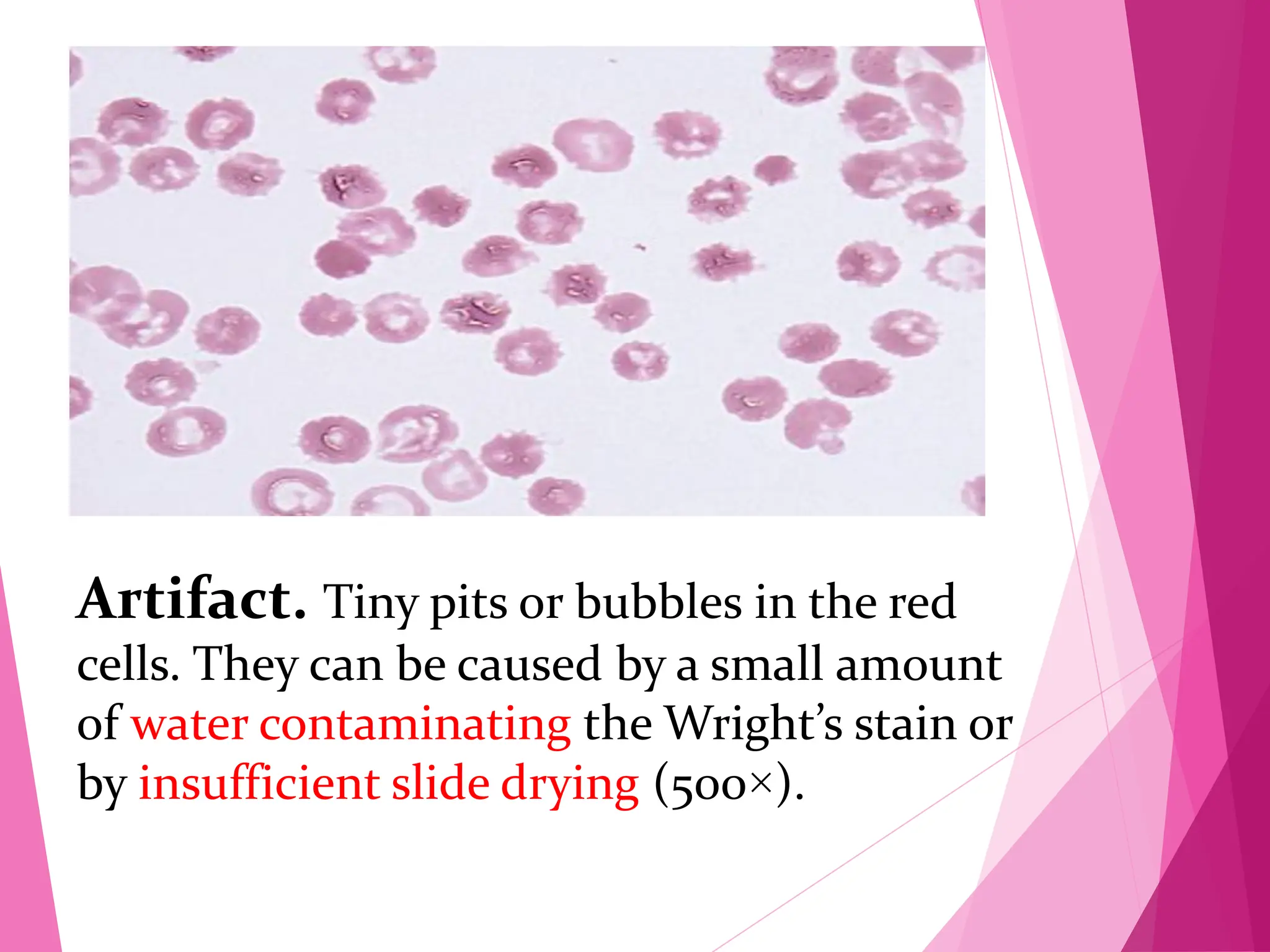
Download as PDF, PPTX
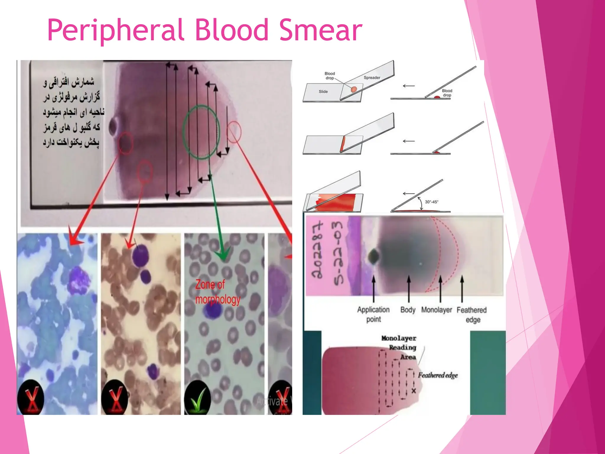
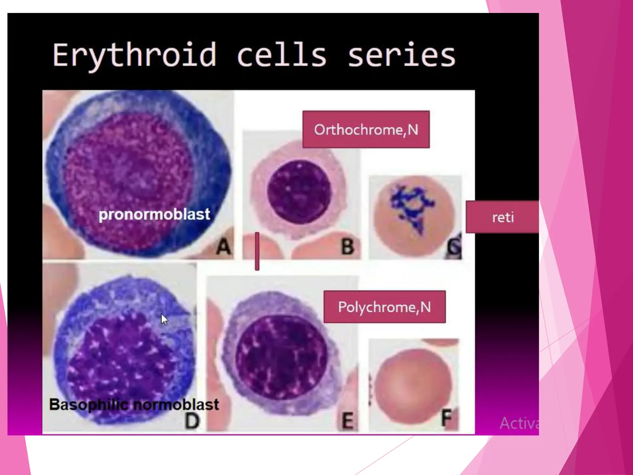
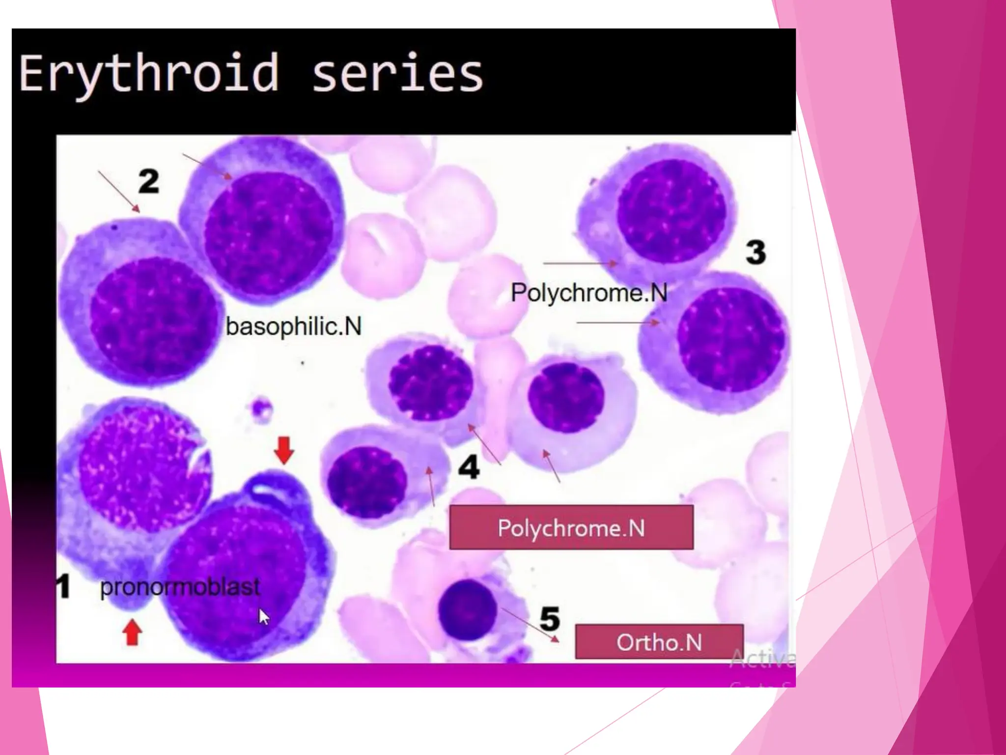
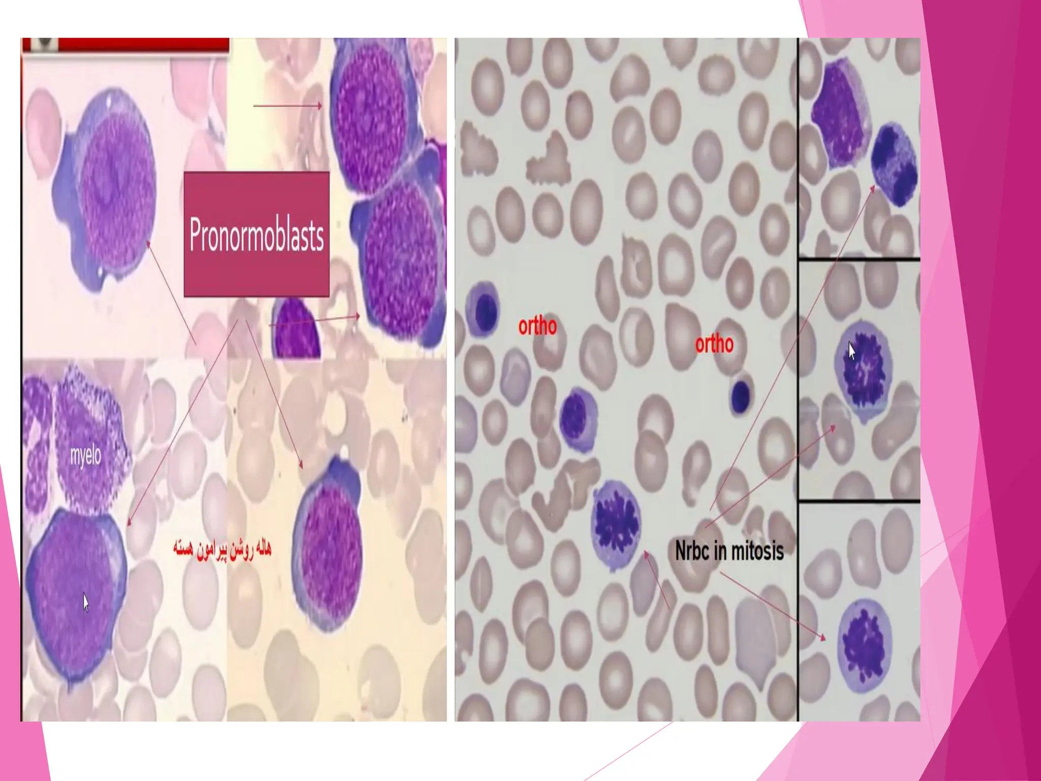
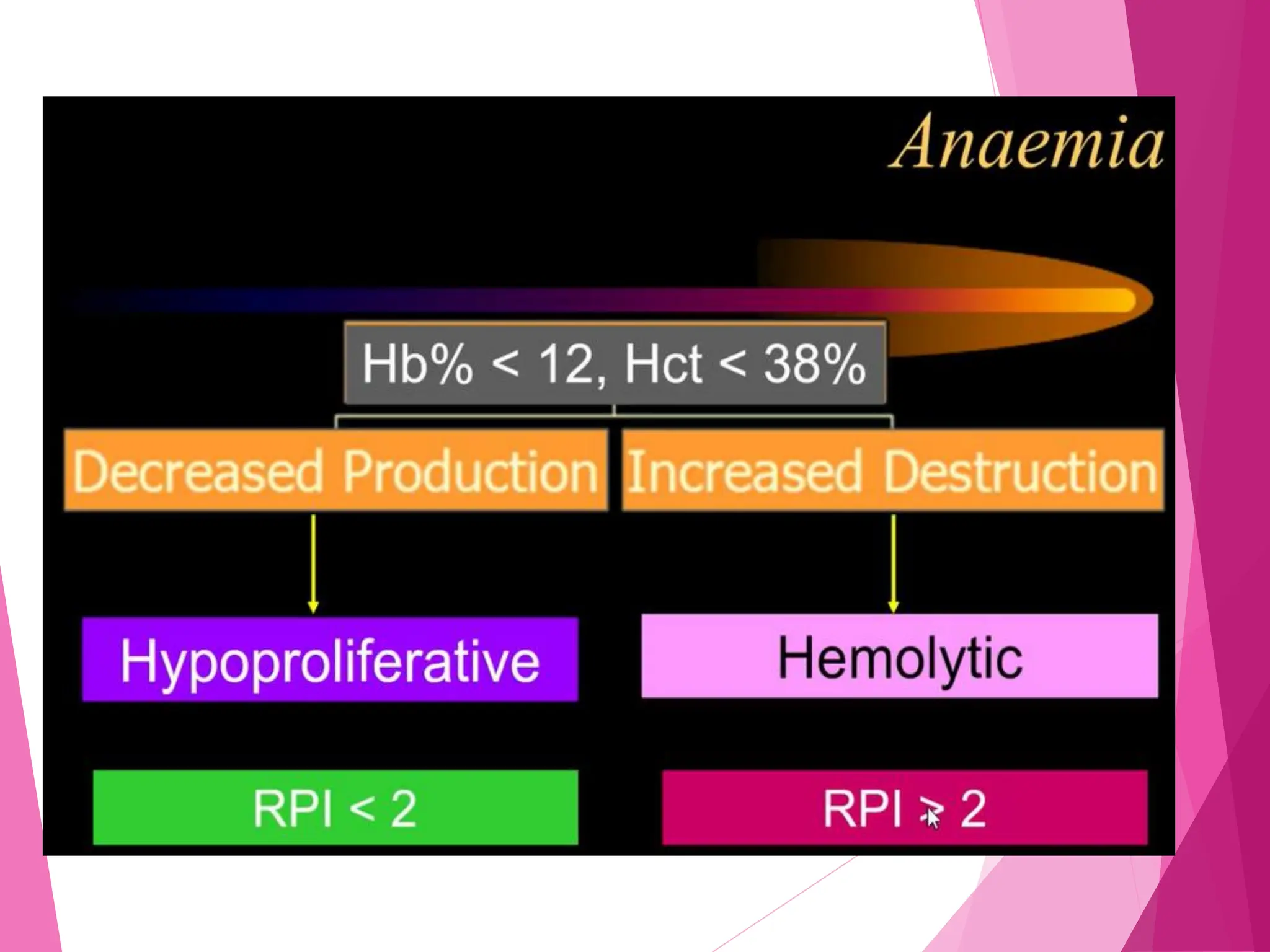
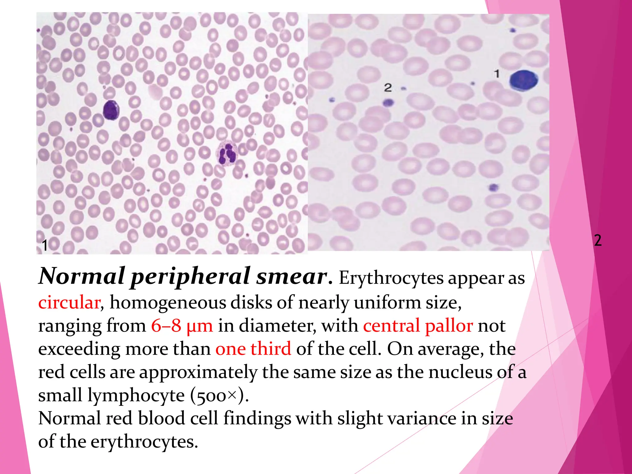
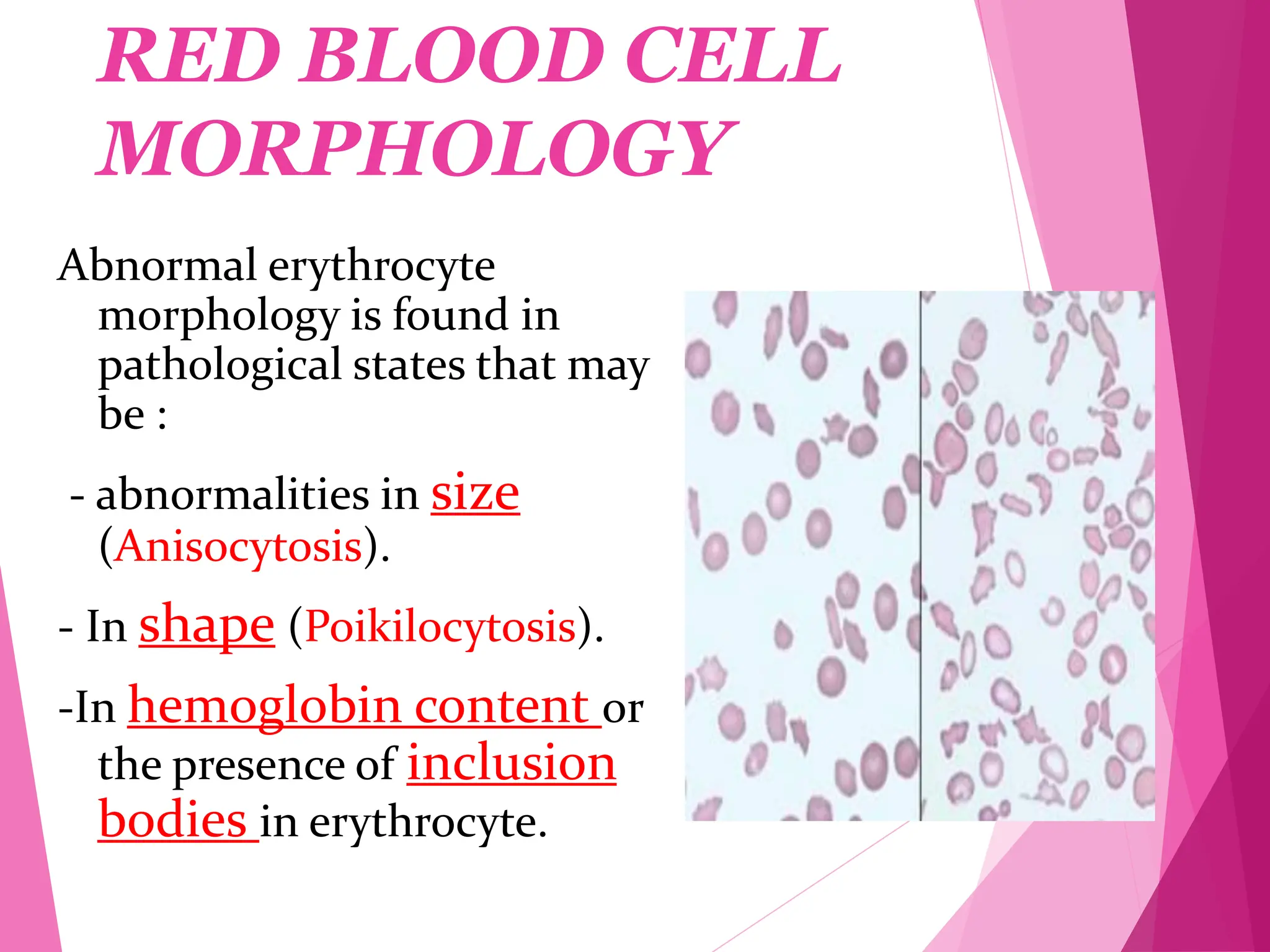
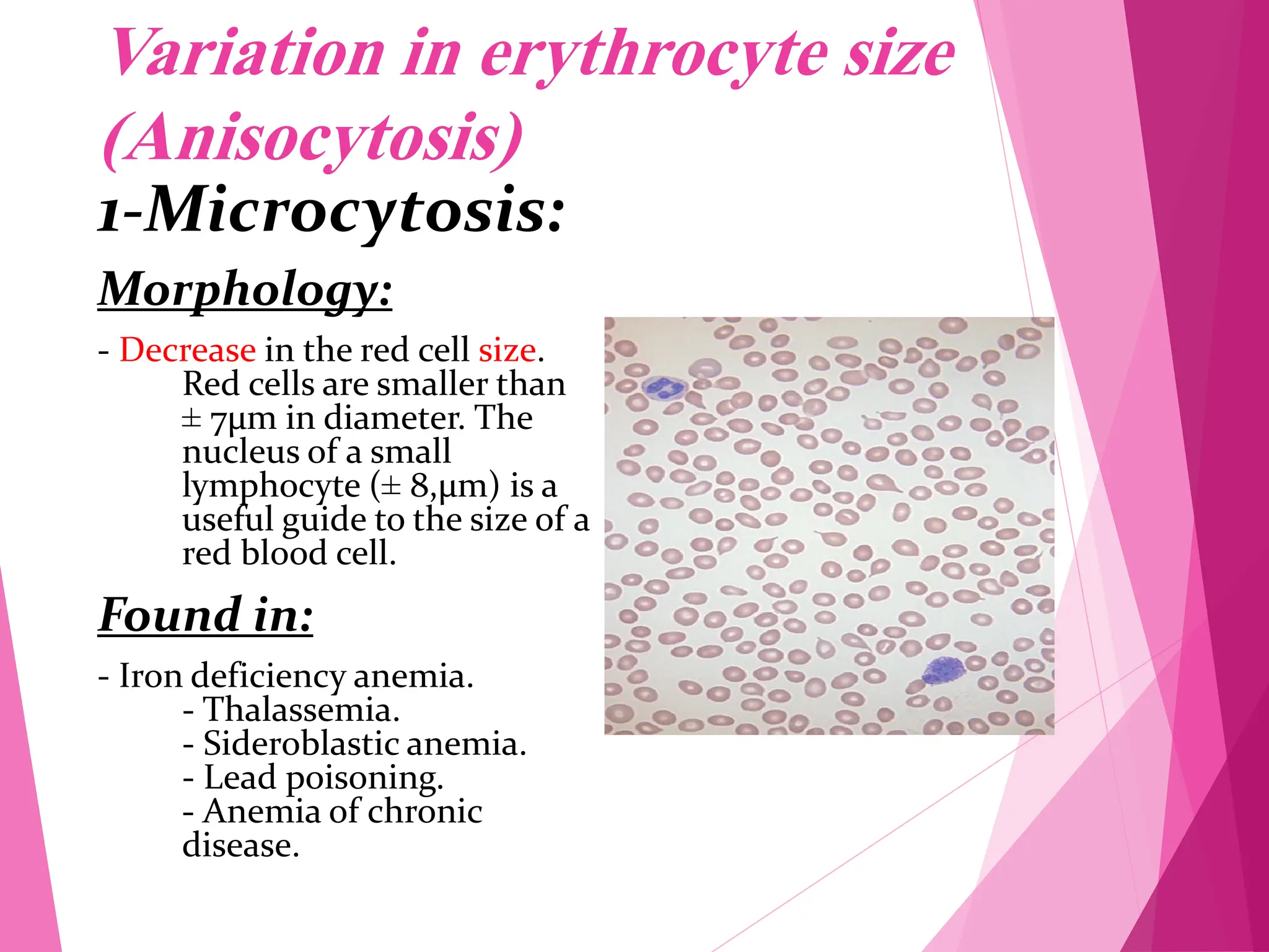
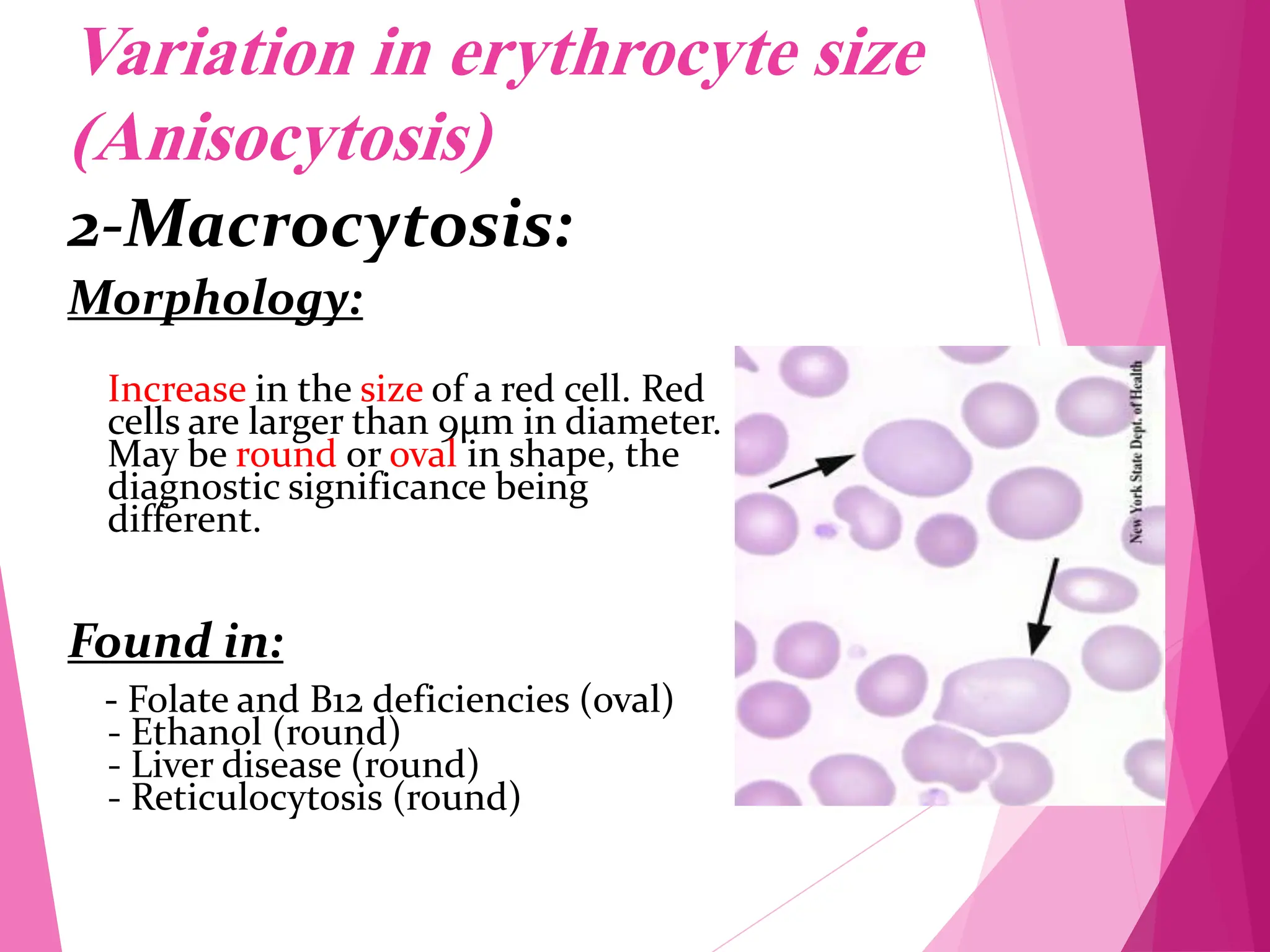
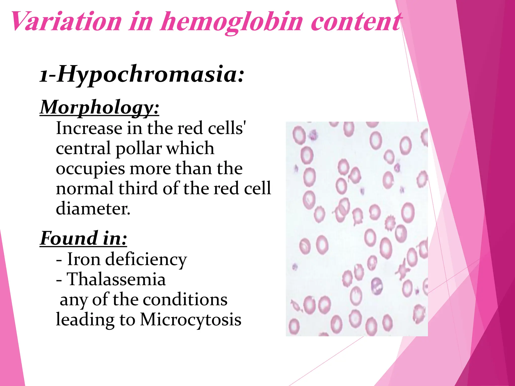
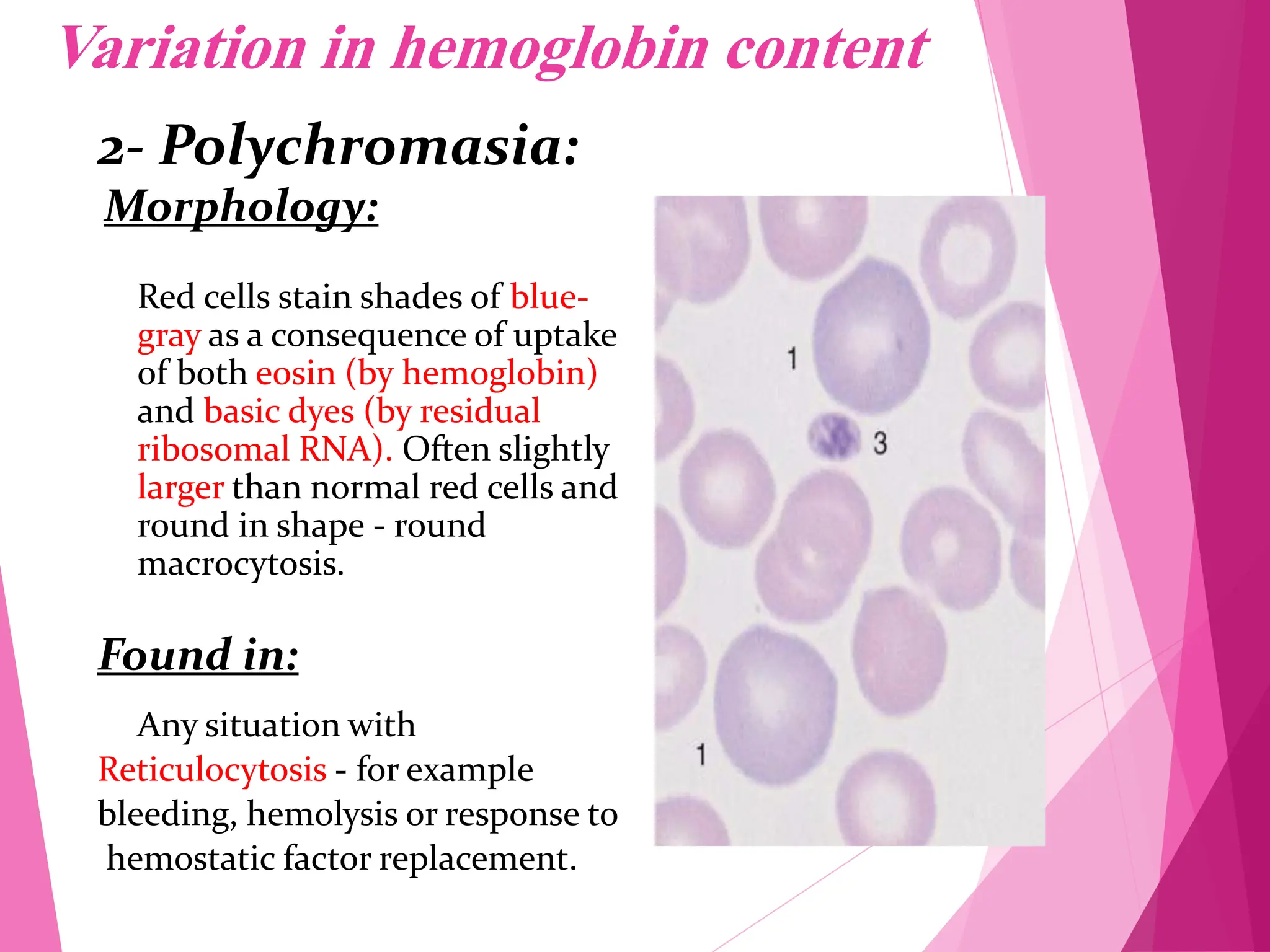
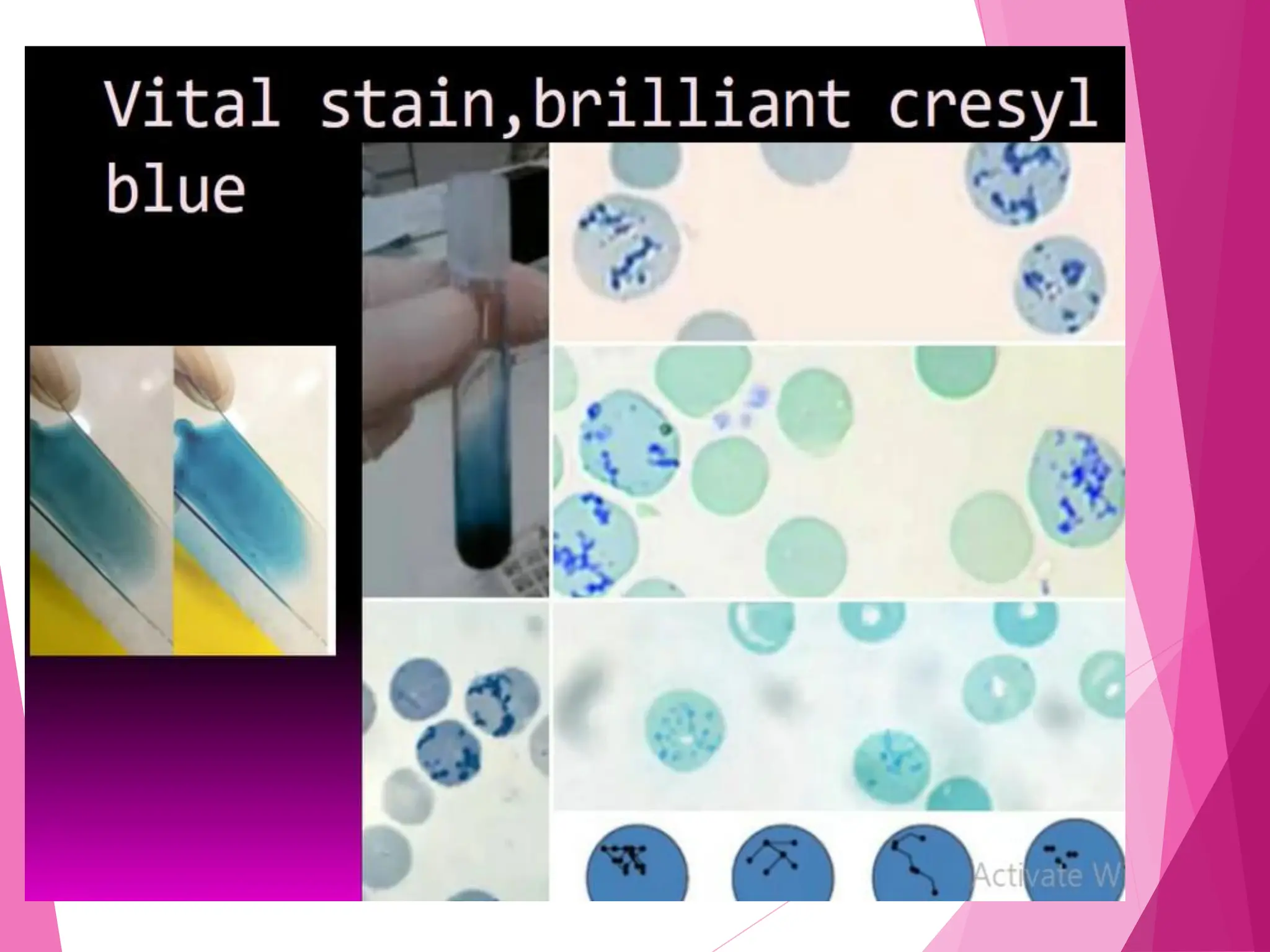
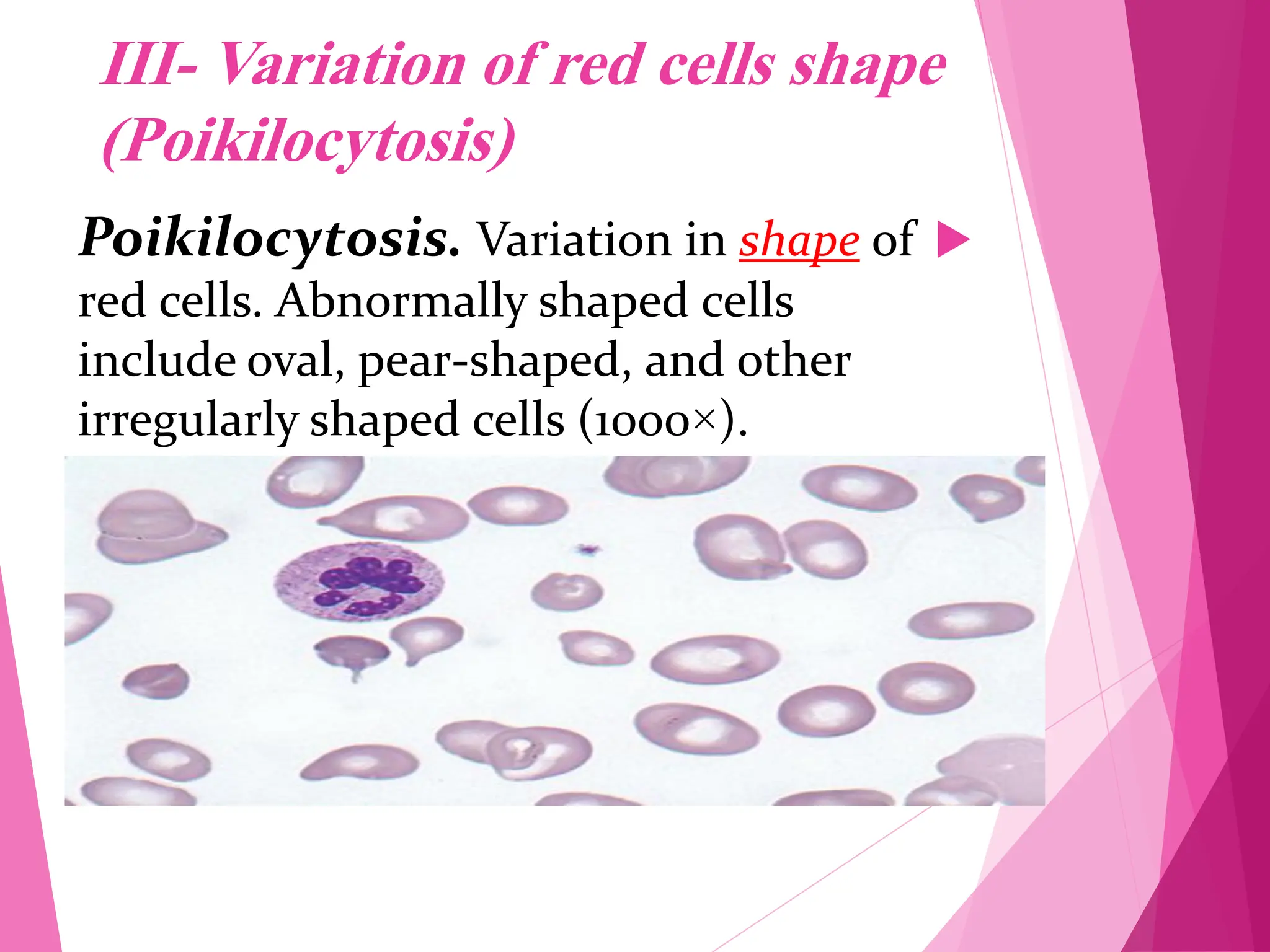
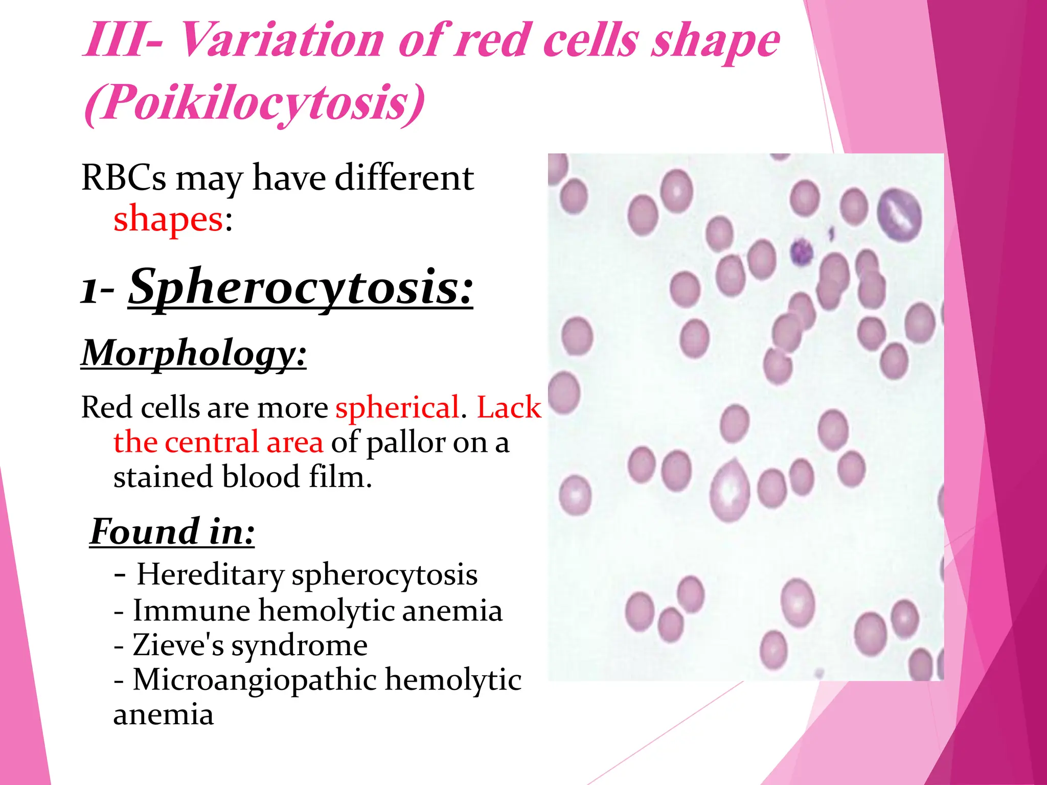
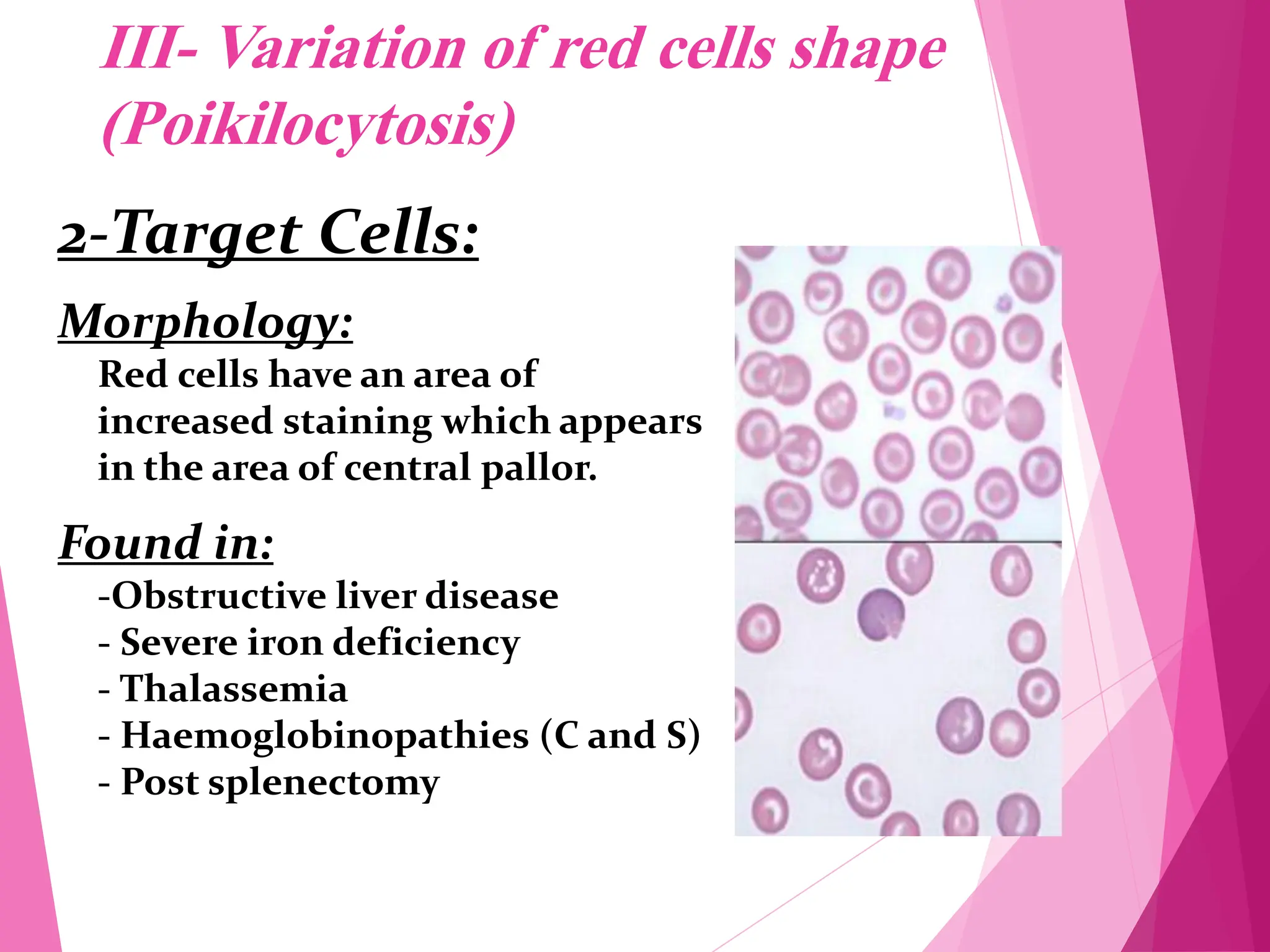
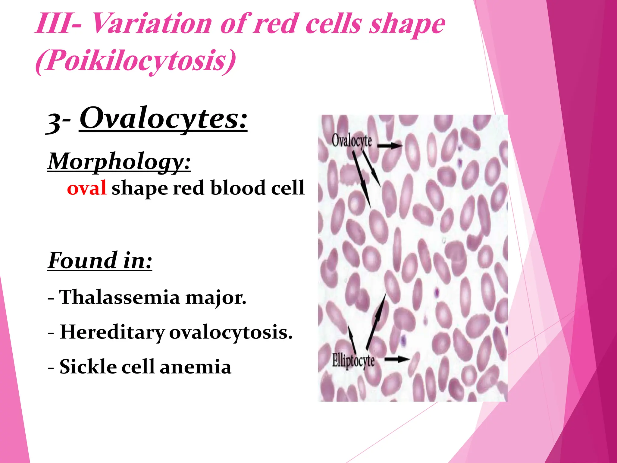
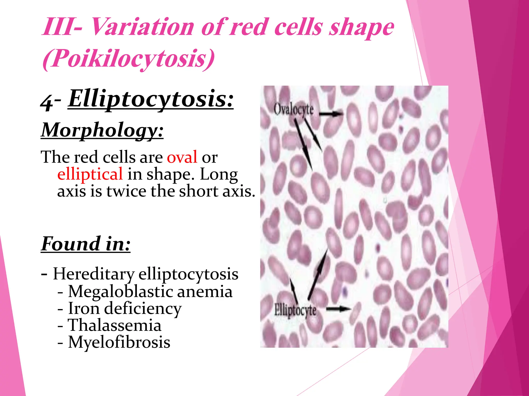


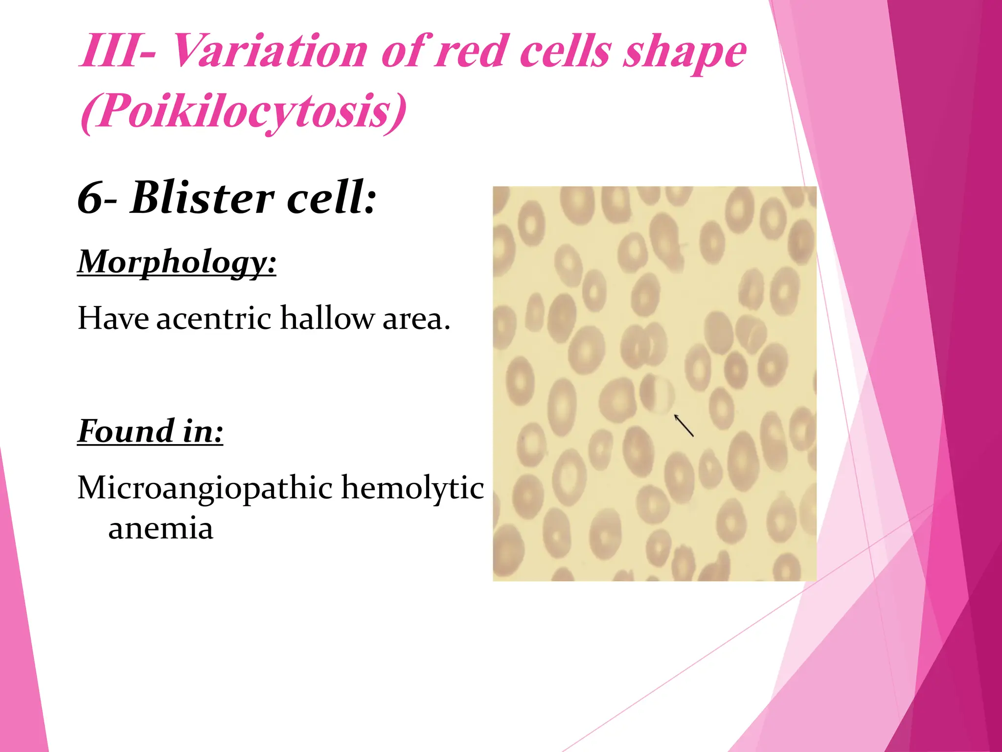
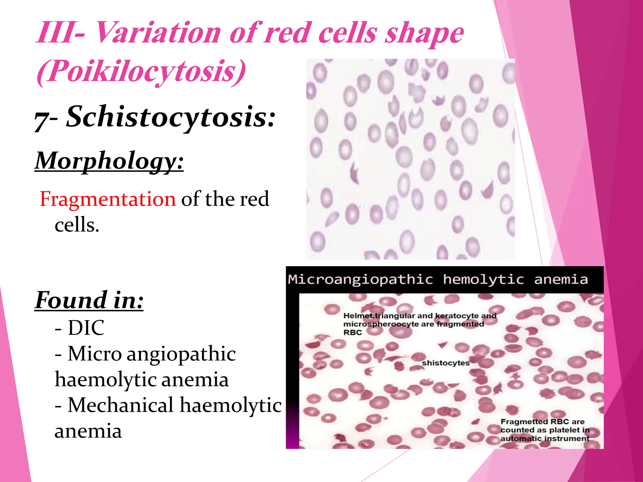
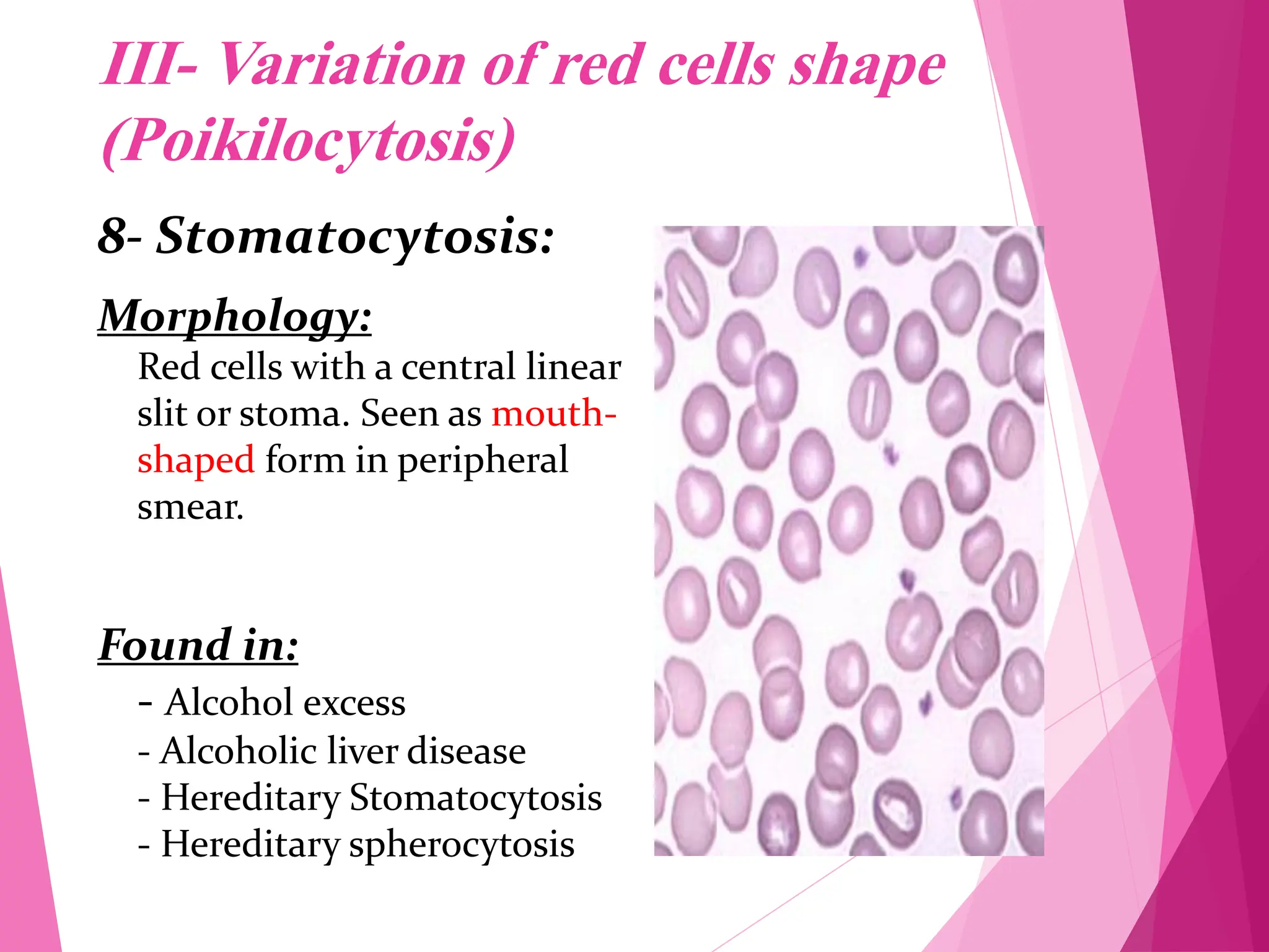

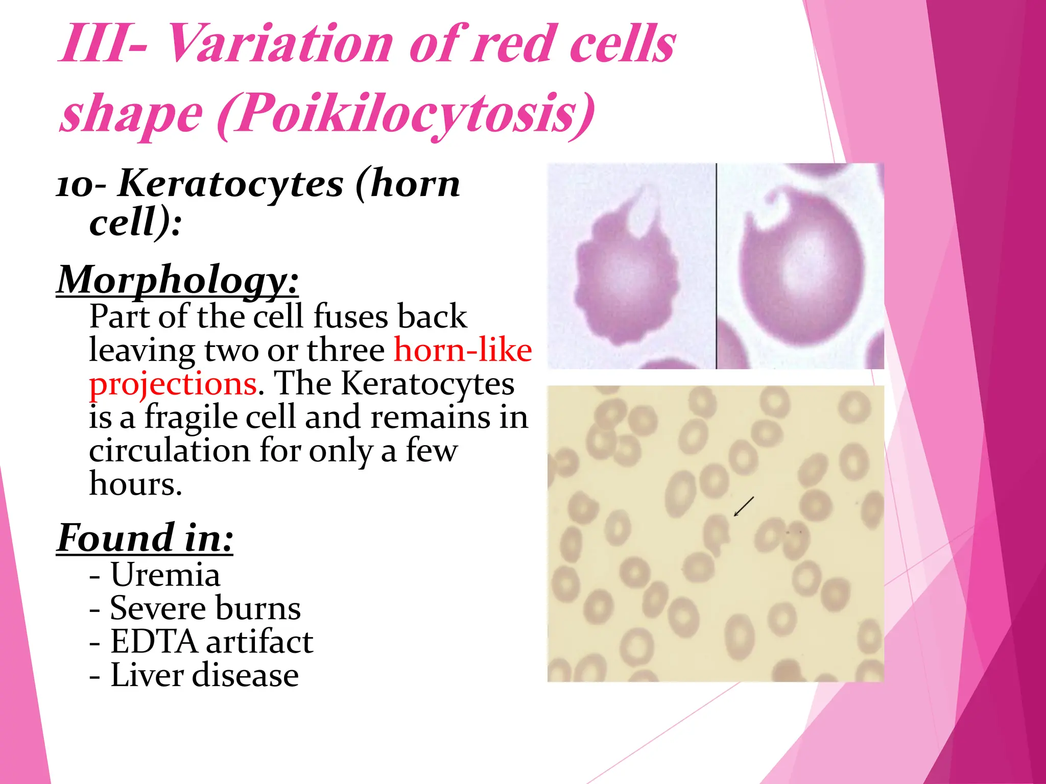
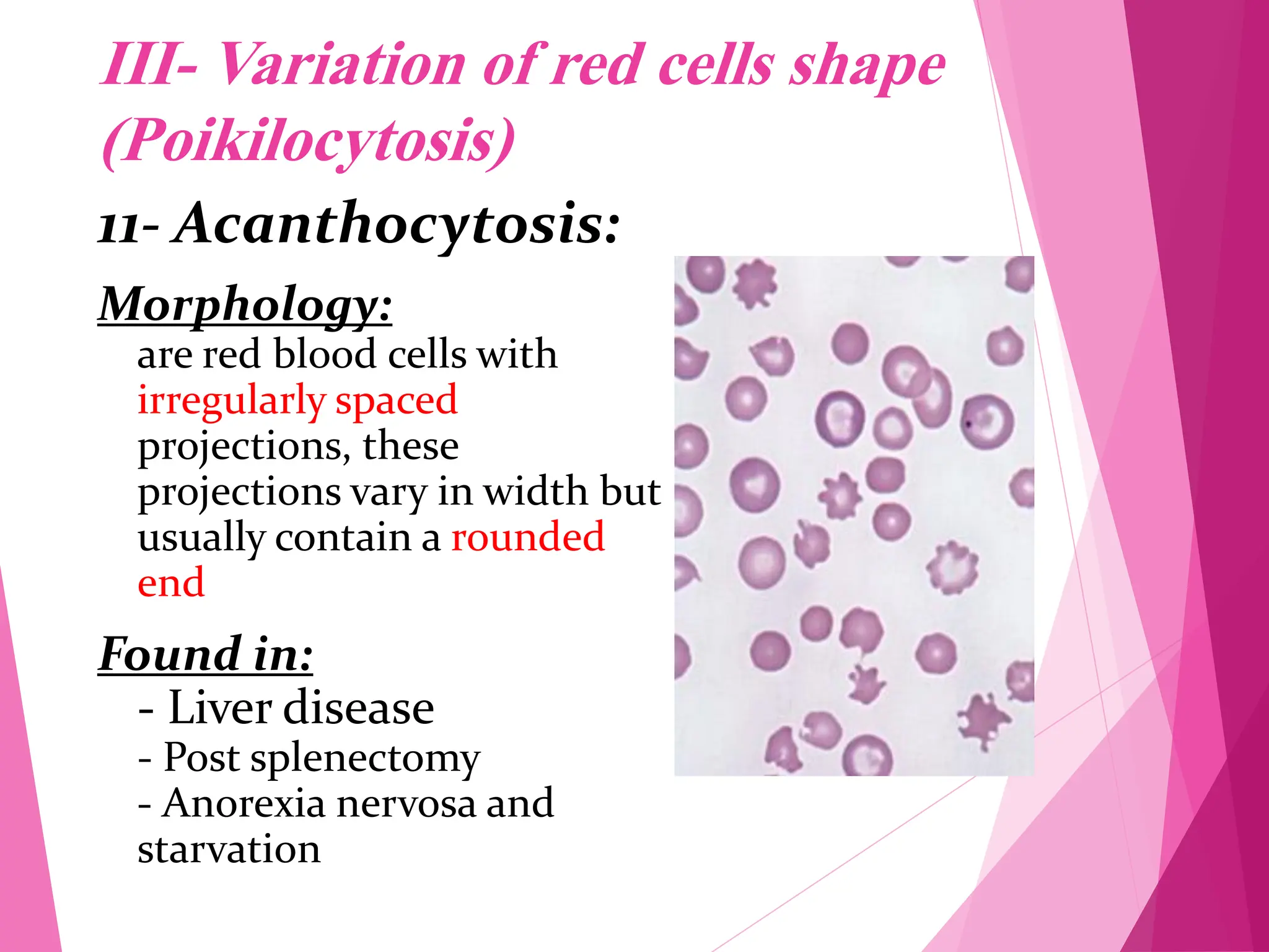

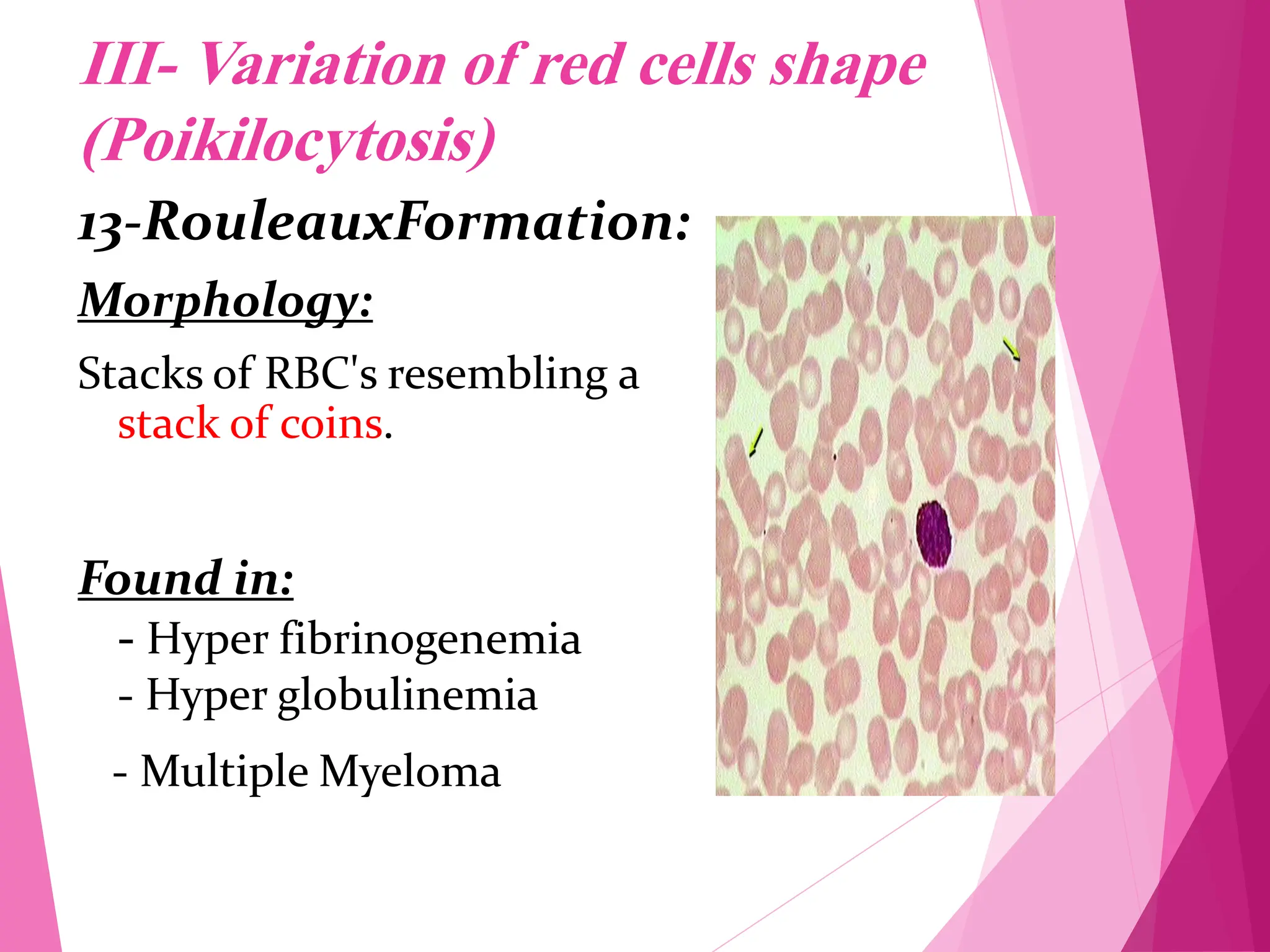
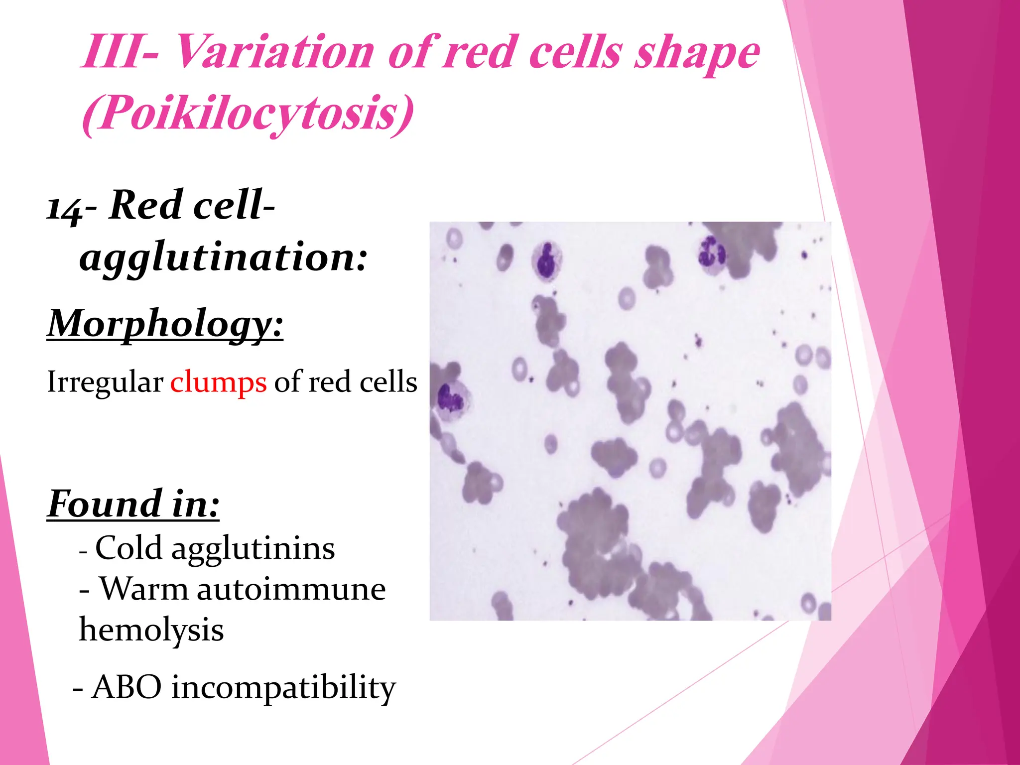

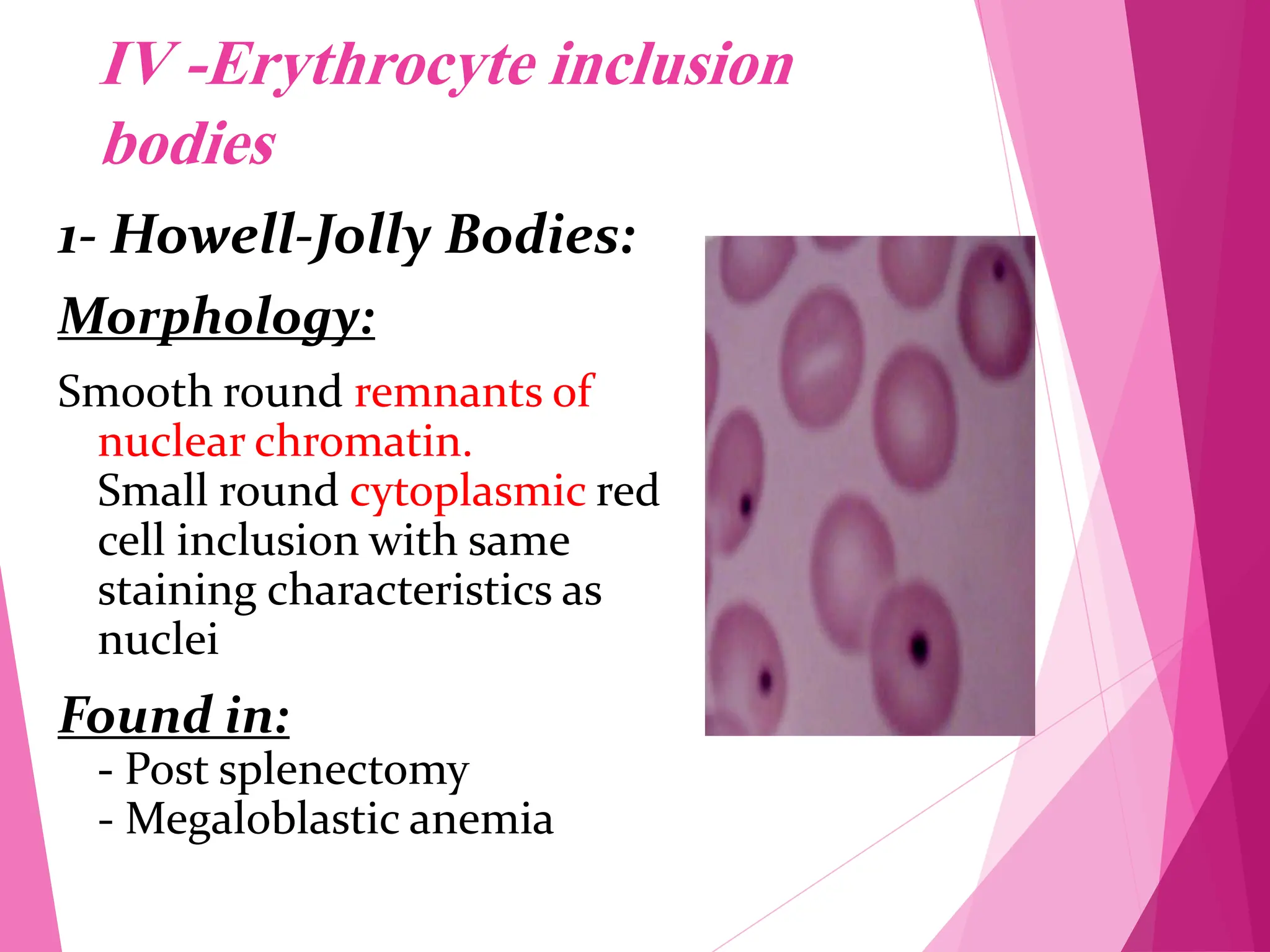
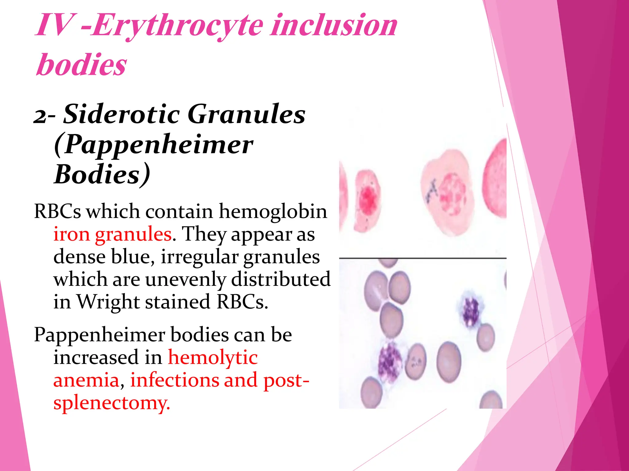
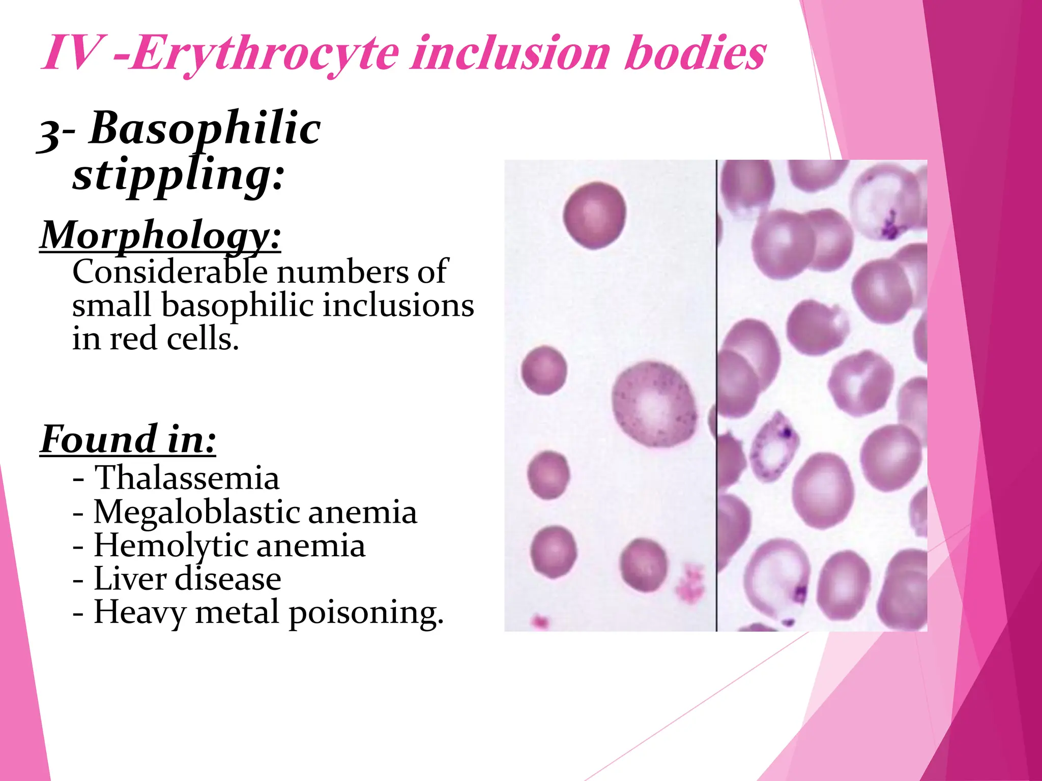
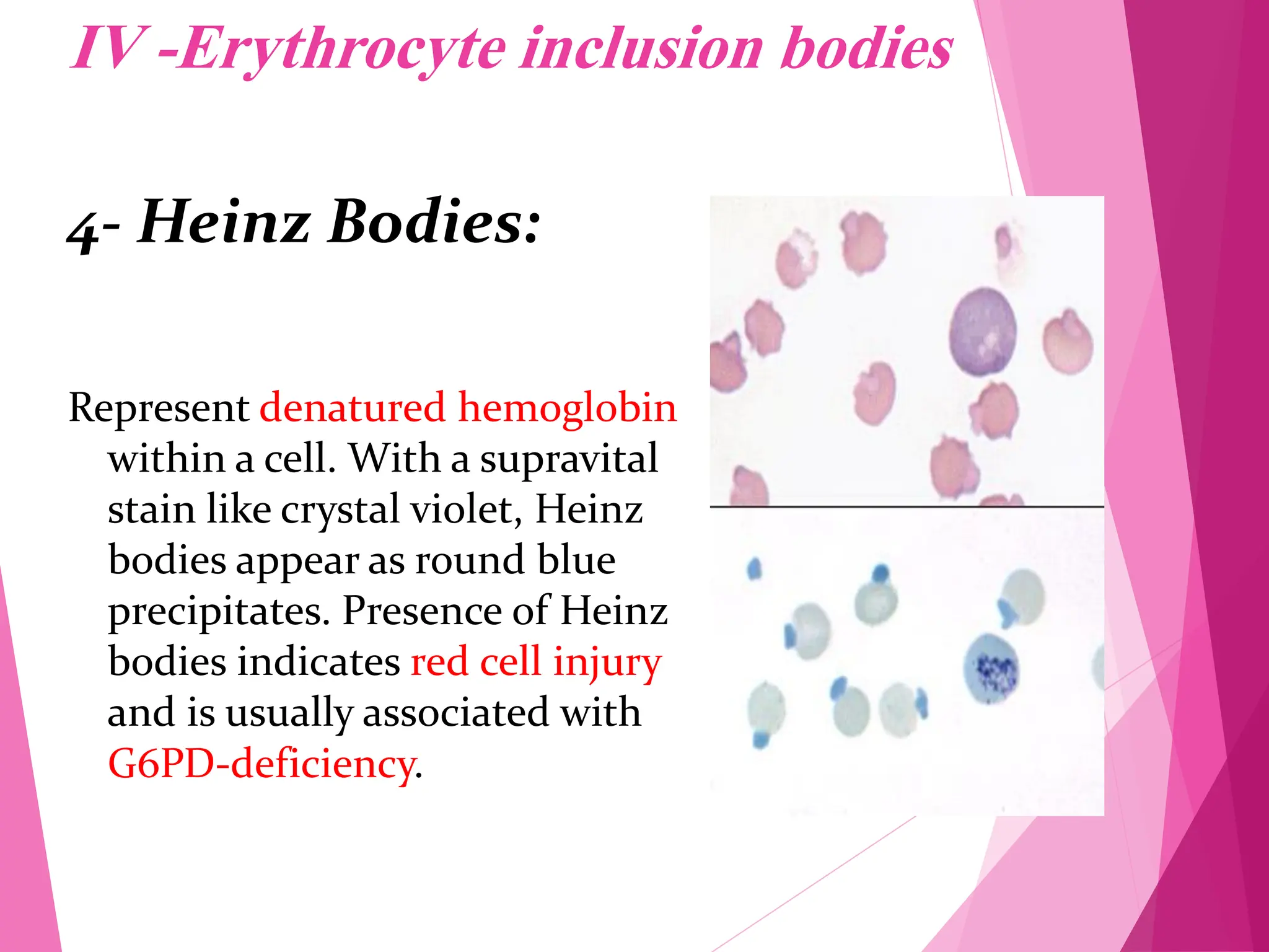

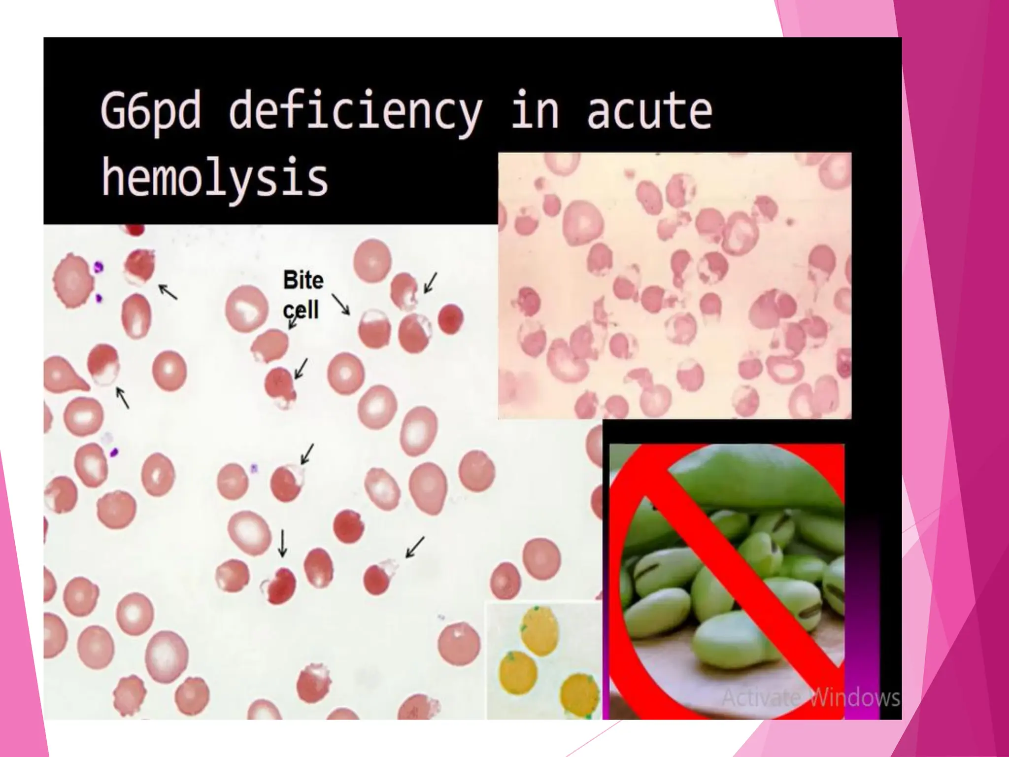


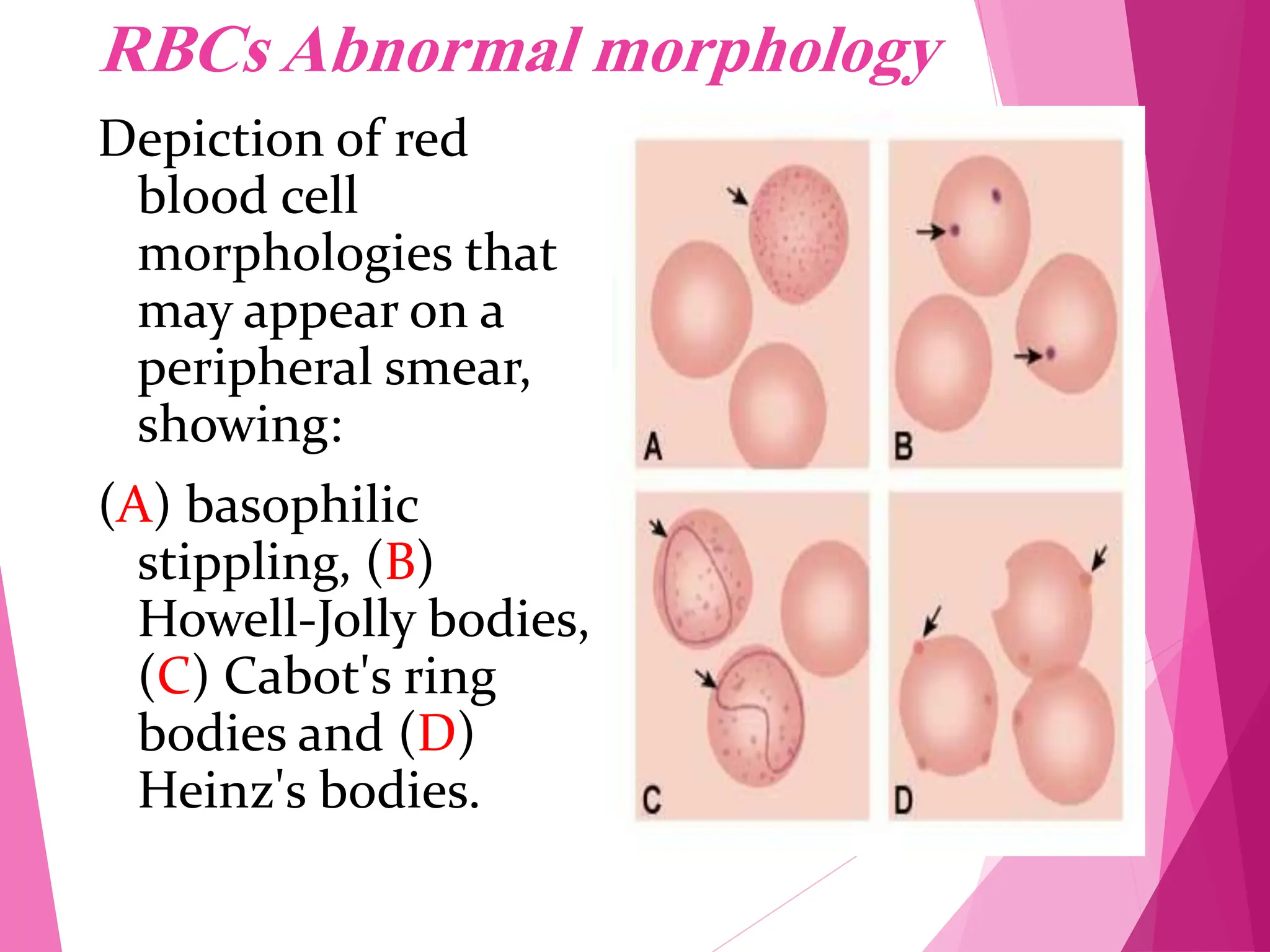
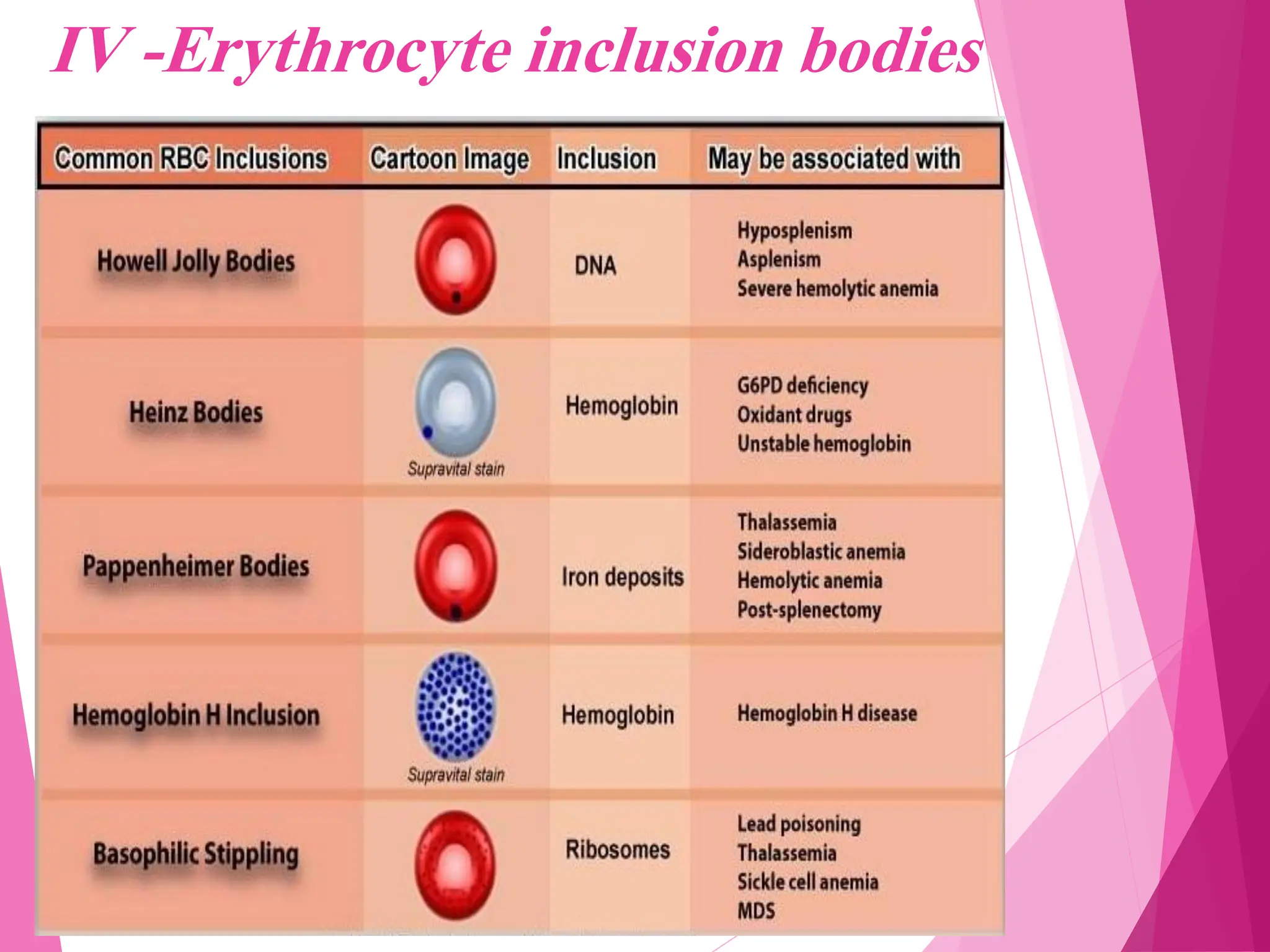
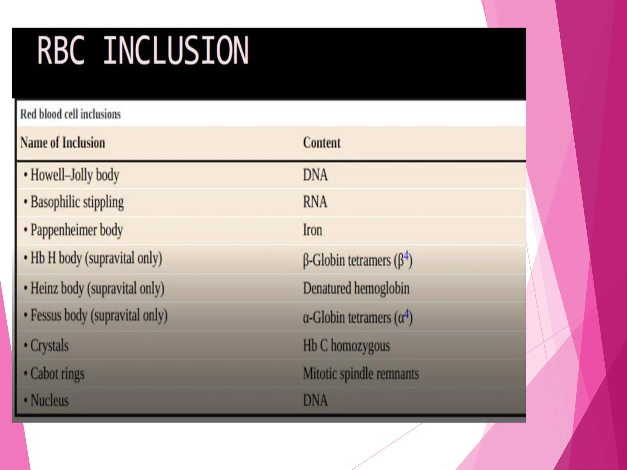


This document provides information on red blood cell morphology from a normal peripheral blood smear and in various pathological states. It describes normal RBC size, shape, and hemoglobin content. Abnormal findings include variations in size (anisocytosis), shape (poikilocytosis), and hemoglobin content. Specific abnormal RBC shapes and inclusions are defined along with potential causes. RBC indexes and rules for estimating relationships between hematological parameters are also presented.


