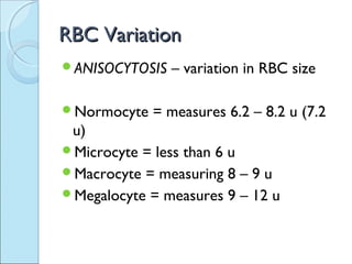
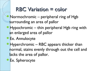
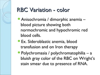
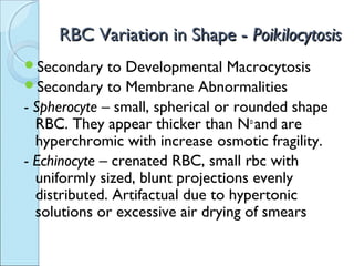
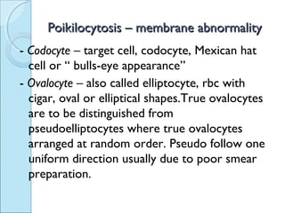
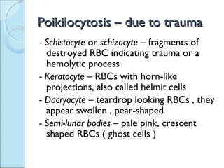
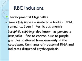
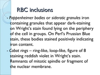
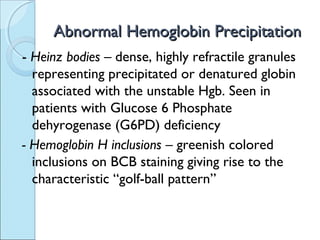
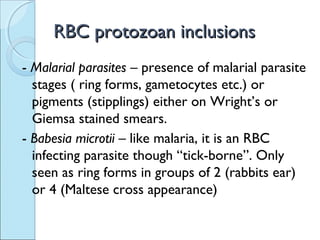
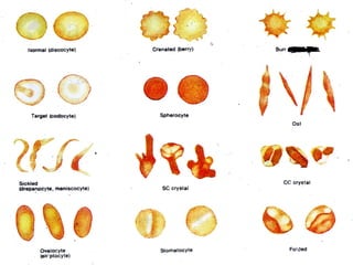
RED BLOOD CELLRED BLOOD CELL
VARIATION & PATHOLOGYVARIATION & PATHOLOGY
RBC VariationRBC Variation
ANISOCYTOSIS – variation in RBC size
Normocyte = measures 6.2 – 8.2 u (7.2
u)
ď‚—Microcyte = less than 6 u
Macrocyte = measuring 8 – 9 u
Megalocyte = measures 9 – 12 u
RBC Variation = colorRBC Variation = color
Normochromic – peripheral ring of Hgb
surrounding an area of pallor
Hypochromic – thin peripheral Hgb ring with
an enlarged area of pallor
ď‚—Ex. Annulocyte
Hyperchromic – RBC appears thicker than
normal, stains evenly through out the cell and
lacks the area of pallor.
ď‚—Ex. Spherocyte
RBC Variation - colorRBC Variation - color
Anisochromia / dimorphic anemia –
blood picture showing both
normochromic and hypochromic red
blood cells.
ď‚—Ex. Sideroblastic anemia, blood
transfusion and on Iron therapy
Polychromasia / polychromatophilia – a
bluish gray color of the RBC on Wright’s
stain smear due to presence of RNA.
ď‚—Residual RNA indicates immaturity of cell
ď‚—On supravital staining with Brilliant Cresyl
Blue or New Methylene Blue, the RNA
combines with the stain to form the dye-
ribonuceloprotein complex which appears as
a lacework pattern called the reticulum and
thus calling the said cell as RETICULOCYTE.
ď‚—Polychromasia = Reticulocyte
Target cell – also called the leptocyte ,the
Mexican hat cell or the codocyte
ď‚—An RBC with a peripheral ring of Hgb
plus a central condensation of Hgb in the
area of pallor or “ bull’s eye appearance “
ď‚—Found in patients with hemoglobinopathy
and thalassemias
RBC Variation in Shape -RBC Variation in Shape - PoikilocytosisPoikilocytosis
ď‚—Secondary to Developmental Macrocytosis
ď‚—Secondary to Membrane Abnormalities
- Spherocyte – small, spherical or rounded shape
RBC. They appear thicker than No
and are
hyperchromic with increase osmotic fragility.
- Echinocyte – crenated RBC, small rbc with
uniformly sized, blunt projections evenly
distributed. Artifactual due to hypertonic
solutions or excessive air drying of smears
Poikilocytosis – membrane abnormalityPoikilocytosis – membrane abnormality
- Burr cell – RBC with unevenly spaced
spicules, more pointed rather than blunt
- Acanthocyte – spur cell, RBC with club-
shaped (swollen) spicules, or prominent
irregular thorn-like projections
- Stomatocyte – slit-shaped or mouth-like
areas of pallor. On wet mounts, appear
to be uniconcave or “rice-bowl”
appearance
Poikilocytosis – membrane abnormalityPoikilocytosis – membrane abnormality
- Codocyte – target cell, codocyte, Mexican hat
cell or “ bulls-eye appearance”
- Ovalocyte – also called elliptocyte, rbc with
cigar, oval or elliptical shapes.True ovalocytes
are to be distinguished from
pseudoelliptocytes where true ovalocytes
arranged at random order. Pseudo follow one
uniform direction usually due to poor smear
preparation.
Poikilocytosis – due to traumaPoikilocytosis – due to trauma
- Schistocyte or schizocyte – fragments of
destroyed RBC indicating trauma or a
hemolytic process
- Keratocyte – RBCs with horn-like
projections, also called helmit cells
- Dacryocyte – teardrop looking RBCs , they
appear swollen , pear-shaped
- Semi-lunar bodies – pale pink, crescent
shaped RBCs ( ghost cells )
Poikilocytosis – abnormal Hgb contentPoikilocytosis – abnormal Hgb content
- Sickle cell – depranocyte or meniscocyte
sickle-shaped or crescent-shaped RBC.
The abnormal Hgb S during decreased
oxygen tension will crystallize or
polymerize causing the cell to sickle
shape
- Hgb CC crystals – hexagonal shaped
crystals with blunt ends
- Hgb SC crystals – “ Washington
monument appearance of crystals”
RBC InclusionsRBC Inclusions
ď‚—Developmental Organelles
- Howell Jolly bodies – single blue bodies, DNA
remnants. Seen in Pernicious anemia
- Basophilic stipplings also known as punctuate
basophilia – fine to coarse, blue to purple
granules scattered homogenously in the
cytoplasm. Remnants of ribosomal RNA and
indicates disturbed erythropoiesis.
RBC inclusionsRBC inclusions
- Pappenheimer bodies or siderotic granules iron
containing granules that appear dark-staining
on Wright’s stain found lying on the periphery
of the cell in groups. On Perl’s Prussian Blue
stain, these bodies stained positively indicating
iron content.
- Cabot rings – ring-like, loop-like, figure of 8
staining reddish violet in Wright’s stain.
Remnants of mitotic spindle or fragment of
the nuclear membrane.
Abnormal Hemoglobin PrecipitationAbnormal Hemoglobin Precipitation
- Heinz bodies – dense, highly refractile granules
representing precipitated or denatured globin
associated with the unstable Hgb. Seen in
patients with Glucose 6 Phosphate
dehyrogenase (G6PD) deficiency
- Hemoglobin H inclusions – greenish colored
inclusions on BCB staining giving rise to the
characteristic “golf-ball pattern”
RBC protozoan inclusionsRBC protozoan inclusions
- Malarial parasites – presence of malarial parasite
stages ( ring forms, gametocytes etc.) or
pigments (stipplings) either on Wright’s or
Giemsa stained smears.
- Babesia microtii – like malaria, it is an RBC
infecting parasite though “tick-borne”. Only
seen as ring forms in groups of 2 (rabbits ear)
or 4 (Maltese cross appearance)
RBC Variation
RBC Variation

RBC Variation

  • 1.
    RED BLOOD CELLREDBLOOD CELL VARIATION & PATHOLOGYVARIATION & PATHOLOGY
  • 2.
    RBC VariationRBC Variation ANISOCYTOSIS– variation in RBC size Normocyte = measures 6.2 – 8.2 u (7.2 u) Microcyte = less than 6 u Macrocyte = measuring 8 – 9 u Megalocyte = measures 9 – 12 u
  • 3.
    RBC Variation =colorRBC Variation = color Normochromic – peripheral ring of Hgb surrounding an area of pallor Hypochromic – thin peripheral Hgb ring with an enlarged area of pallor Ex. Annulocyte Hyperchromic – RBC appears thicker than normal, stains evenly through out the cell and lacks the area of pallor. Ex. Spherocyte
  • 4.
    RBC Variation -colorRBC Variation - color Anisochromia / dimorphic anemia – blood picture showing both normochromic and hypochromic red blood cells. Ex. Sideroblastic anemia, blood transfusion and on Iron therapy Polychromasia / polychromatophilia – a bluish gray color of the RBC on Wright’s stain smear due to presence of RNA.
  • 5.
    ď‚—Residual RNA indicatesimmaturity of cell ď‚—On supravital staining with Brilliant Cresyl Blue or New Methylene Blue, the RNA combines with the stain to form the dye- ribonuceloprotein complex which appears as a lacework pattern called the reticulum and thus calling the said cell as RETICULOCYTE. ď‚—Polychromasia = Reticulocyte
  • 6.
    Target cell –also called the leptocyte ,the Mexican hat cell or the codocyte An RBC with a peripheral ring of Hgb plus a central condensation of Hgb in the area of pallor or “ bull’s eye appearance “ Found in patients with hemoglobinopathy and thalassemias
  • 7.
    RBC Variation inShape -RBC Variation in Shape - PoikilocytosisPoikilocytosis Secondary to Developmental Macrocytosis Secondary to Membrane Abnormalities - Spherocyte – small, spherical or rounded shape RBC. They appear thicker than No and are hyperchromic with increase osmotic fragility. - Echinocyte – crenated RBC, small rbc with uniformly sized, blunt projections evenly distributed. Artifactual due to hypertonic solutions or excessive air drying of smears
  • 8.
    Poikilocytosis – membraneabnormalityPoikilocytosis – membrane abnormality - Burr cell – RBC with unevenly spaced spicules, more pointed rather than blunt - Acanthocyte – spur cell, RBC with club- shaped (swollen) spicules, or prominent irregular thorn-like projections - Stomatocyte – slit-shaped or mouth-like areas of pallor. On wet mounts, appear to be uniconcave or “rice-bowl” appearance
  • 9.
    Poikilocytosis – membraneabnormalityPoikilocytosis – membrane abnormality - Codocyte – target cell, codocyte, Mexican hat cell or “ bulls-eye appearance” - Ovalocyte – also called elliptocyte, rbc with cigar, oval or elliptical shapes.True ovalocytes are to be distinguished from pseudoelliptocytes where true ovalocytes arranged at random order. Pseudo follow one uniform direction usually due to poor smear preparation.
  • 10.
    Poikilocytosis – dueto traumaPoikilocytosis – due to trauma - Schistocyte or schizocyte – fragments of destroyed RBC indicating trauma or a hemolytic process - Keratocyte – RBCs with horn-like projections, also called helmit cells - Dacryocyte – teardrop looking RBCs , they appear swollen , pear-shaped - Semi-lunar bodies – pale pink, crescent shaped RBCs ( ghost cells )
  • 11.
    Poikilocytosis – abnormalHgb contentPoikilocytosis – abnormal Hgb content - Sickle cell – depranocyte or meniscocyte sickle-shaped or crescent-shaped RBC. The abnormal Hgb S during decreased oxygen tension will crystallize or polymerize causing the cell to sickle shape - Hgb CC crystals – hexagonal shaped crystals with blunt ends - Hgb SC crystals – “ Washington monument appearance of crystals”
  • 12.
    RBC InclusionsRBC Inclusions DevelopmentalOrganelles - Howell Jolly bodies – single blue bodies, DNA remnants. Seen in Pernicious anemia - Basophilic stipplings also known as punctuate basophilia – fine to coarse, blue to purple granules scattered homogenously in the cytoplasm. Remnants of ribosomal RNA and indicates disturbed erythropoiesis.
  • 13.
    RBC inclusionsRBC inclusions -Pappenheimer bodies or siderotic granules iron containing granules that appear dark-staining on Wright’s stain found lying on the periphery of the cell in groups. On Perl’s Prussian Blue stain, these bodies stained positively indicating iron content. - Cabot rings – ring-like, loop-like, figure of 8 staining reddish violet in Wright’s stain. Remnants of mitotic spindle or fragment of the nuclear membrane.
  • 14.
    Abnormal Hemoglobin PrecipitationAbnormalHemoglobin Precipitation - Heinz bodies – dense, highly refractile granules representing precipitated or denatured globin associated with the unstable Hgb. Seen in patients with Glucose 6 Phosphate dehyrogenase (G6PD) deficiency - Hemoglobin H inclusions – greenish colored inclusions on BCB staining giving rise to the characteristic “golf-ball pattern”
  • 15.
    RBC protozoan inclusionsRBCprotozoan inclusions - Malarial parasites – presence of malarial parasite stages ( ring forms, gametocytes etc.) or pigments (stipplings) either on Wright’s or Giemsa stained smears. - Babesia microtii – like malaria, it is an RBC infecting parasite though “tick-borne”. Only seen as ring forms in groups of 2 (rabbits ear) or 4 (Maltese cross appearance)