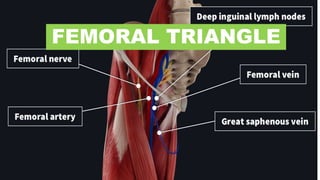
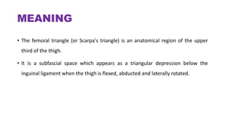
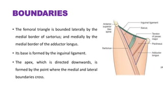
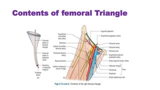
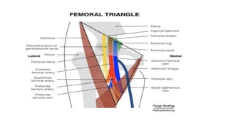
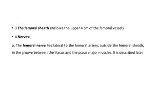
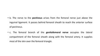
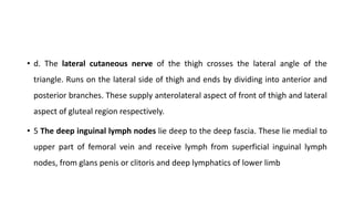
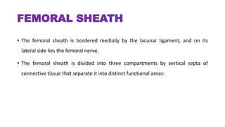
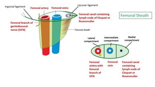
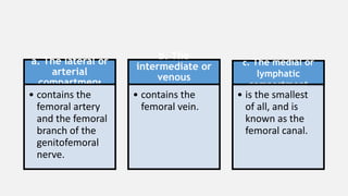
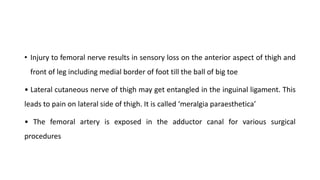
The femoral triangle is an anatomical region in the upper thigh bounded laterally by the sartorius muscle and medially by the adductor longus muscle. Its base is formed by the inguinal ligament and its apex points downwards. The femoral triangle contains the femoral artery and vein, branches of the femoral nerve, and deep inguinal lymph nodes. The femoral sheath encloses the upper part of the femoral vessels and is divided into three compartments. The contents of the femoral triangle are clinically relevant to femoral hernias, nerve injuries, and vascular procedures.
2. MEANING
• The femoral triangle (or Scarpa's triangle) is an anatomical region of the upper
third of the thigh.
• It is a subfascial space which appears as a triangular depression below the
inguinal ligament when the thigh is flexed, abducted and laterally rotated.
3. BOUNDARIES
• The femoral triangle is bounded laterally by the
medial border of sartorius; and medially by the
medial border of the adductor longus.
• Its base is formed by the inguinal ligament.
• The apex, which is directed downwards, is
formed by the point where the medial and lateral
boundaries cross.
4. • The apex is continuous, below with the adductor canal
• The roof of the femoral triangle is formed by:
• a. Skin.
• b. Superficial fascia containing the superficial inguinal lymph nodes, the femoral branch
of the genitofemoral nerve, branches of the ilioinguinal nerve, superficial branches of the
femoral artery with accompanying veins, and the upper part of the great saphenous vein.
• c. Deep fascia, including the cribriform fascia covering the saphenous opening.
5. • The floor of the triangle is formed medially by the adductor longus
and pectineus, and laterally by the psoas major and iliacus
6. CONTENTS
The contents of the femoral are as follows:
• 1 Femoral artery and its branches: The femoral artery traverses the triangle from
its base at the midinguinal point to the apex. In the triangle, it gives off six
branches, three superficial and three deep.
9. • 2 Femoral vein and its tributaries: The femoral vein accompanies the femoral
artery. The vein is medial to the artery at base of triangle, but posteromedial to
artery at the apex.
• The femoral vein receives the great saphenous vein, circumflex veins and veins
corresponding to the branches of femoral artery
10. • 3 The femoral sheath encloses the upper 4 cm of the femoral vessels
• 4 Nerves:
a. The femoral nerve lies lateral to the femoral artery, outside the femoral sheath,
in the groove between the iliacus and the psoas major muscles. It is described later
11. • b. The nerve to the pectineus arises from the femoral nerve just above the
inguinal ligament. It passes behind femoral sheath to reach the anterior surface
of pectineus.
• c. The femoral branch of the genitofemoral nerve occupies the lateral
compartment of the femoral sheath along with the femoral artery. It supplies
most of the skin over the femoral triangle.
12. • d. The lateral cutaneous nerve of the thigh crosses the lateral angle of the
triangle. Runs on the lateral side of thigh and ends by dividing into anterior and
posterior branches. These supply anterolateral aspect of front of thigh and lateral
aspect of gluteal region respectively.
• 5 The deep inguinal lymph nodes lie deep to the deep fascia. These lie medial to
upper part of femoral vein and receive lymph from superficial inguinal lymph
nodes, from glans penis or clitoris and deep lymphatics of lower limb
13. FEMORAL SHEATH
• The femoral sheath is bordered medially by the lacunar ligament, and on its
lateral side lies the femoral nerve.
• The femoral sheath is divided into three compartments by vertical septa of
connective tissue that separate it into distinct functional areas:
14.
15. a. The lateral or
arterial
compartment
• contains the
femoral artery
and the femoral
branch of the
genitofemoral
nerve.
b. The
intermediate or
venous
compartment
• contains the
femoral vein.
c. The medial or
lymphatic
compartment
• is the smallest
of all, and is
known as the
femoral canal.
16. CLINICAL ANATOMY/APPLIED ANATOMY
• Femoral hernia: The femoral canal is an area of potential weakness in the
abdominal wall through which abdominal contents may bulge out forming a
femoral hernia. A femoral hernia is more common in females because the
femoral canal is wider. This is associated with the wider pelvis, and the smaller
size of the femoral vessels, in the female It is never congenital.
17.
18. • Injury to femoral nerve results in sensory loss on the anterior aspect of thigh and
front of leg including medial border of foot till the ball of big toe
• Lateral cutaneous nerve of thigh may get entangled in the inguinal ligament. This
leads to pain on lateral side of thigh. It is called ‘meralgia paraesthetica’
• The femoral artery is exposed in the adductor canal for various surgical
procedures