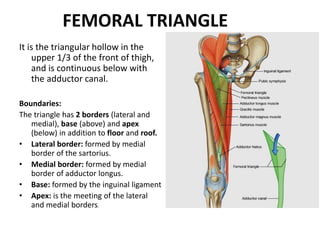
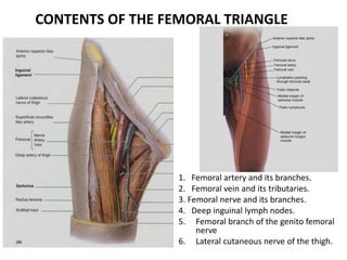
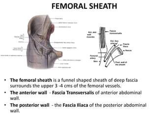
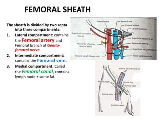
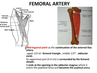
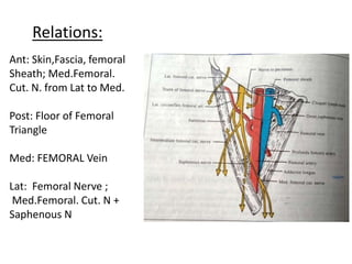
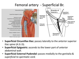
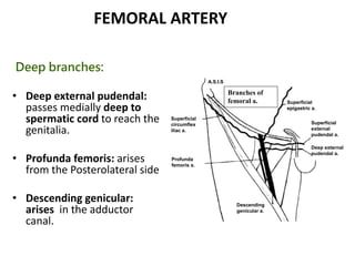
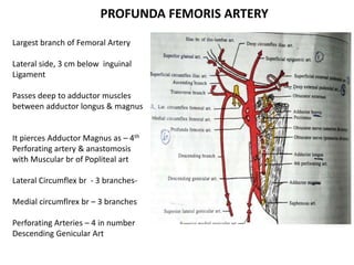
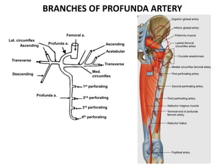
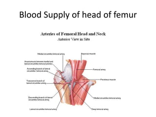
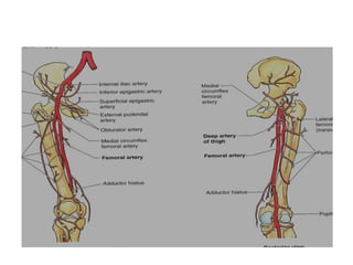
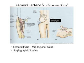
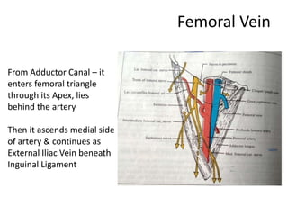
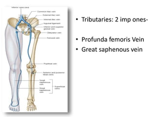
Femoral Vessels
Dr. SOMESH M.S.
PROFESSOR & HOD
FEMORAL TRIANGLE
It is the triangular hollow in the
upper 1/3 of the front of thigh,
and is continuous below with
the adductor canal.
Boundaries:
The triangle has 2 borders (lateral and
medial), base (above) and apex
(below) in addition to floor and roof.
• Lateral border: formed by medial
border of the sartorius.
• Medial border: formed by medial
border of adductor longus.
• Base: formed by the inguinal ligament
• Apex: is the meeting of the lateral
and medial borders.
ROOF OF
THE
FEMORAL
TRIANGLE
Roof: Is formed by skin superficial fascia & its contents
and the deep fascia.
CONTENTS OF THE FEMORAL TRIANGLE
1. Femoral artery and its branches.
2. Femoral vein and its tributaries.
3. Femoral nerve and its branches.
4. Deep inguinal lymph nodes.
5. Femoral branch of the genito femoral
nerve
6. Lateral cutaneous nerve of the thigh.
Fascia
transversalis
Ant. abd.
wall
muscles
Ext. iliac
artery Fascia
iliaca
Iliacus
Post. wall of
the sheath
Skin
Femoral
artery
• The femoral sheath is a funnel shaped sheath of deep fascia
surrounds the upper 3 -4 cms of the femoral vessels.
• The anterior wall - Fascia Transversalis of anterior abdominal
wall.
• The posterior wall - the Fascia Iliaca of the posterior abdominal
wall.
FEMORAL SHEATH
FEMORAL SHEATH
The sheath is divided by two septa
into three compartments:
1. Lateral compartment: contains
the Femoral artery and
Femoral branch of Genito-
femoral nerve.
2. Intermediate compartment:
contains the Femoral vein.
3. Medial compartment: Called
the Femoral canal, contains
lymph node + some fat.
FEMORAL ARTERY
Mid-Inguinal point as the continuation of the external iliac
artery.
upper 1/3 rd - femoral triangle , middle 1/3rd adductor
canal.
Its uppermost part (3-4 cm) is surrounded by the femoral
sheath.
It ends at the opening in the adductor magnus where it
enters the popliteal fossa and becomes the popliteal artery.
A.S.I.S
Inguinal lig.
Pubic tubercle
Apex
Add. longus
Sartorius
Femoral
triangle
te of sub.
artorial canal
Relations:
Ant: Skin,Fascia, femoral
Sheath; Med.Femoral.
Cut. N. from Lat to Med.
Post: Floor of Femoral
Triangle
Med: FEMORAL Vein
Lat: Femoral Nerve ;
Med.Femoral. Cut. N +
Saphenous N
Femoral artery - Superficial Br.
• Superficial Circumflex iliac: passes laterally to the anterior superior
iliac spine (A.S.I.S).
• Superficial Epigastric: ascends to the lower part of anterior
abdominal wall.
• Superficial External Pudendal: passes medially to the genitalia &
superficial to spermatic cord.
Branches of
femoral a.
A.S.I.S
Descending
genicular a.
Superficial
epigastric a.
Superficial
circumflex
iliac a.
Superficial
external
pudendal a.
Deep external
pudendal a.
Profunda
femoris a.
FEMORAL ARTERY
Deep branches:
• Deep external pudendal:
passes medially deep to
spermatic cord to reach the
genitalia.
• Profunda femoris: arises
from the Posterolateral side
• Descending genicular:
arises in the adductor
canal.
Branches of
femoral a.
A.S.I.S
Descending
genicular a.
Superficial
epigastric a.
Superficial
circumflex
iliac a.
Superficial
external
pudendal a.
Deep external
pudendal a.
Profunda
femoris a.
PROFUNDA FEMORIS ARTERY
Largest branch of Femoral Artery
Lateral side, 3 cm below inguinal
Ligament
Passes deep to adductor muscles
between adductor longus & magnus
It pierces Adductor Magnus as – 4th
Perforating artery & anastomosis
with Muscular br of Popliteal art
Lateral Circumflex br - 3 branches-
Medial circumflrex br – 3 branches
Perforating Arteries – 4 in number
Descending Genicular Art
BRANCHES OF PROFUNDA ARTERY
Femoral a.
Ascending
Acetabular
Transverse
Med.
circumflex
1st perforating
2nd perforating
3rd perforating
4th perforating
Profunda a.
Descending
Transverse
Lat. circumflex
Ascending
Profunda a.
Blood Supply of head of femur
Femoral artery (surface marking)
symphysis
• Femoral Pulse – Mid-Inguinal Point
• Angiographic Studies
Femoral Vein
From Adductor Canal – it
enters femoral triangle
through its Apex, lies
behind the artery
Then it ascends medial side
of artery & continues as
External Iliac Vein beneath
Inguinal Ligament
• Tributaries: 2 imp ones-
• Profunda femoris Vein
• Great saphenous vein
Summary
• Femoral triangle is situated at the upper part
of the anterior aspect the thigh
• Main contents of the triangle are femoral
artery, femoral vein, femoral nerve and its
branches.
• Femoral artery and femoral vein are enclosed
by the femoral sheath.
Summary contd…..
• Superficial branches arises from the femoral
artery
• Superficial veins do not drain directly into the
femoral vein, these drain into great saphenous
vein.
• The femoral canal which forms the medial
compartment of the femoral sheath is prone
for femoral hernia.
MCQ:
1. The pulsation of Femoral Artery can be best
felt at:
a. The Apex of Femoral Triangle
b. the Adductor canal
c. the mid inguinal Point
d. The popliteal Fossa
• 2. A serious complication of fractures of the
femoral neck is avascular necrosis of the
femoral head. This usually occurs from rupture
of which artery?
• A. Deep Circumflex
• b. Descending br. Of Lat. Circumflex A
• C. Medial circumflex Femoral
• d. 2nd Peforatoring br. Of Lat. circumflex

Femoral Vessels_RD.pptx

  • 1.
    Femoral Vessels Dr. SOMESHM.S. PROFESSOR & HOD
  • 2.
    FEMORAL TRIANGLE It isthe triangular hollow in the upper 1/3 of the front of thigh, and is continuous below with the adductor canal. Boundaries: The triangle has 2 borders (lateral and medial), base (above) and apex (below) in addition to floor and roof. • Lateral border: formed by medial border of the sartorius. • Medial border: formed by medial border of adductor longus. • Base: formed by the inguinal ligament • Apex: is the meeting of the lateral and medial borders.
  • 3.
    ROOF OF THE FEMORAL TRIANGLE Roof: Isformed by skin superficial fascia & its contents and the deep fascia.
  • 4.
    CONTENTS OF THEFEMORAL TRIANGLE 1. Femoral artery and its branches. 2. Femoral vein and its tributaries. 3. Femoral nerve and its branches. 4. Deep inguinal lymph nodes. 5. Femoral branch of the genito femoral nerve 6. Lateral cutaneous nerve of the thigh.
  • 5.
    Fascia transversalis Ant. abd. wall muscles Ext. iliac arteryFascia iliaca Iliacus Post. wall of the sheath Skin Femoral artery • The femoral sheath is a funnel shaped sheath of deep fascia surrounds the upper 3 -4 cms of the femoral vessels. • The anterior wall - Fascia Transversalis of anterior abdominal wall. • The posterior wall - the Fascia Iliaca of the posterior abdominal wall. FEMORAL SHEATH
  • 6.
    FEMORAL SHEATH The sheathis divided by two septa into three compartments: 1. Lateral compartment: contains the Femoral artery and Femoral branch of Genito- femoral nerve. 2. Intermediate compartment: contains the Femoral vein. 3. Medial compartment: Called the Femoral canal, contains lymph node + some fat.
  • 7.
    FEMORAL ARTERY Mid-Inguinal pointas the continuation of the external iliac artery. upper 1/3 rd - femoral triangle , middle 1/3rd adductor canal. Its uppermost part (3-4 cm) is surrounded by the femoral sheath. It ends at the opening in the adductor magnus where it enters the popliteal fossa and becomes the popliteal artery. A.S.I.S Inguinal lig. Pubic tubercle Apex Add. longus Sartorius Femoral triangle te of sub. artorial canal
  • 8.
    Relations: Ant: Skin,Fascia, femoral Sheath;Med.Femoral. Cut. N. from Lat to Med. Post: Floor of Femoral Triangle Med: FEMORAL Vein Lat: Femoral Nerve ; Med.Femoral. Cut. N + Saphenous N
  • 9.
    Femoral artery -Superficial Br. • Superficial Circumflex iliac: passes laterally to the anterior superior iliac spine (A.S.I.S). • Superficial Epigastric: ascends to the lower part of anterior abdominal wall. • Superficial External Pudendal: passes medially to the genitalia & superficial to spermatic cord. Branches of femoral a. A.S.I.S Descending genicular a. Superficial epigastric a. Superficial circumflex iliac a. Superficial external pudendal a. Deep external pudendal a. Profunda femoris a.
  • 10.
    FEMORAL ARTERY Deep branches: •Deep external pudendal: passes medially deep to spermatic cord to reach the genitalia. • Profunda femoris: arises from the Posterolateral side • Descending genicular: arises in the adductor canal. Branches of femoral a. A.S.I.S Descending genicular a. Superficial epigastric a. Superficial circumflex iliac a. Superficial external pudendal a. Deep external pudendal a. Profunda femoris a.
  • 11.
    PROFUNDA FEMORIS ARTERY Largestbranch of Femoral Artery Lateral side, 3 cm below inguinal Ligament Passes deep to adductor muscles between adductor longus & magnus It pierces Adductor Magnus as – 4th Perforating artery & anastomosis with Muscular br of Popliteal art Lateral Circumflex br - 3 branches- Medial circumflrex br – 3 branches Perforating Arteries – 4 in number Descending Genicular Art
  • 12.
    BRANCHES OF PROFUNDAARTERY Femoral a. Ascending Acetabular Transverse Med. circumflex 1st perforating 2nd perforating 3rd perforating 4th perforating Profunda a. Descending Transverse Lat. circumflex Ascending Profunda a.
  • 13.
    Blood Supply ofhead of femur
  • 15.
    Femoral artery (surfacemarking) symphysis • Femoral Pulse – Mid-Inguinal Point • Angiographic Studies
  • 16.
    Femoral Vein From AdductorCanal – it enters femoral triangle through its Apex, lies behind the artery Then it ascends medial side of artery & continues as External Iliac Vein beneath Inguinal Ligament
  • 17.
    • Tributaries: 2imp ones- • Profunda femoris Vein • Great saphenous vein
  • 18.
    Summary • Femoral triangleis situated at the upper part of the anterior aspect the thigh • Main contents of the triangle are femoral artery, femoral vein, femoral nerve and its branches. • Femoral artery and femoral vein are enclosed by the femoral sheath.
  • 19.
    Summary contd….. • Superficialbranches arises from the femoral artery • Superficial veins do not drain directly into the femoral vein, these drain into great saphenous vein. • The femoral canal which forms the medial compartment of the femoral sheath is prone for femoral hernia.
  • 20.
    MCQ: 1. The pulsationof Femoral Artery can be best felt at: a. The Apex of Femoral Triangle b. the Adductor canal c. the mid inguinal Point d. The popliteal Fossa
  • 21.
    • 2. Aserious complication of fractures of the femoral neck is avascular necrosis of the femoral head. This usually occurs from rupture of which artery? • A. Deep Circumflex • b. Descending br. Of Lat. Circumflex A • C. Medial circumflex Femoral • d. 2nd Peforatoring br. Of Lat. circumflex