The document discusses microscopes and their uses in biology. It defines different types of microscopes, including electron microscopes and optical light microscopes. It also covers microscopic units of measurement from meters down to nanometers. The document provides examples of images from light microscopes and electron microscopes, asking students to identify which type of microscope was used. It demonstrates how to calculate magnification and use scale bars to determine actual sizes of microscopic images.
Overview of microscopy as a topic in Regents Biology.
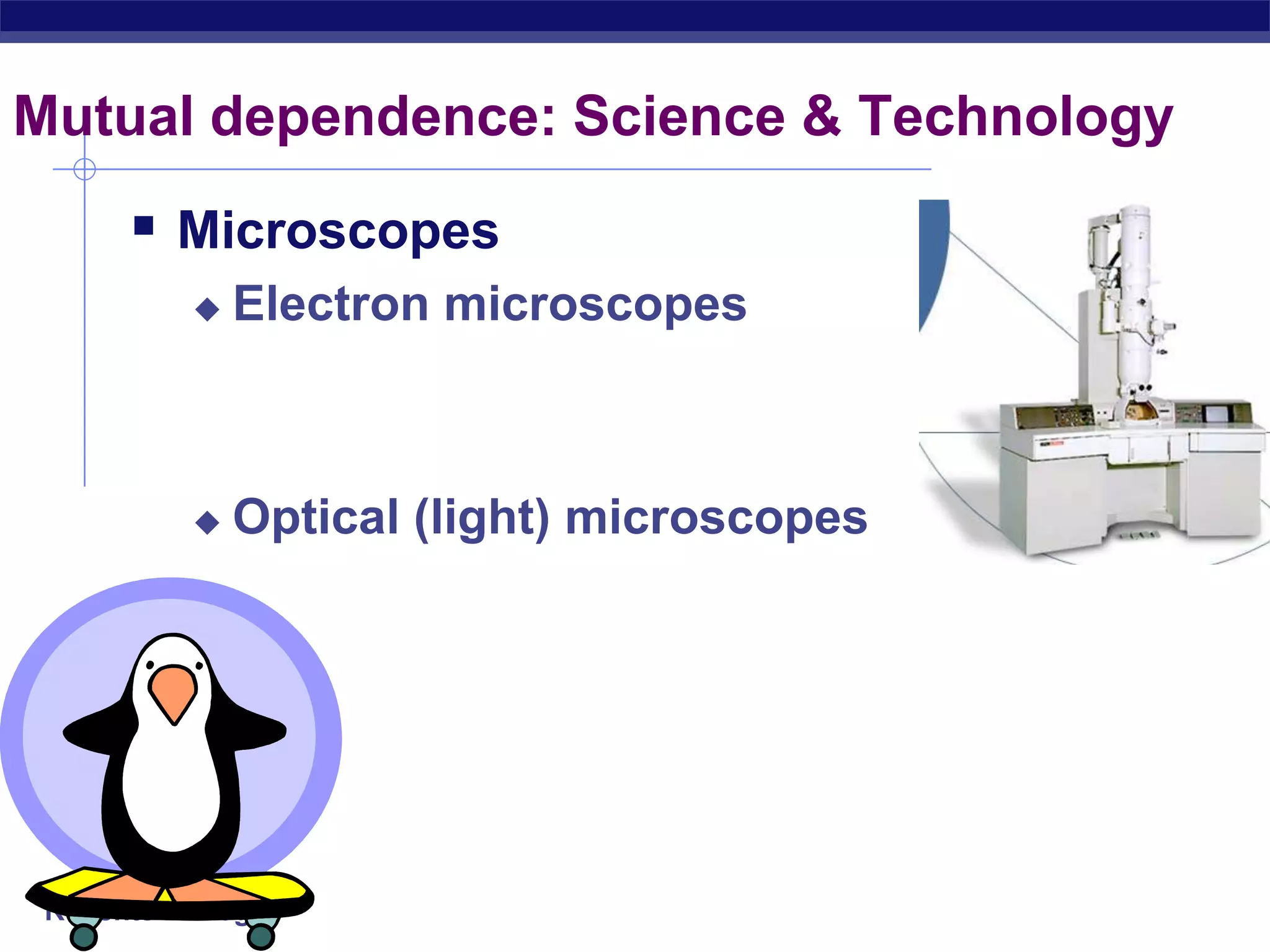
Explores the mutual dependence of science and technology, highlighting microscopes as a key tool.
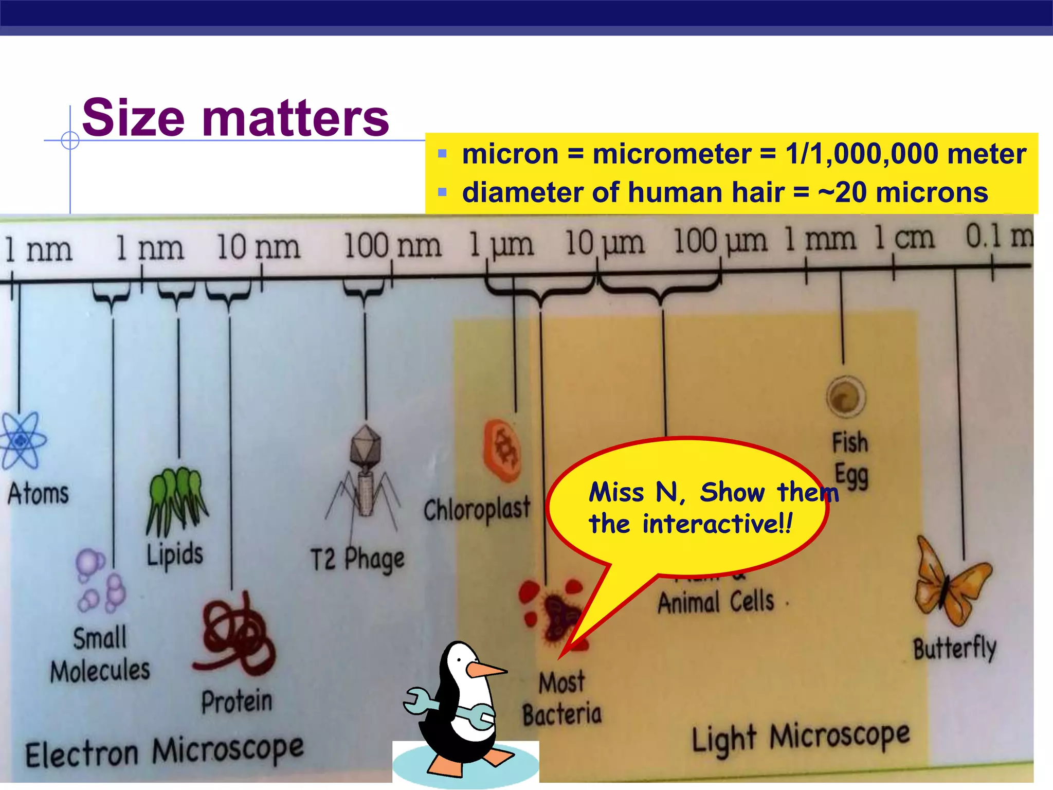
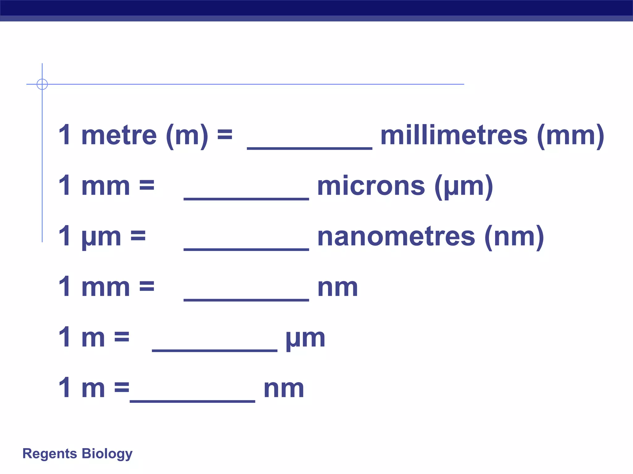
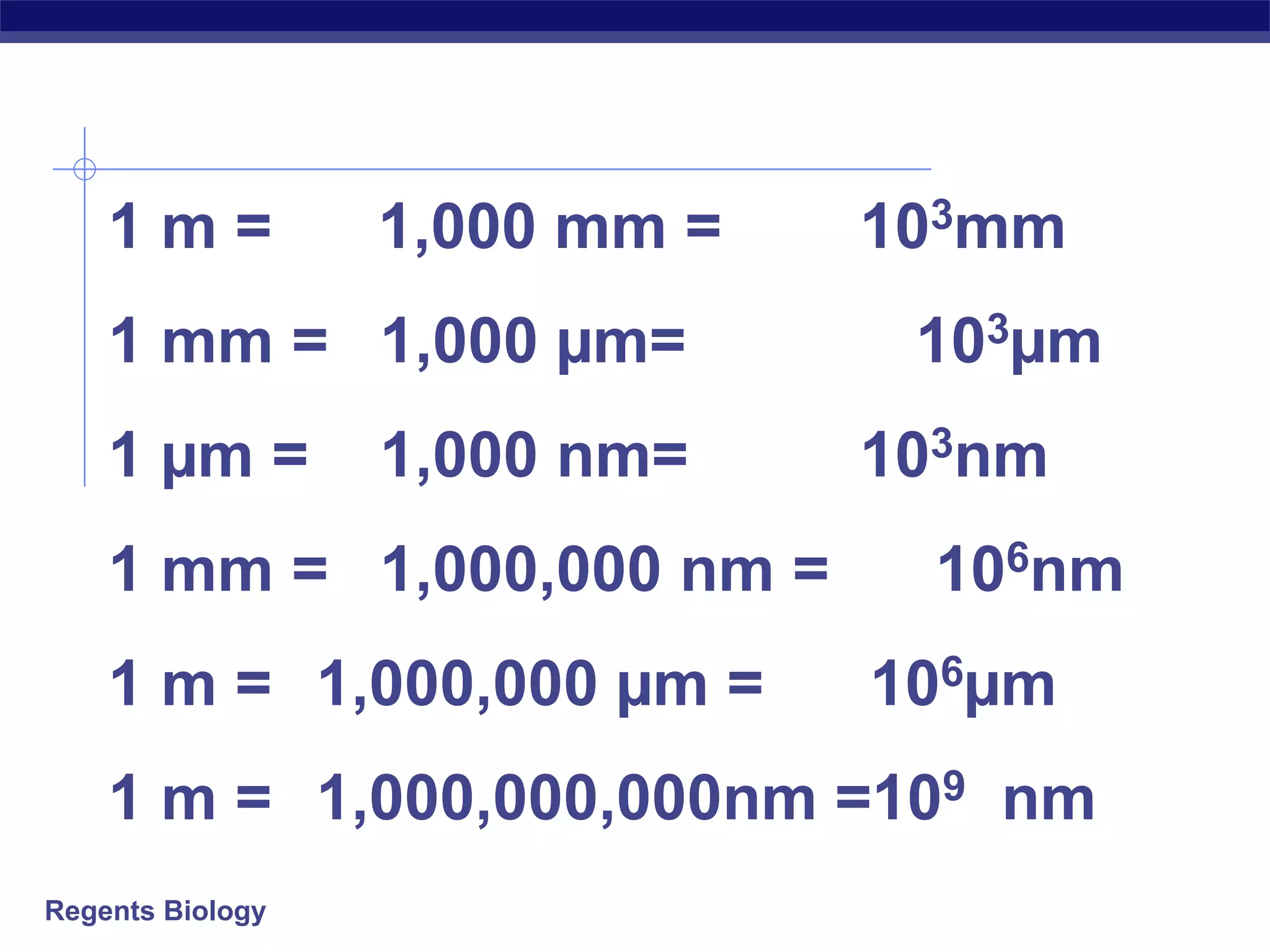
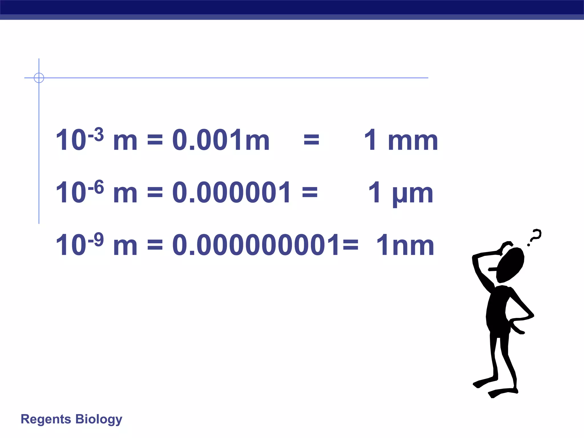
Details the concept of microns as a unit of measurement, with comparisons to human hair and conversions between different metric units.
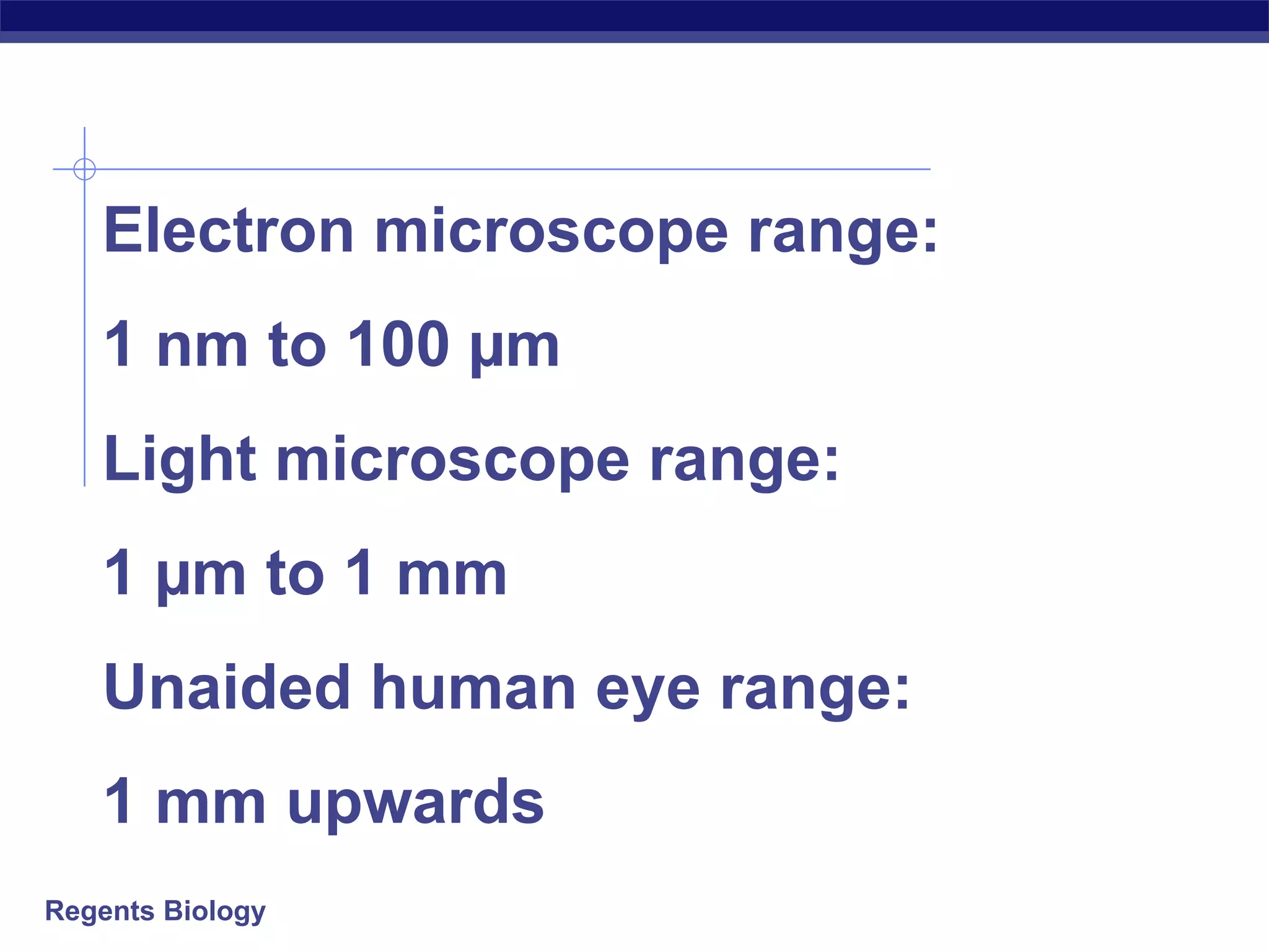
Sets the ranges for electron, light microscopes, and unaided human eye, emphasizing their distinct capabilities.
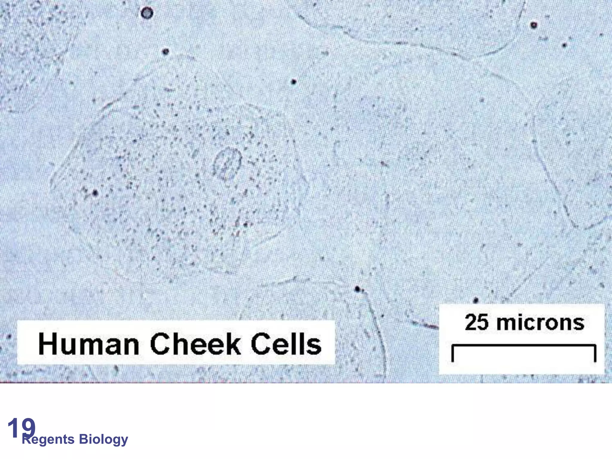
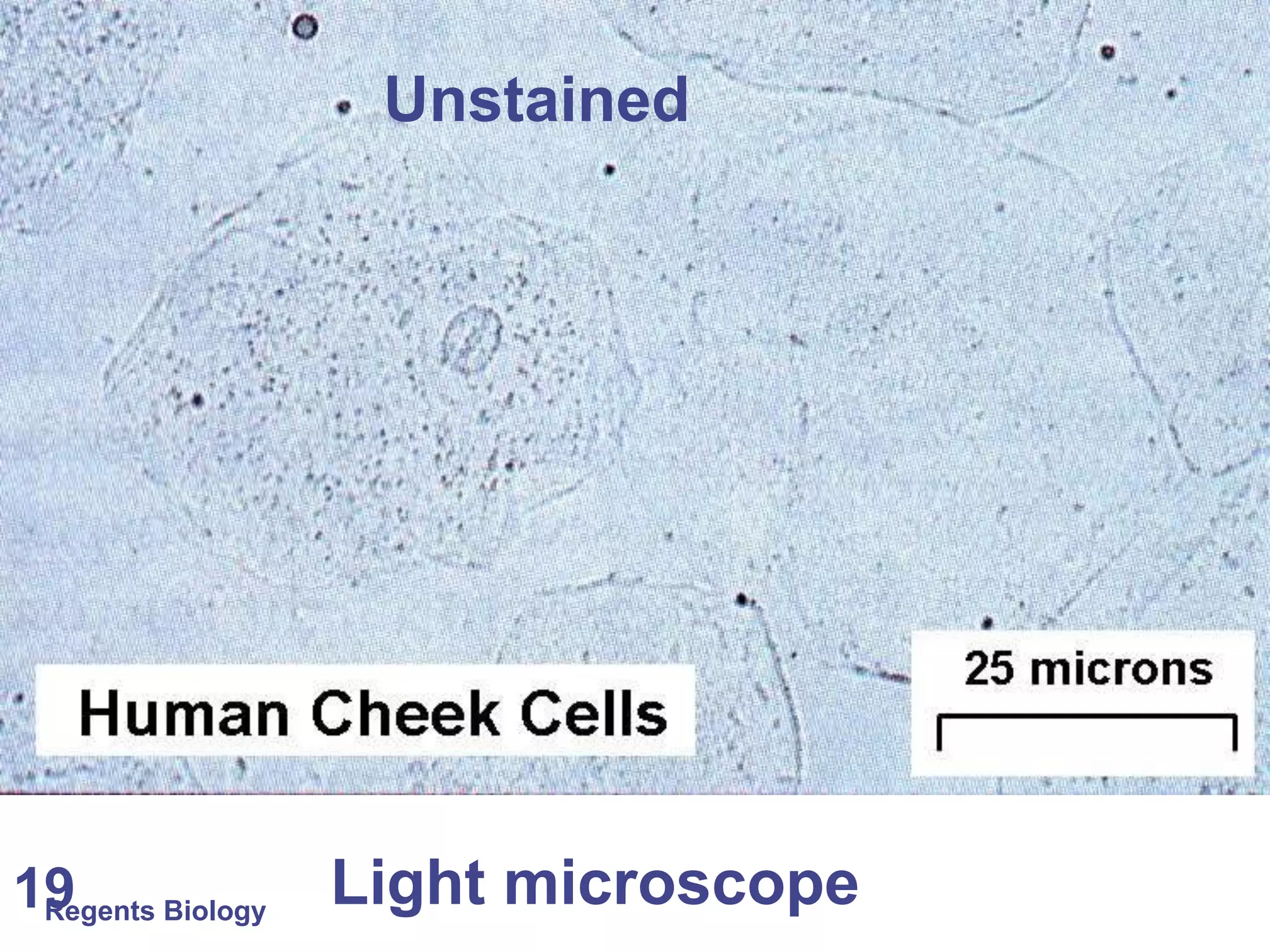
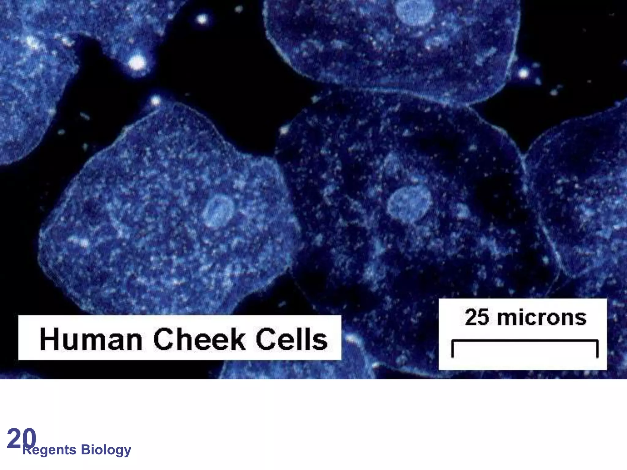
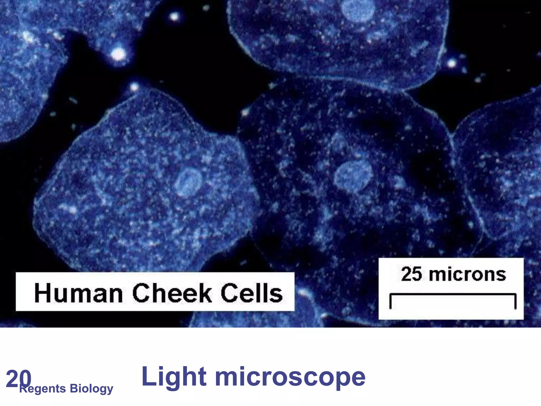
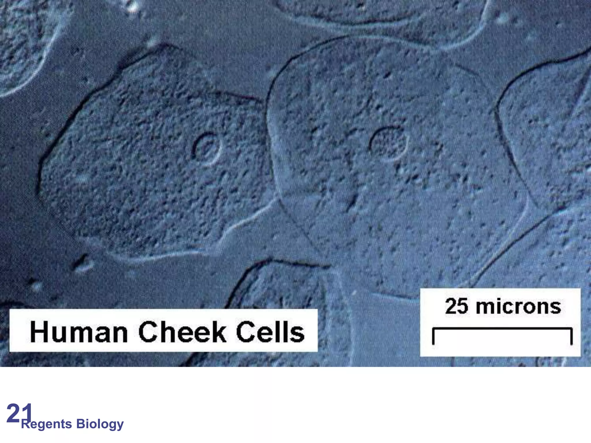
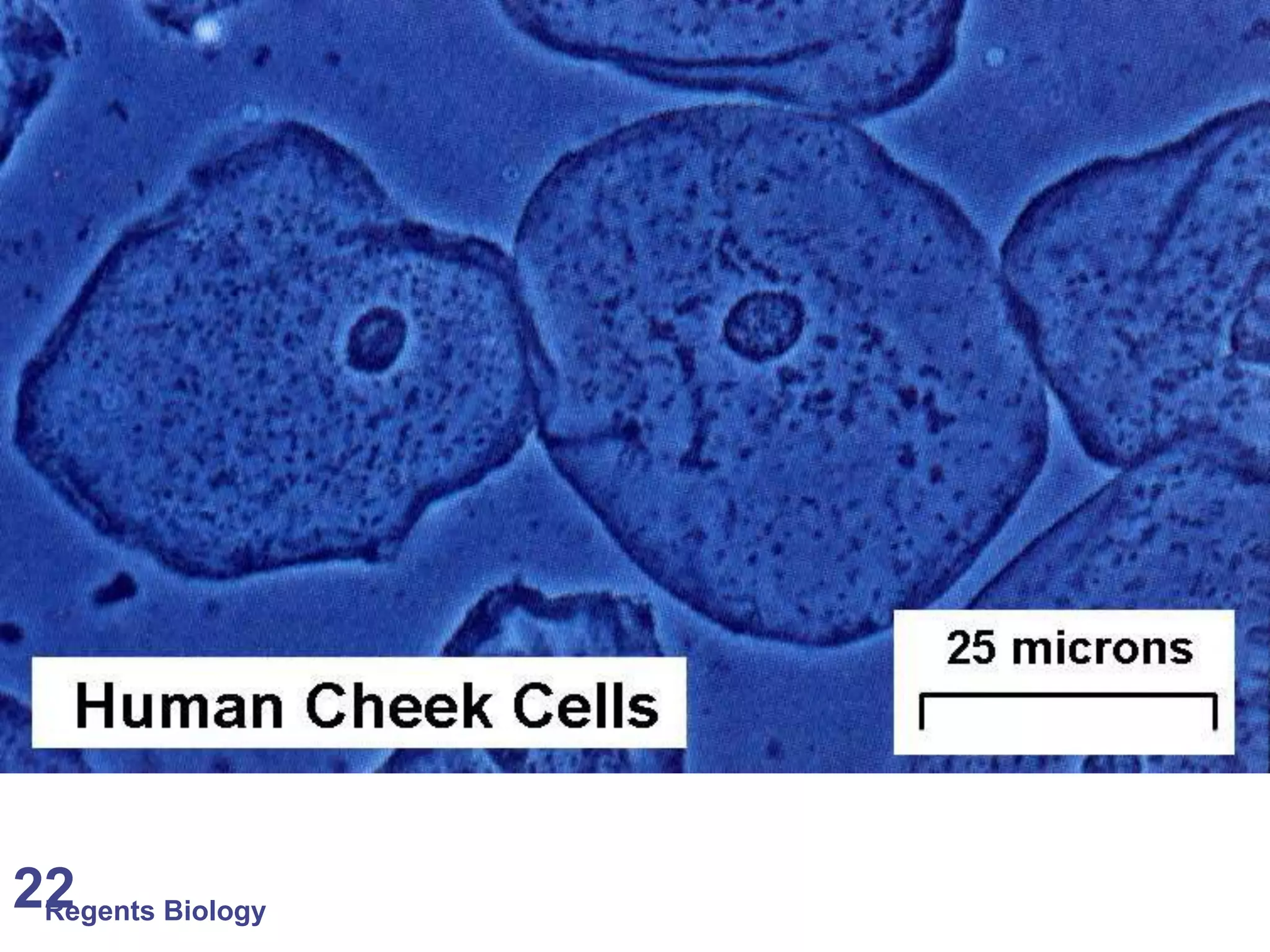
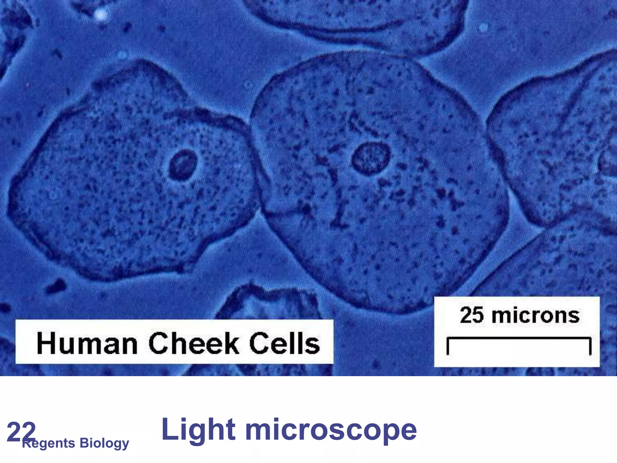
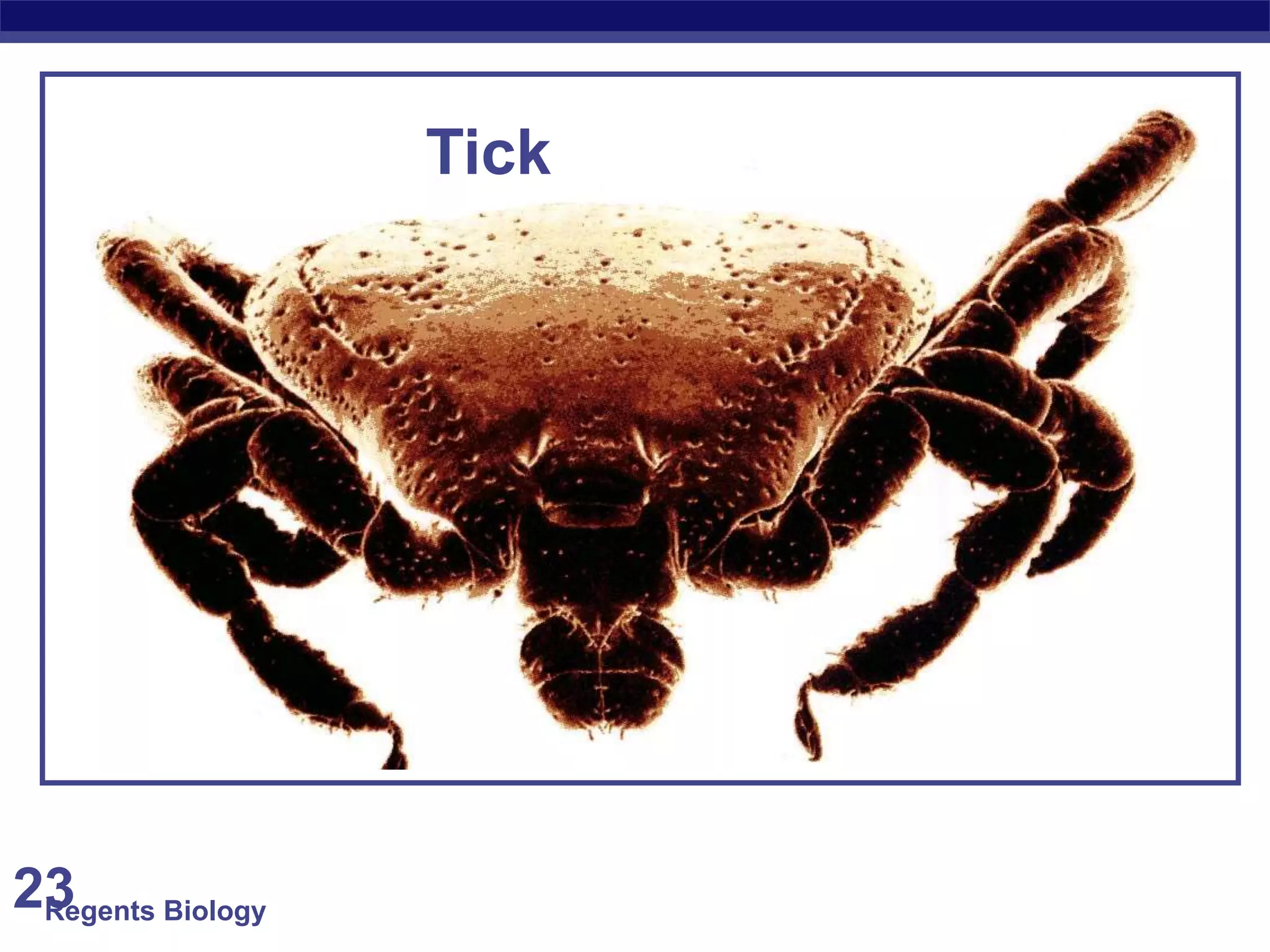
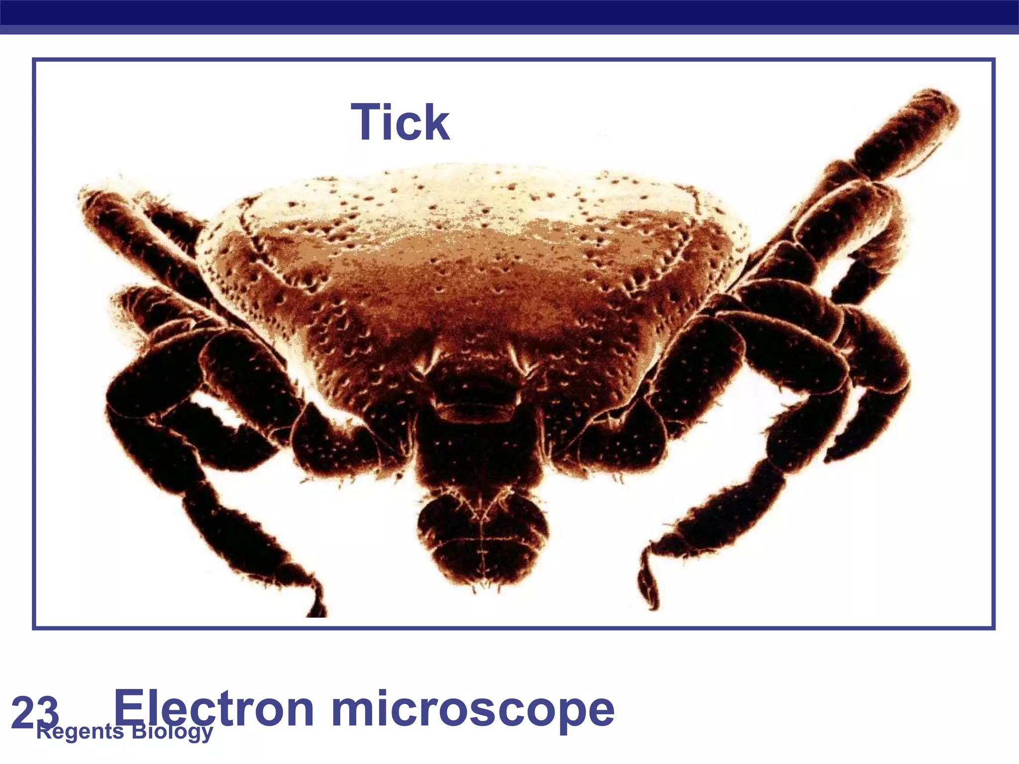
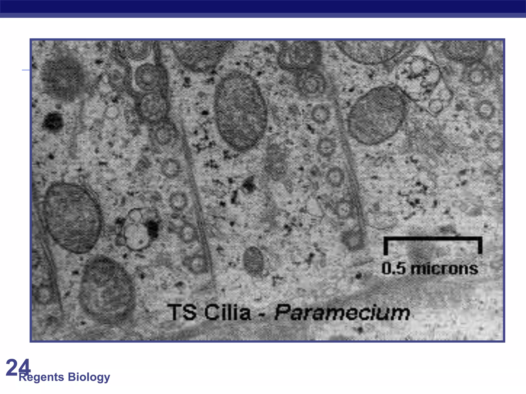
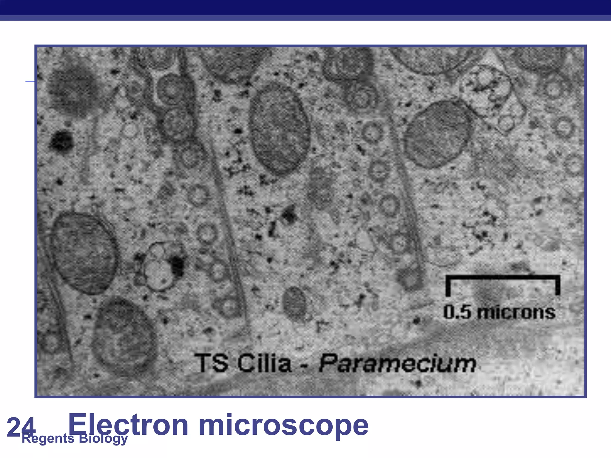
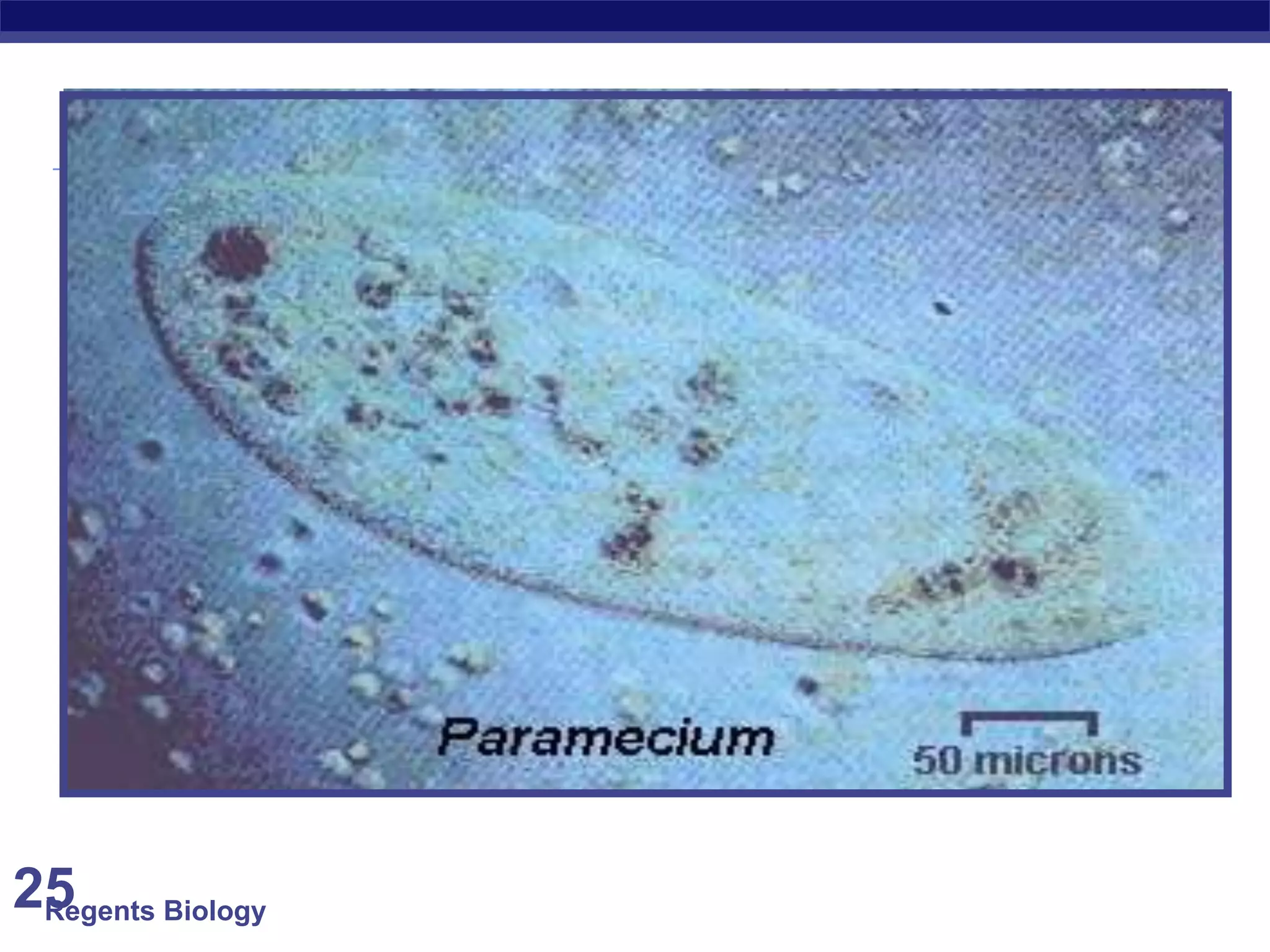
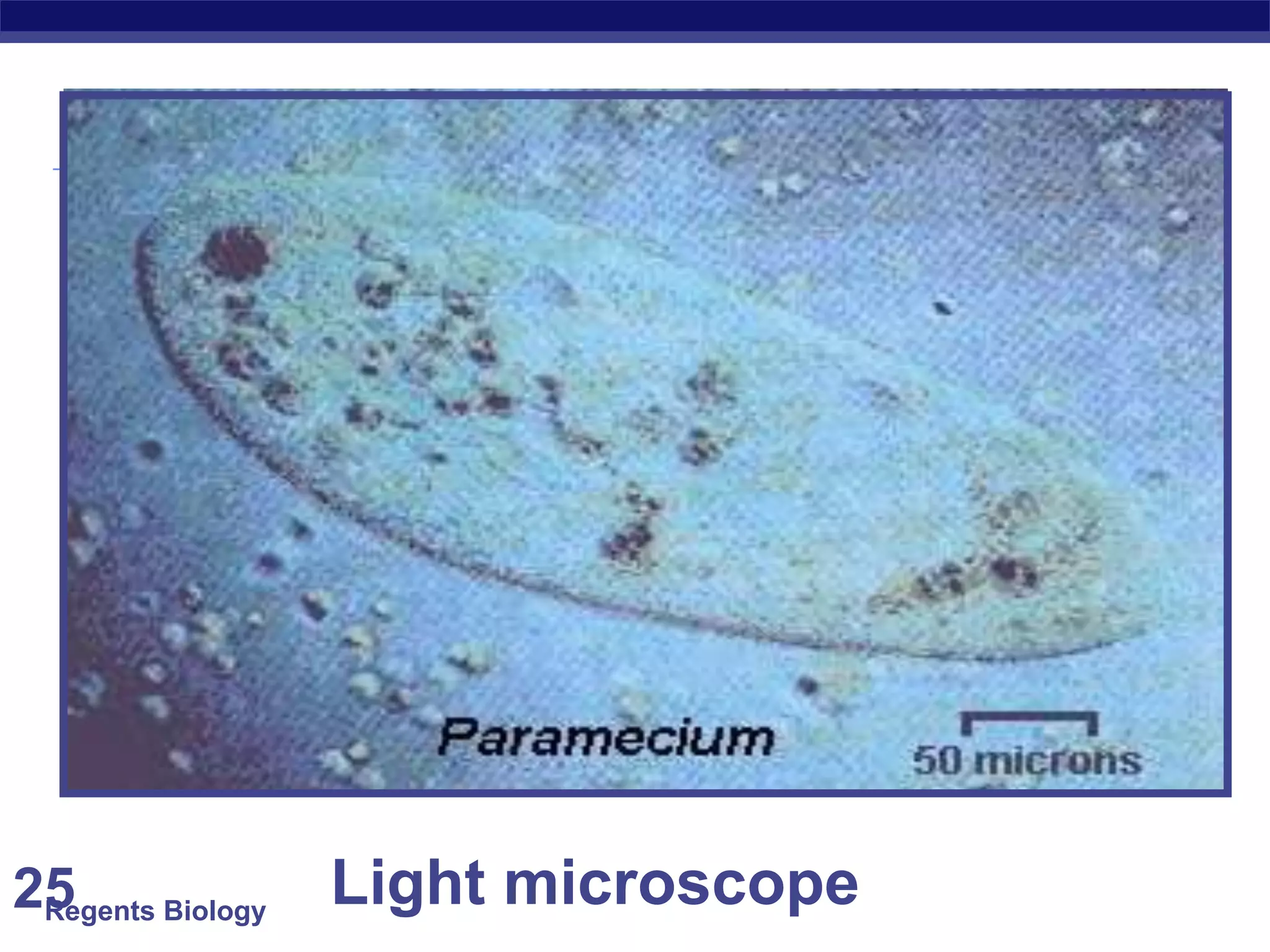
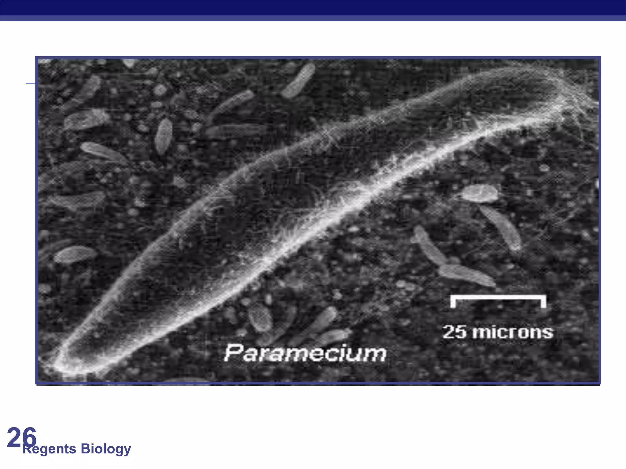
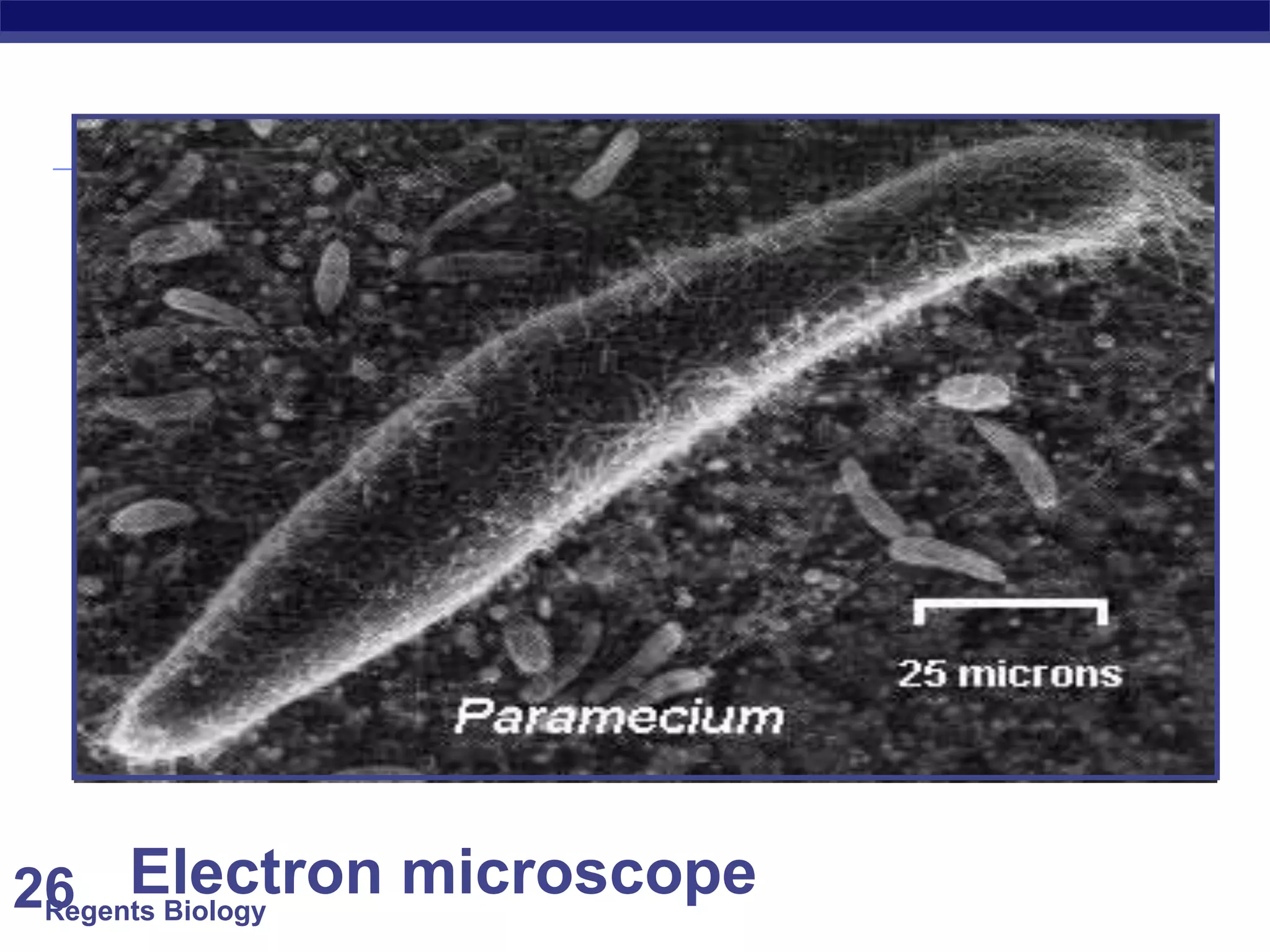
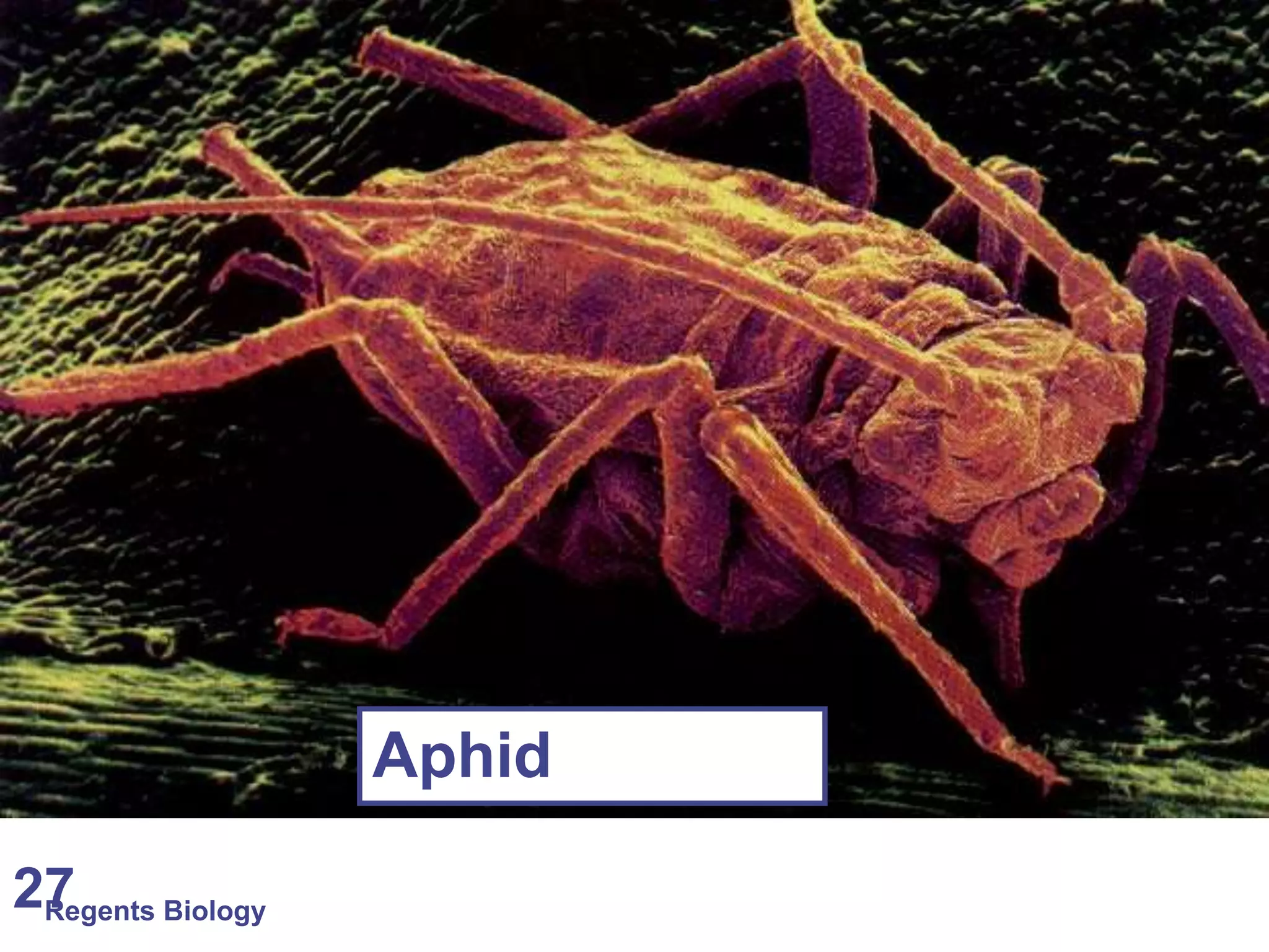
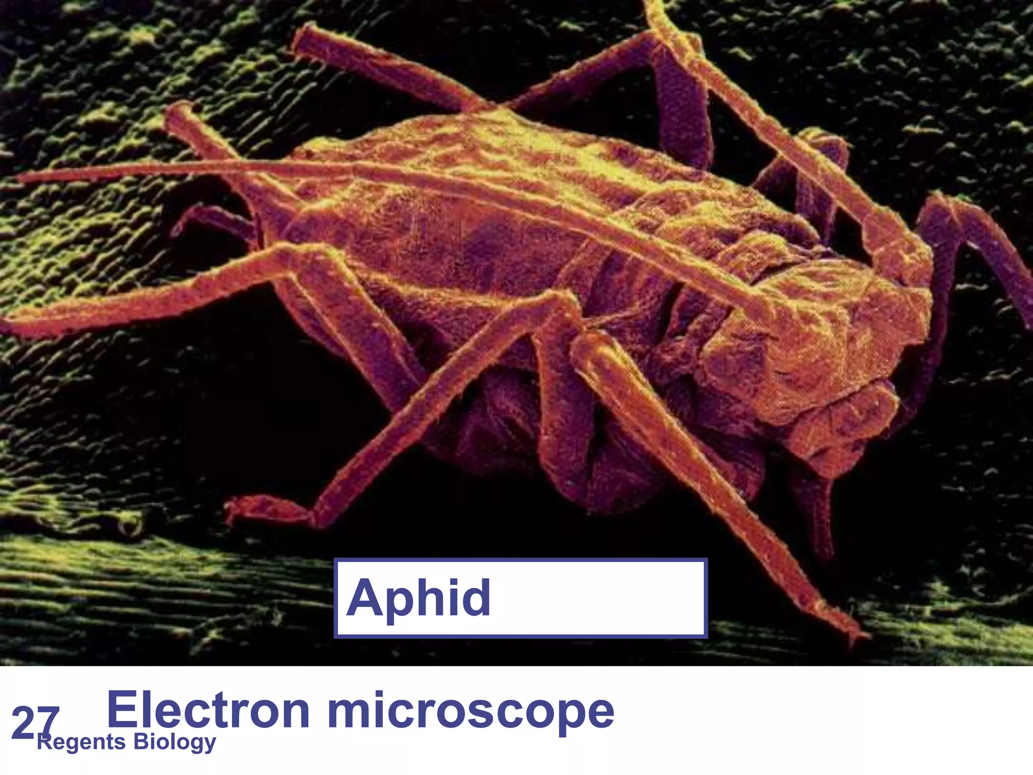
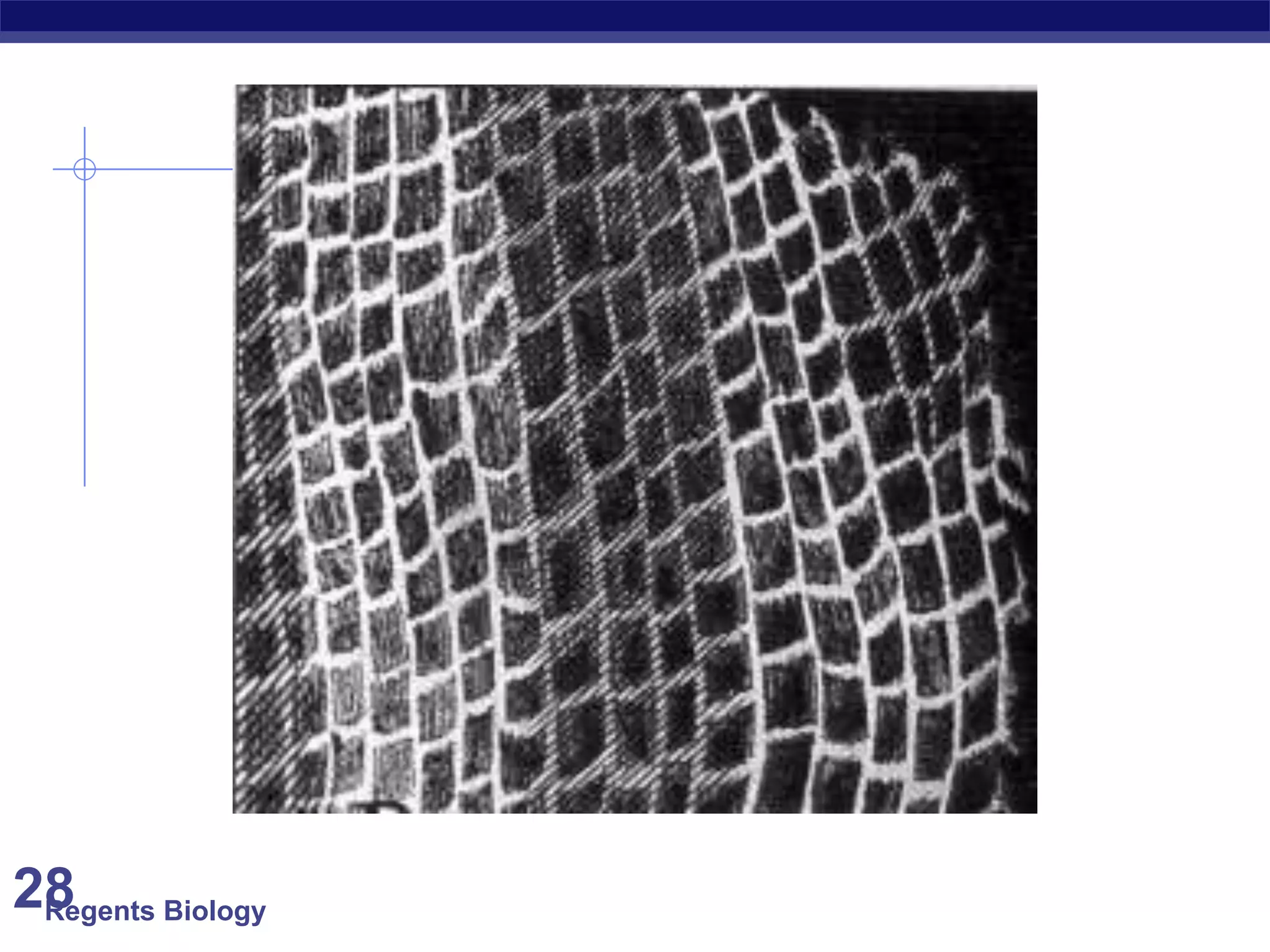
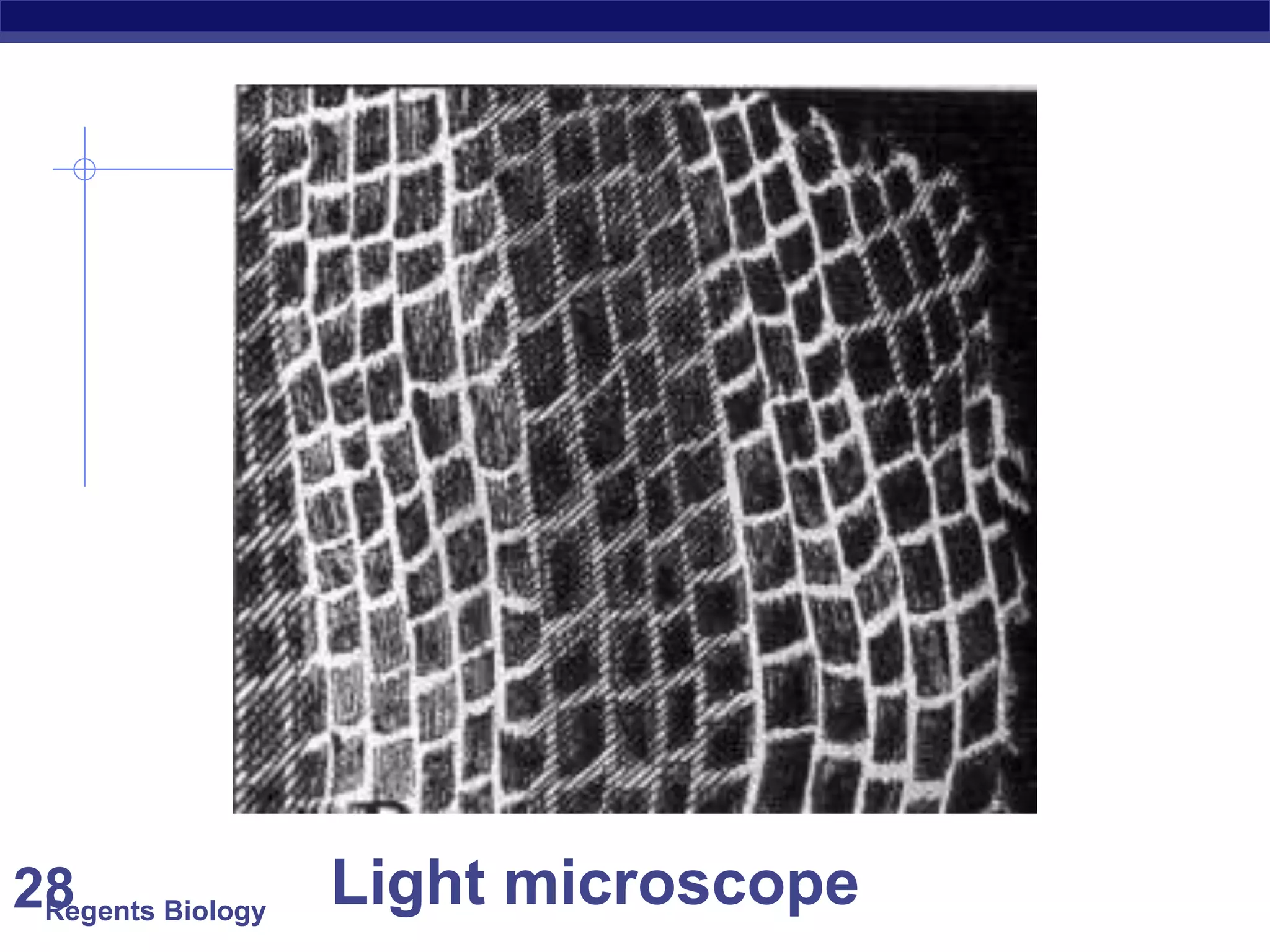
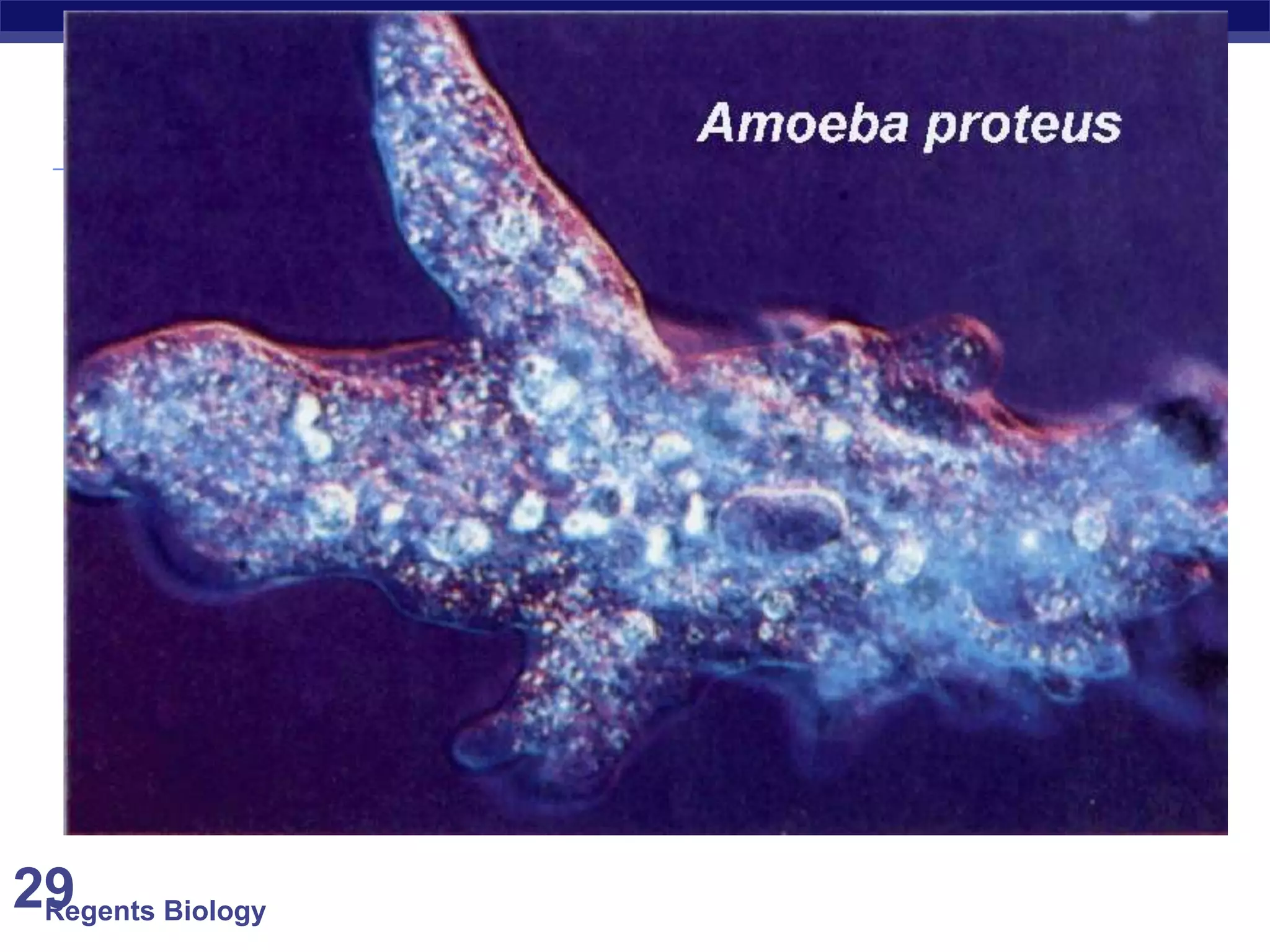
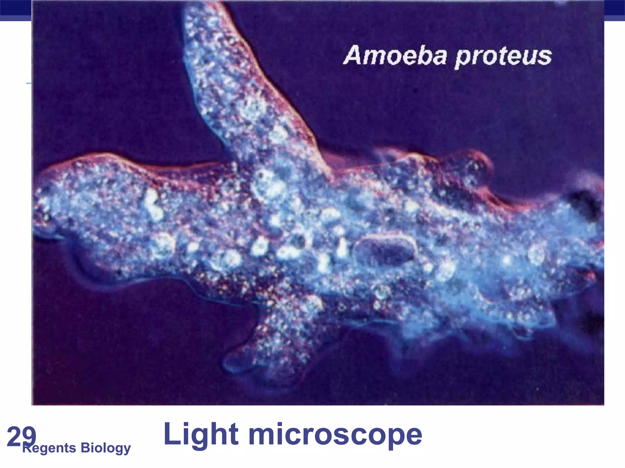
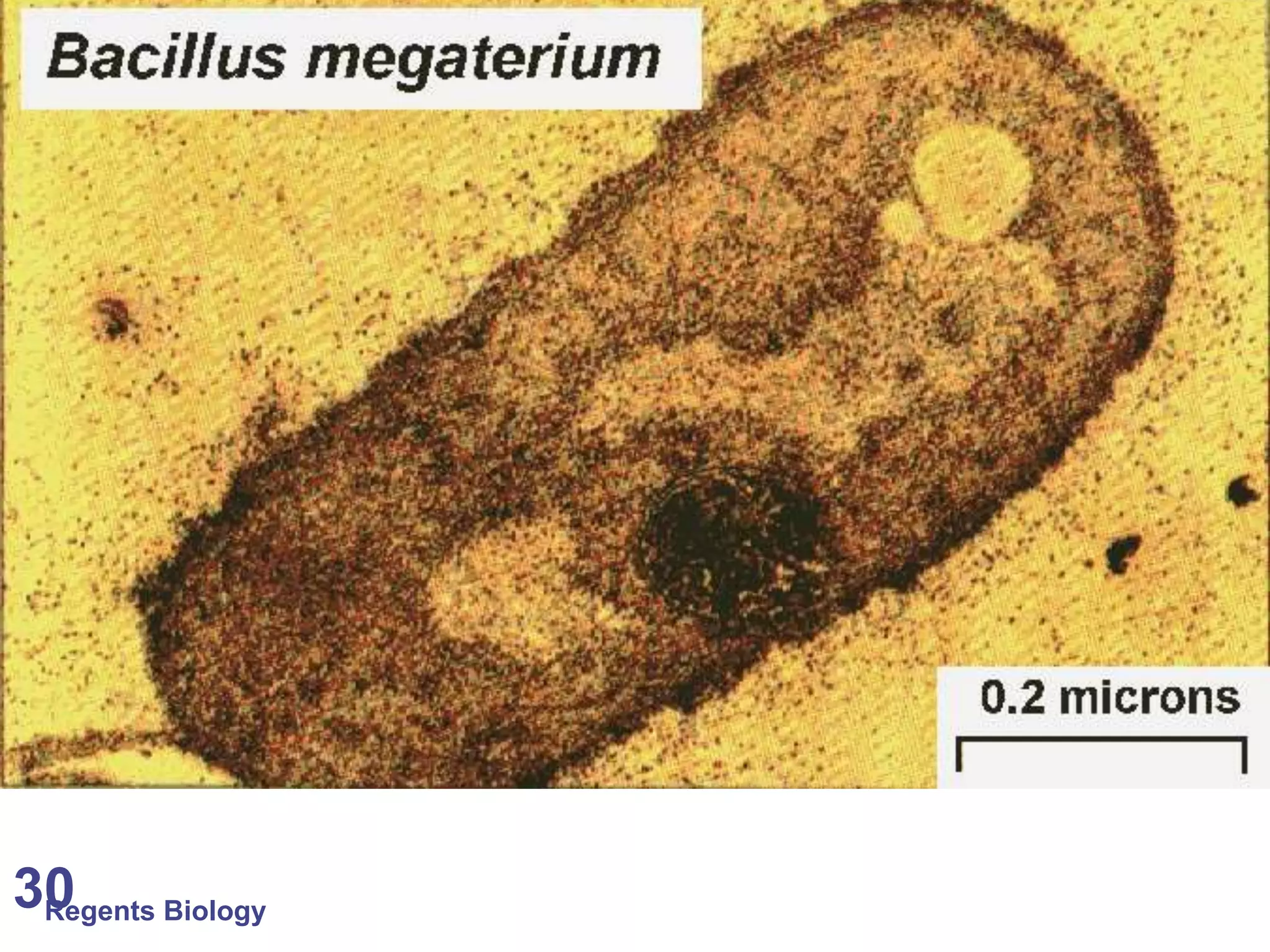
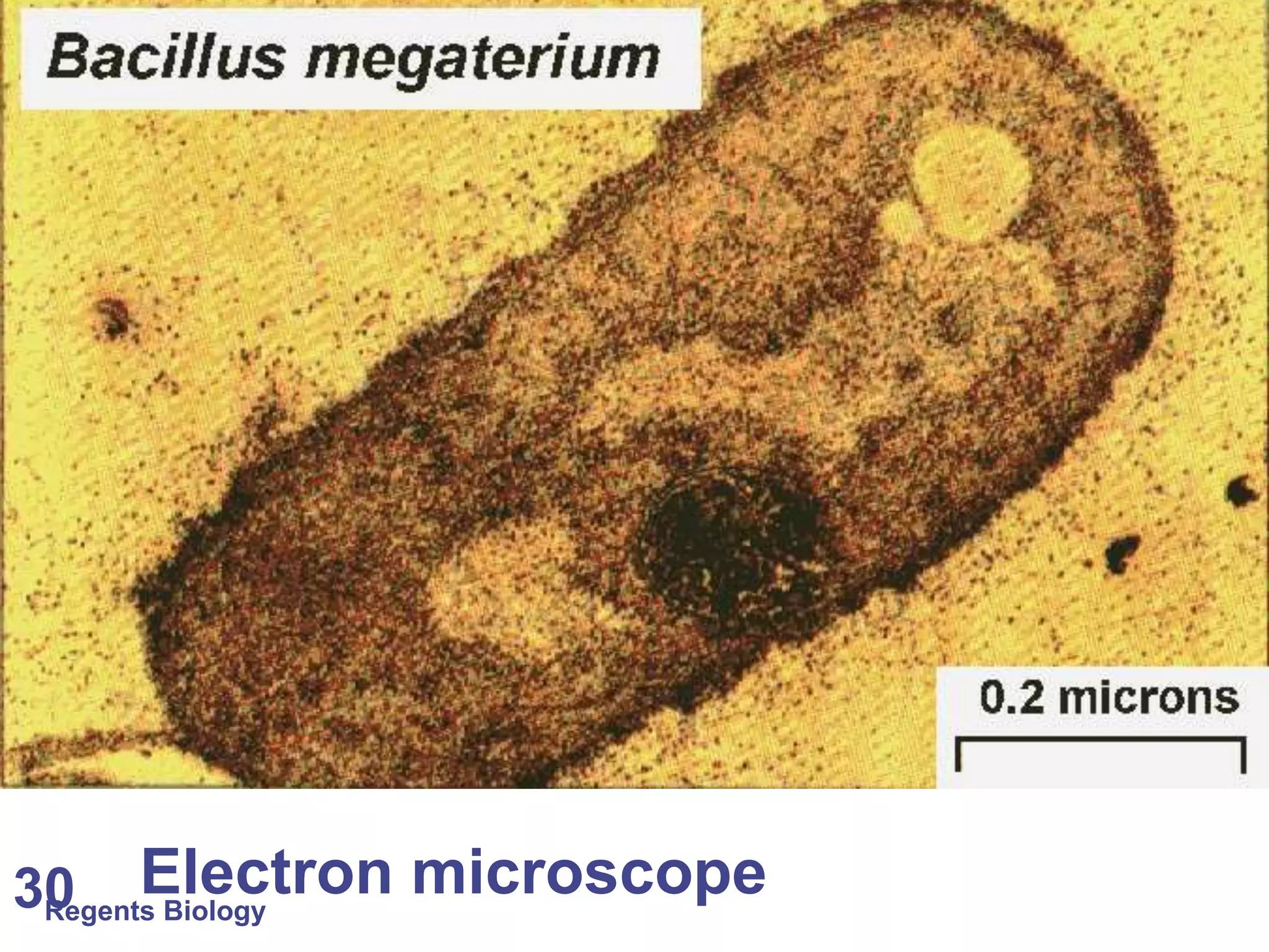
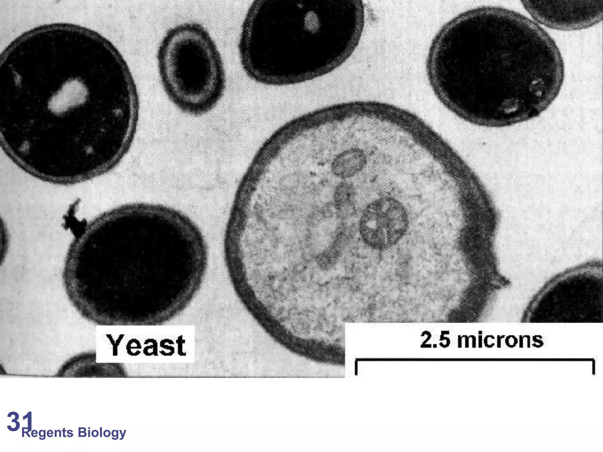
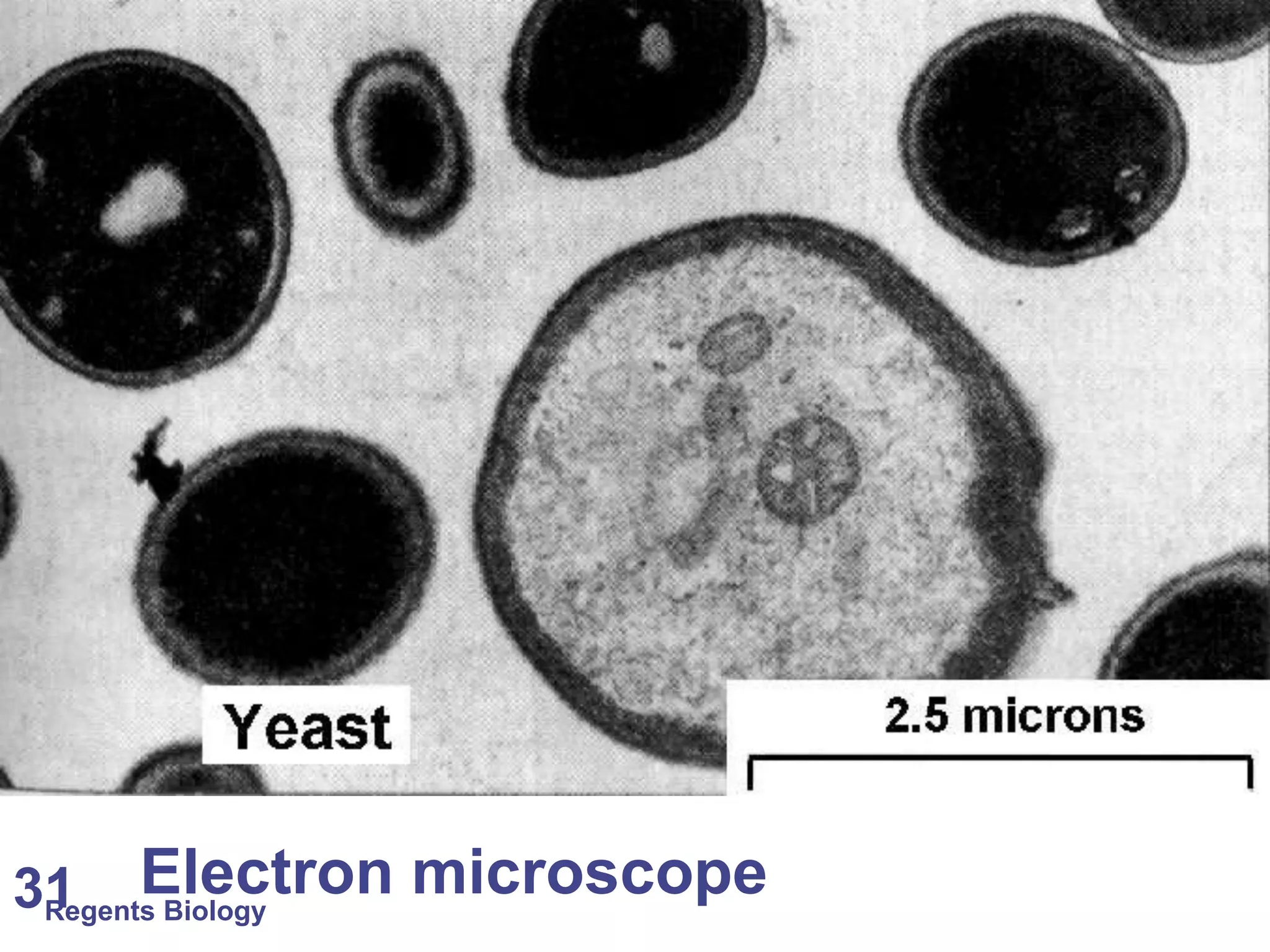
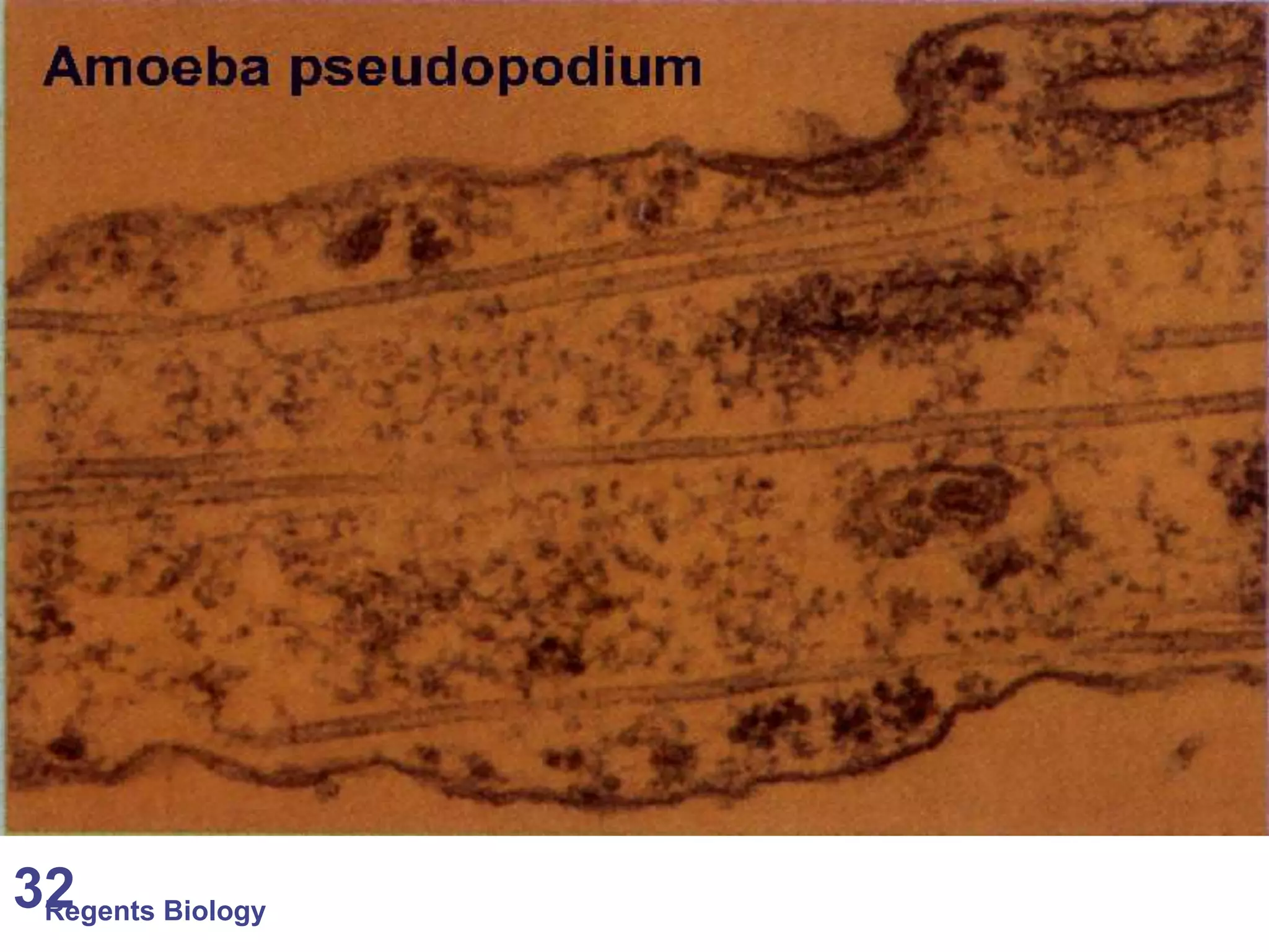
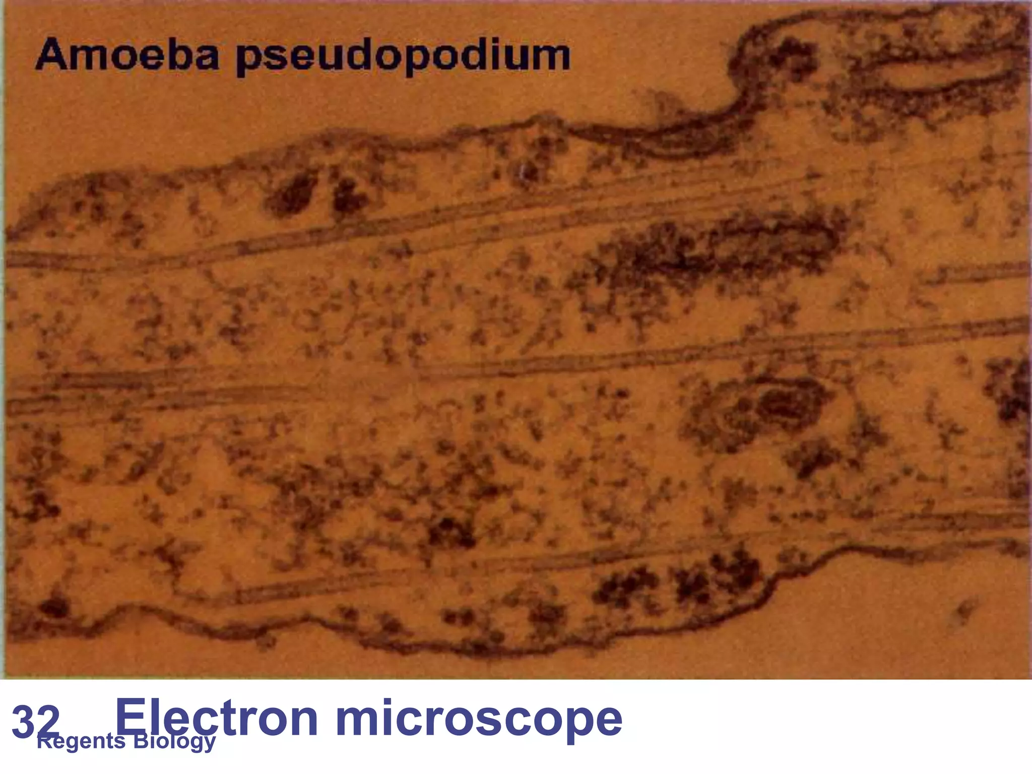
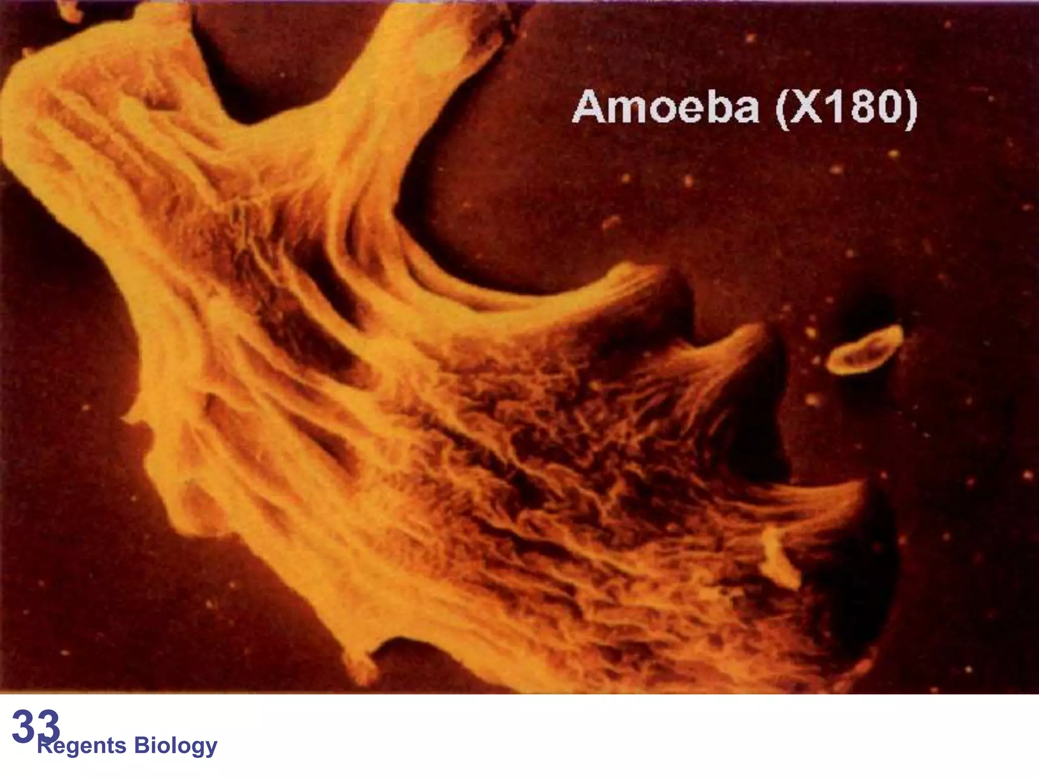
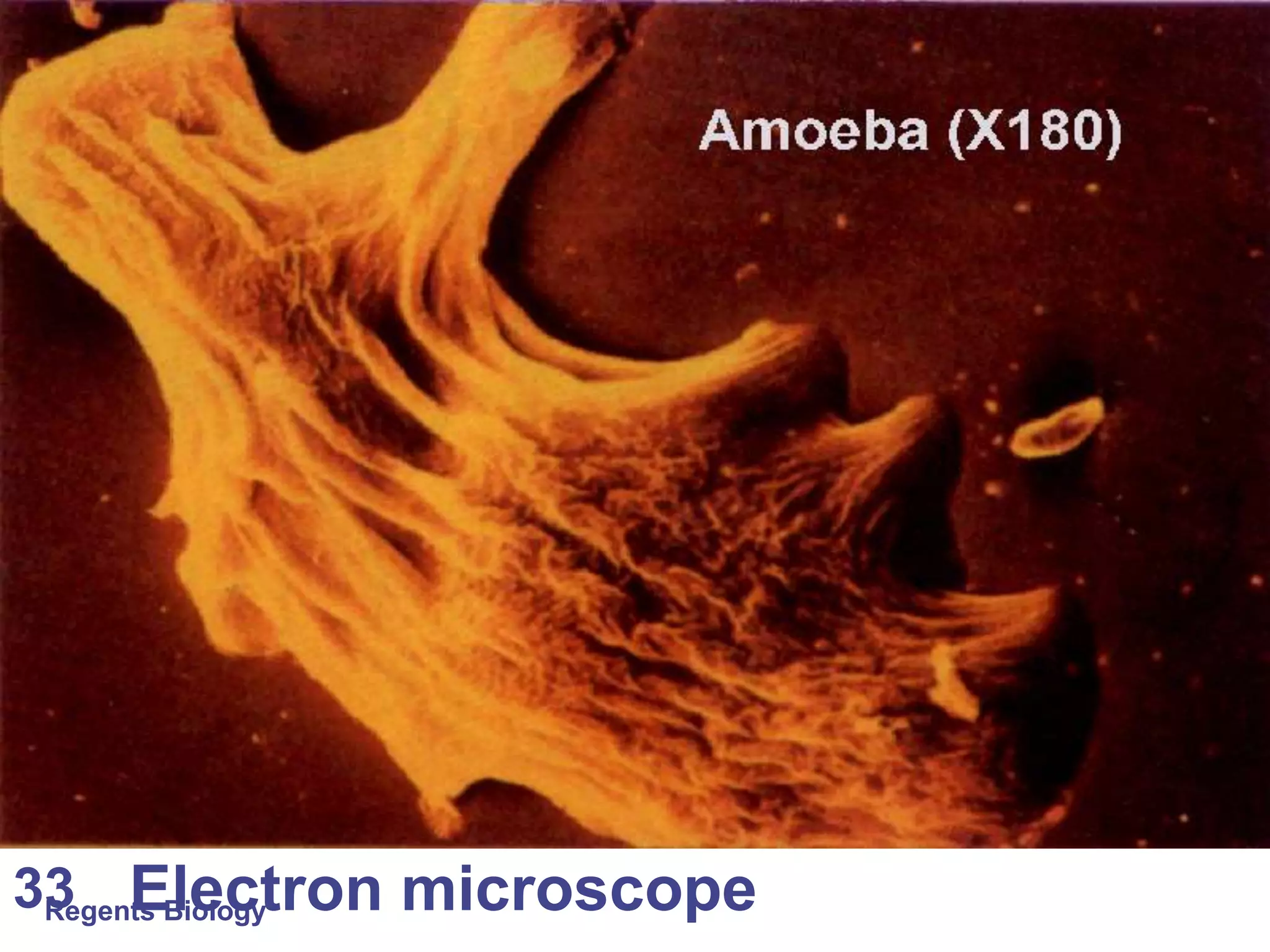
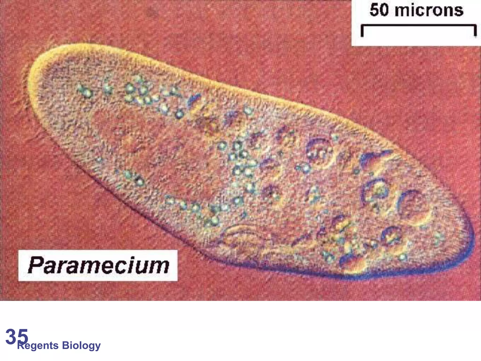
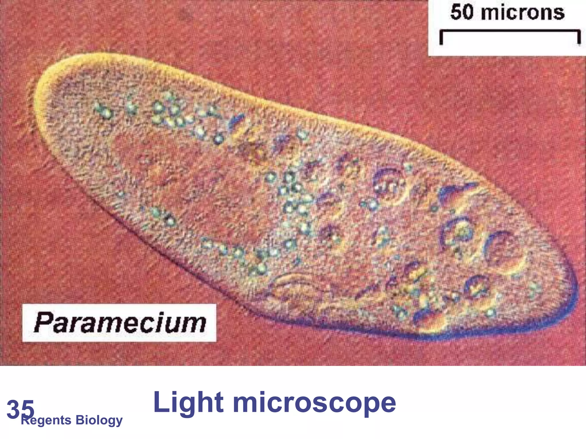
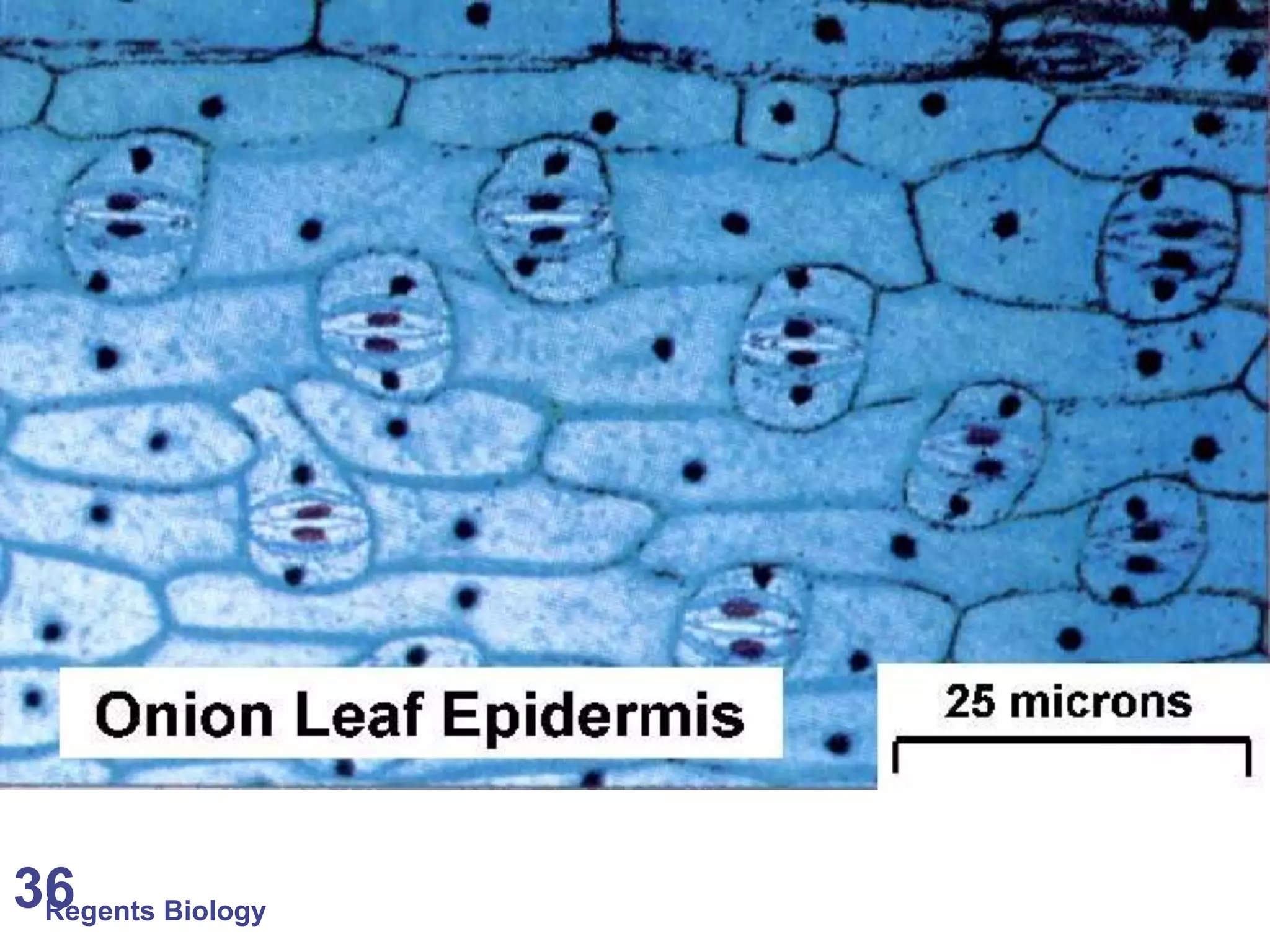
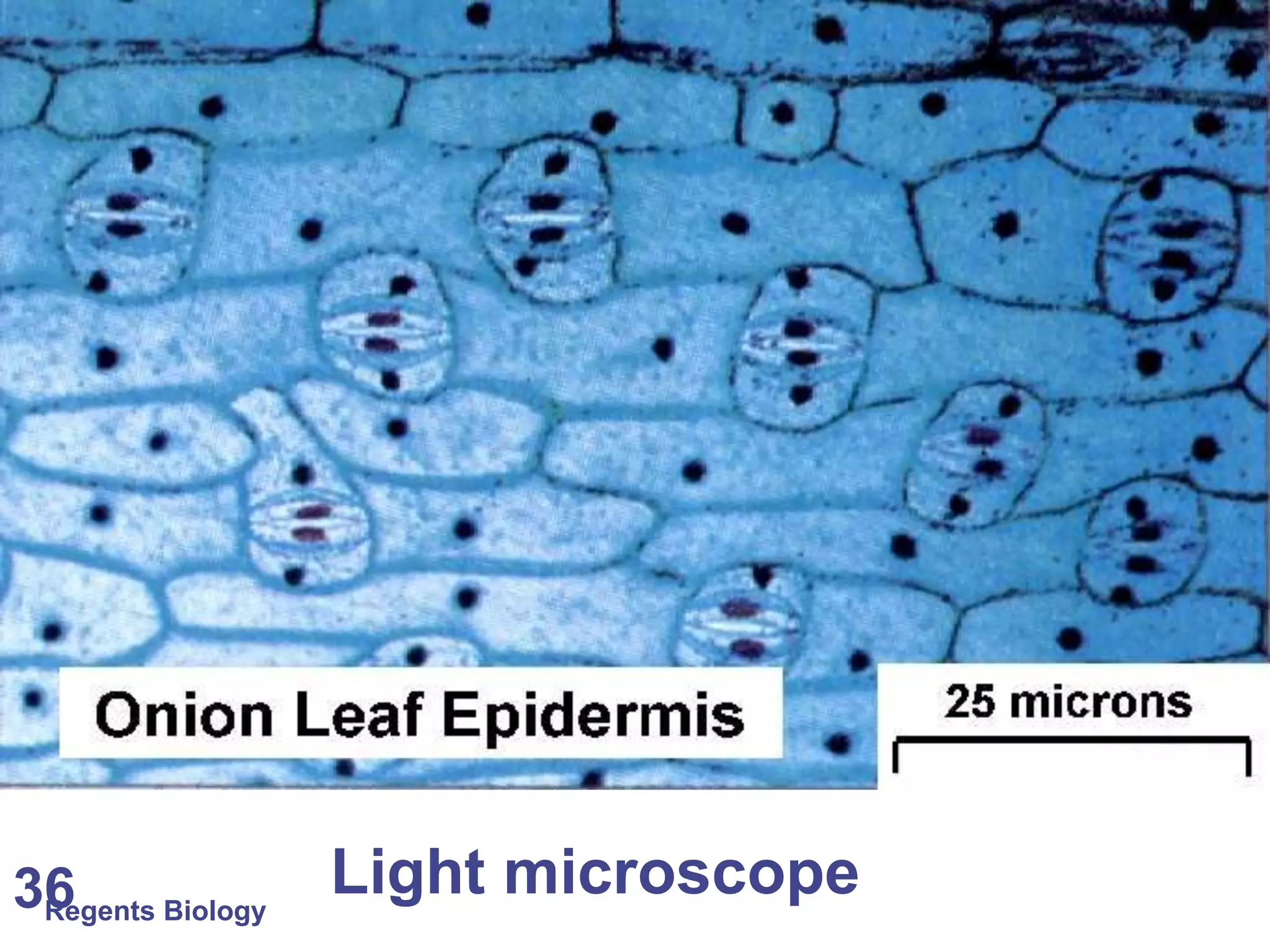
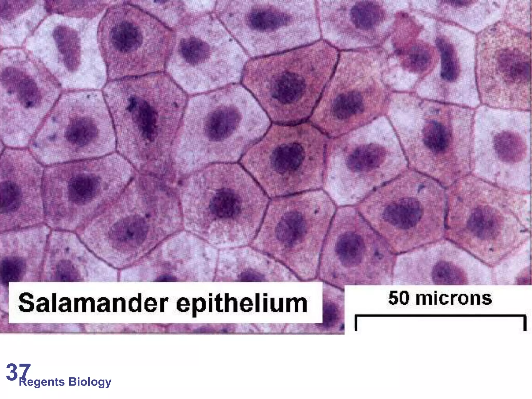
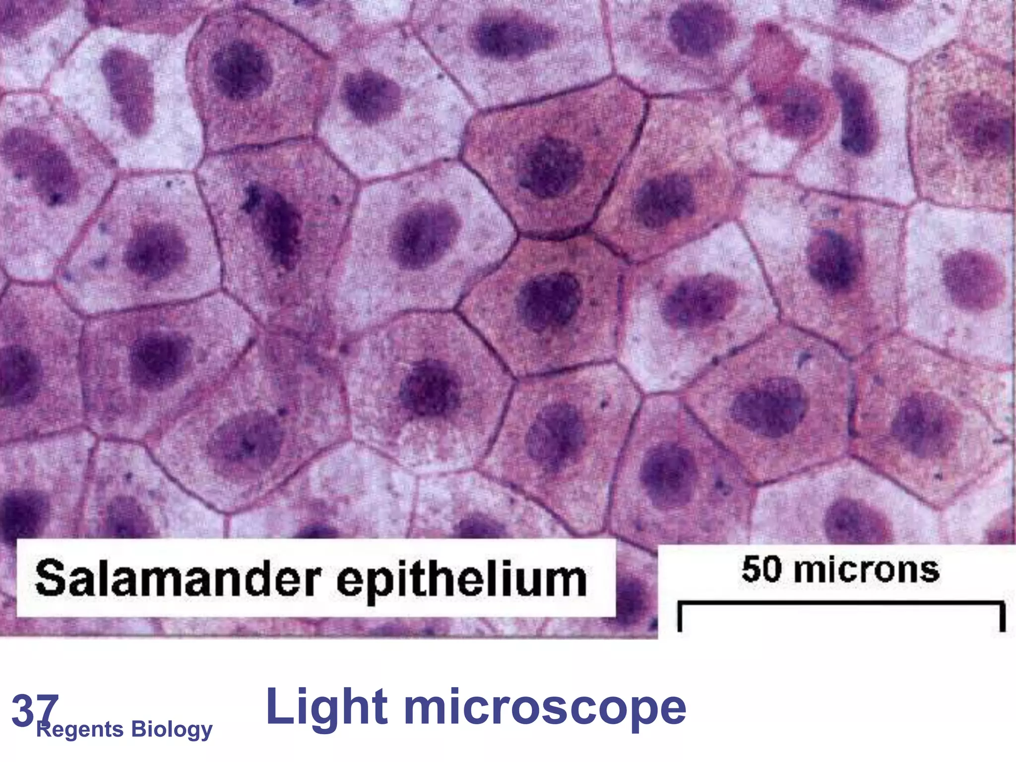
Interactive identification of images as seen through light and electron microscopes with visual examples.
Concluding slides indicating the end of the presentation, transitioning into the topic of magnification.
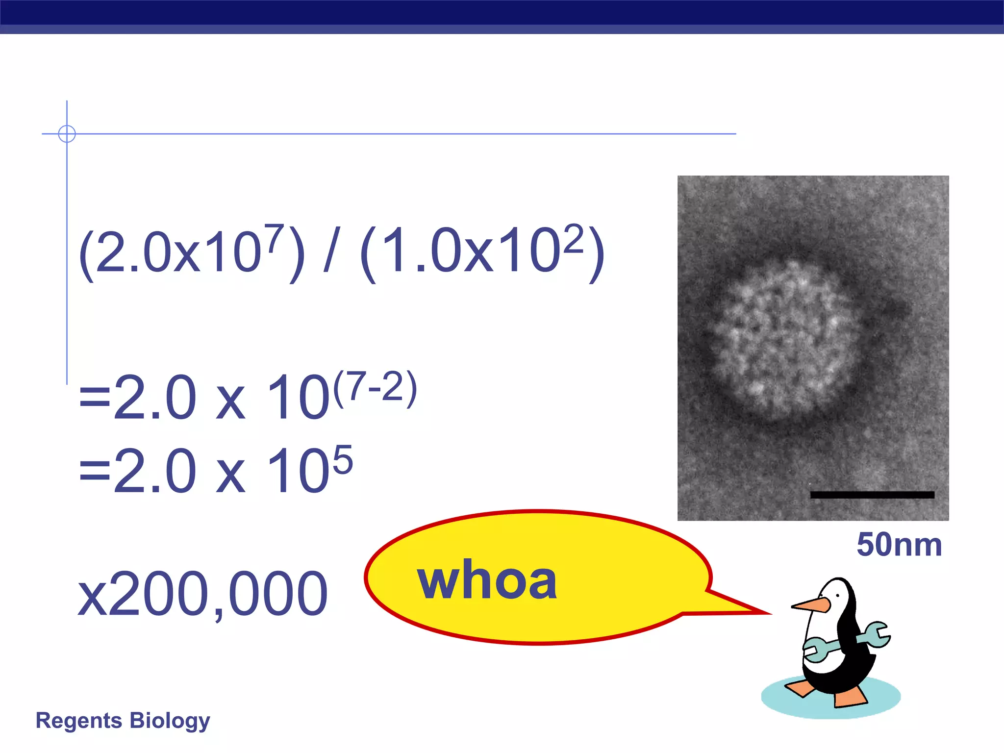
Explains magnification formulas, calculations, and the importance of units in microscopy. Demonstrates how to calculate actual sizes and magnification of samples using scale bars.
Regents Biology
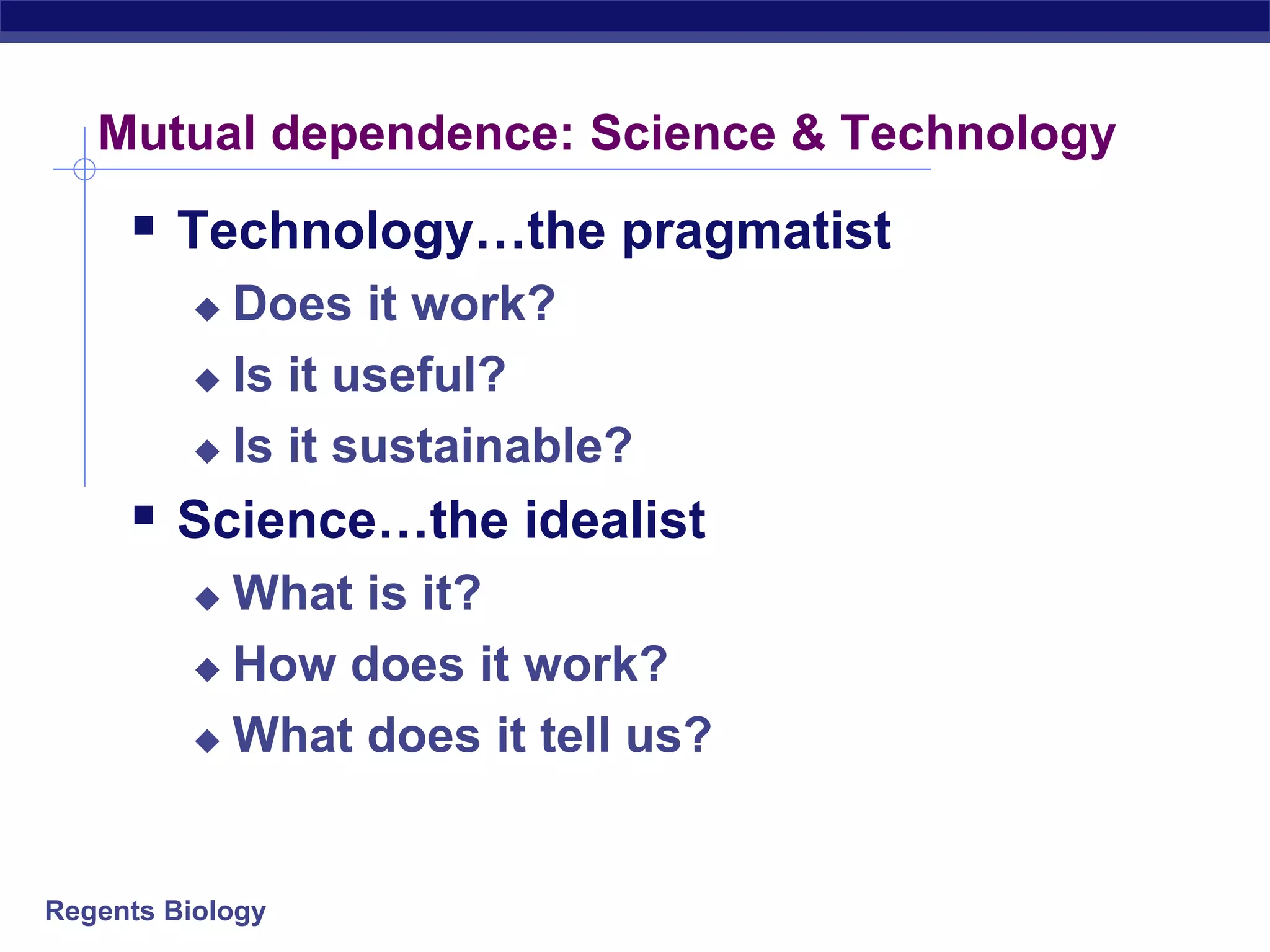
Mutual dependence:Science & Technology
Technology…the pragmatist
Does it work?
Is it useful?
Is it sustainable?
Science…the idealist
What is it?
How does it work?
What does it tell us?
Regents Biology
Magnification
Magnification =ocular lens x objective lens
A microscope has an eyepiece lens with a power
of 20X. The objective lens being used has a
power of 10X. What is the total magnification?
50.
Regents Biology
Using ScaleBars
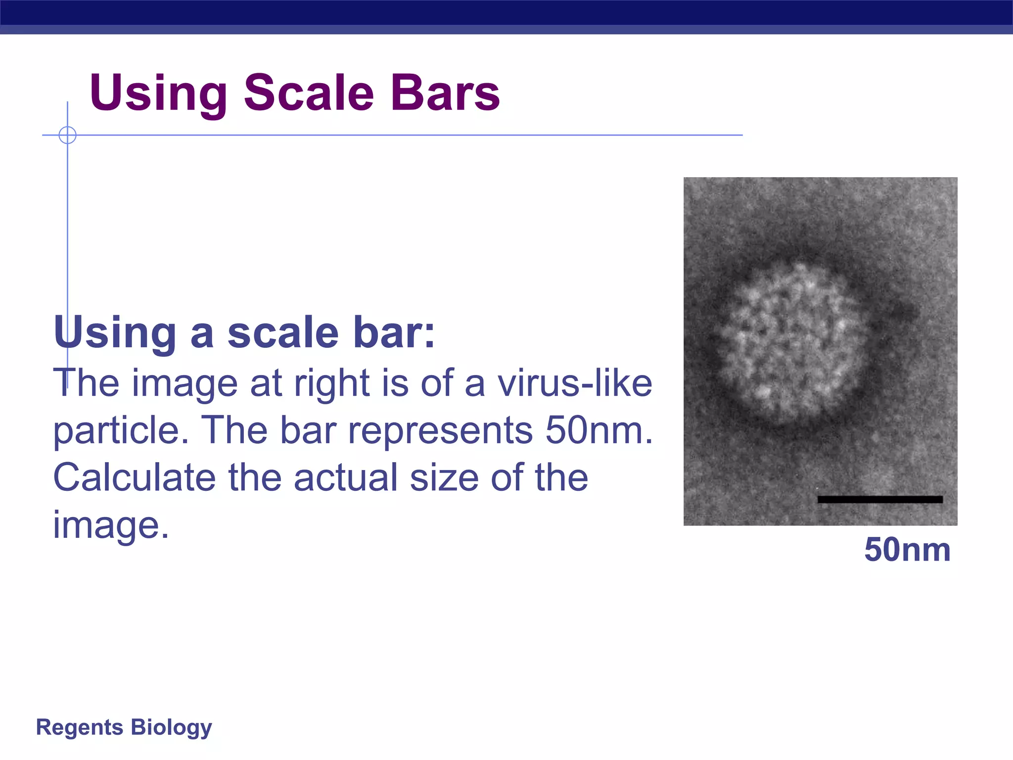
Using a scale bar:
The image at right is of a virus-like
particle. The bar represents 50nm.
Calculate the actual size of the
image.
50nm
51.
Regents Biology
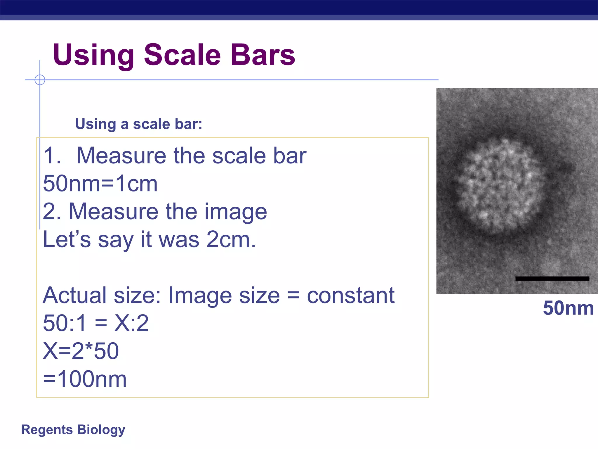
1. Measurethe scale bar
50nm=1cm
2. Measure the image
Let’s say it was 2cm.
Actual size: Image size = constant
50:1 = X:2
X=2*50
=100nm
Using a scale bar:
50nm
Using Scale Bars
52.
Regents Biology
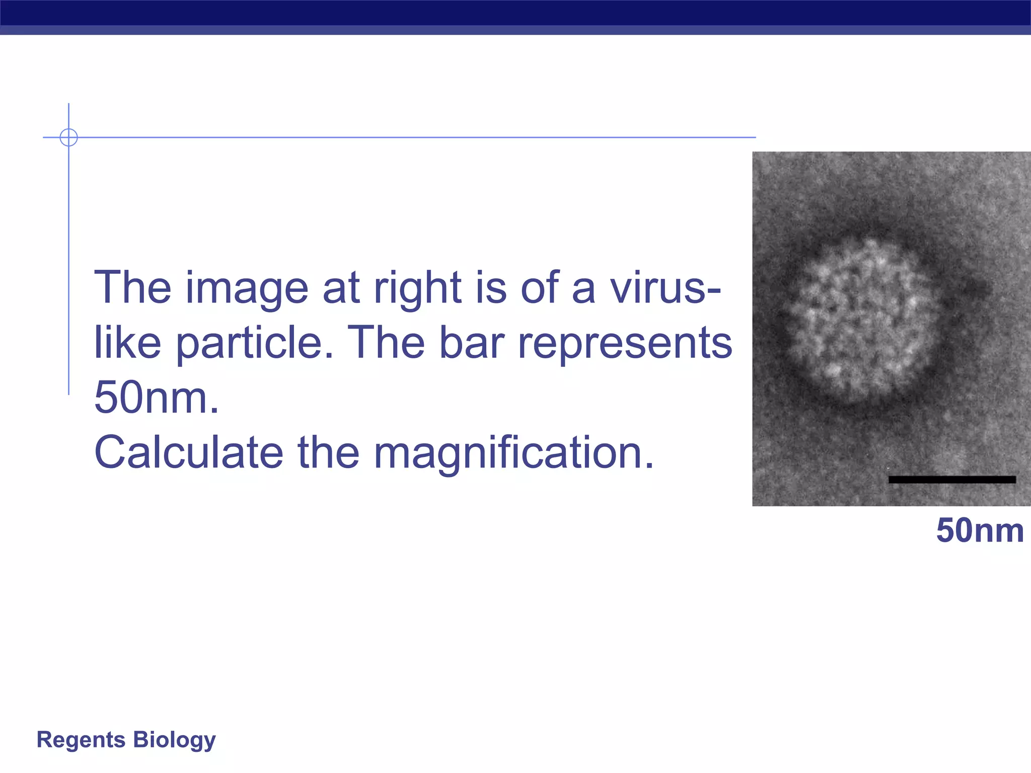
The imageat right is of a virus-
like particle. The bar represents
50nm.
Calculate the magnification.
50nm
53.
Regents Biology
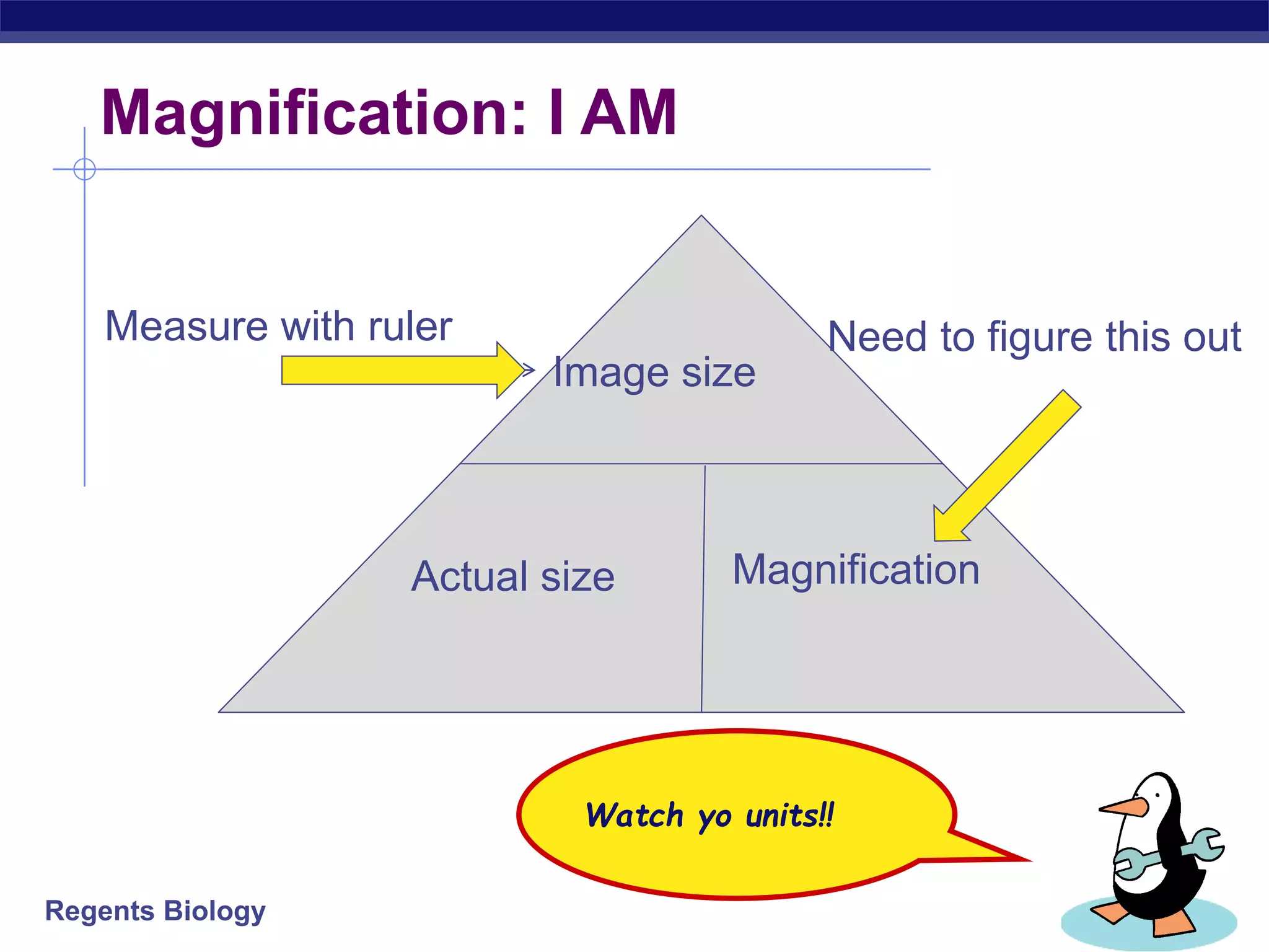
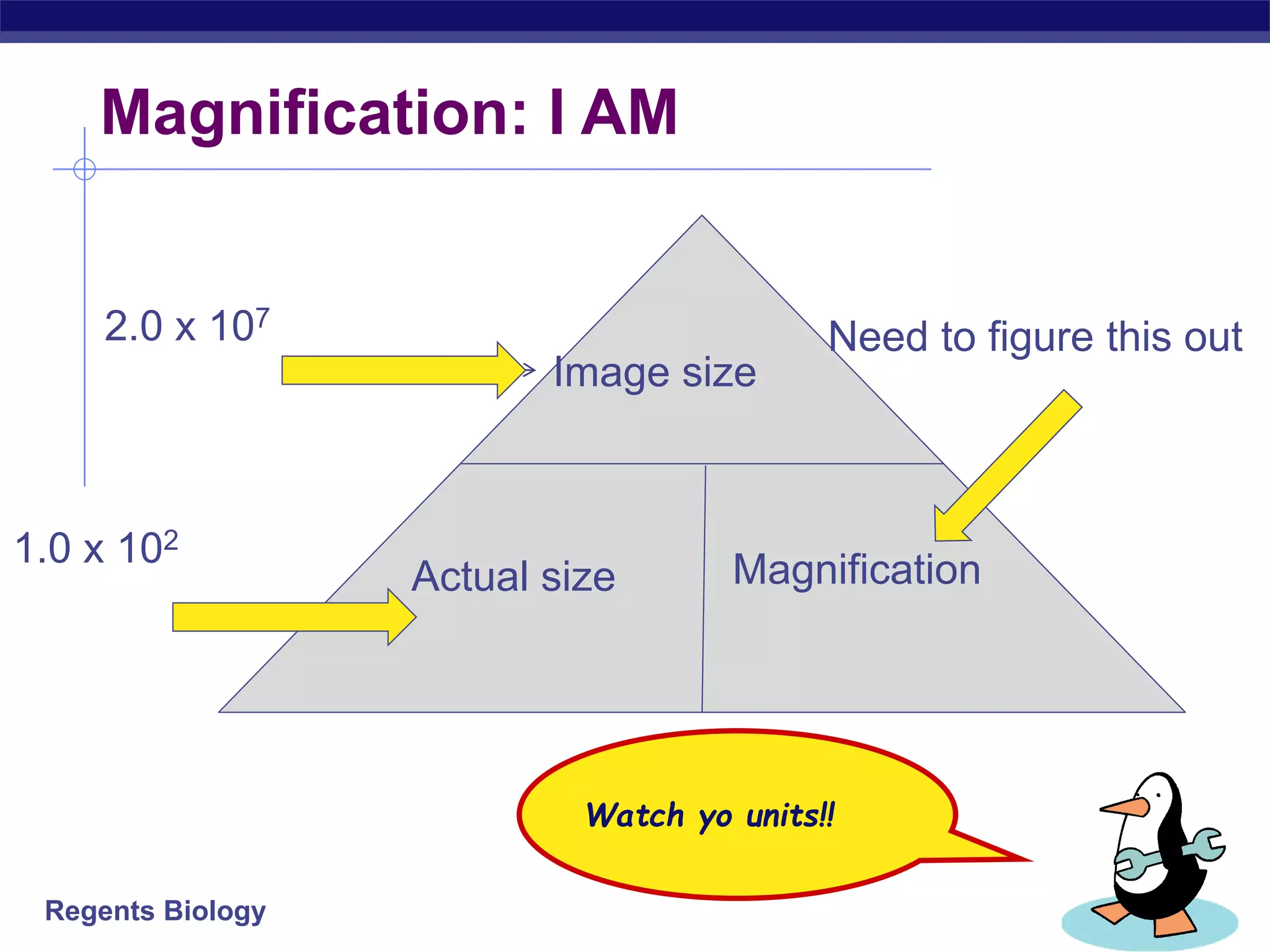
Magnification: IAM
Watch yo units!!
Image size
Actual size Magnification
Measure with ruler Need to figure this out
54.
Regents Biology
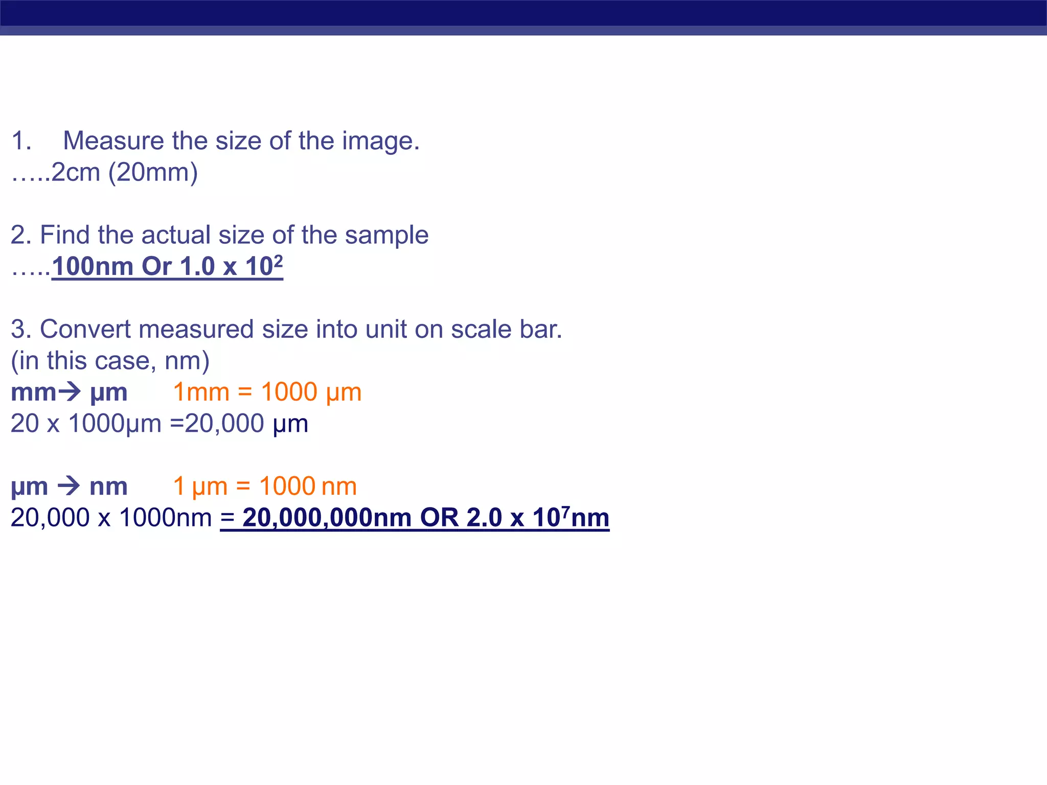
1. Measurethe size of the image.
…..2cm (20mm)
2. Find the actual size of the sample
…..100nm Or 1.0 x 10²
3. Convert measured size into unit on scale bar.
(in this case, nm)
mm µm 1mm = 1000 µm
20 x 1000µm =20,000 µm
µm nm 1 µm = 1000 nm
20,000 x 1000nm = 20,000,000nm OR 2.0 x 10⁷nm
55.
Regents Biology
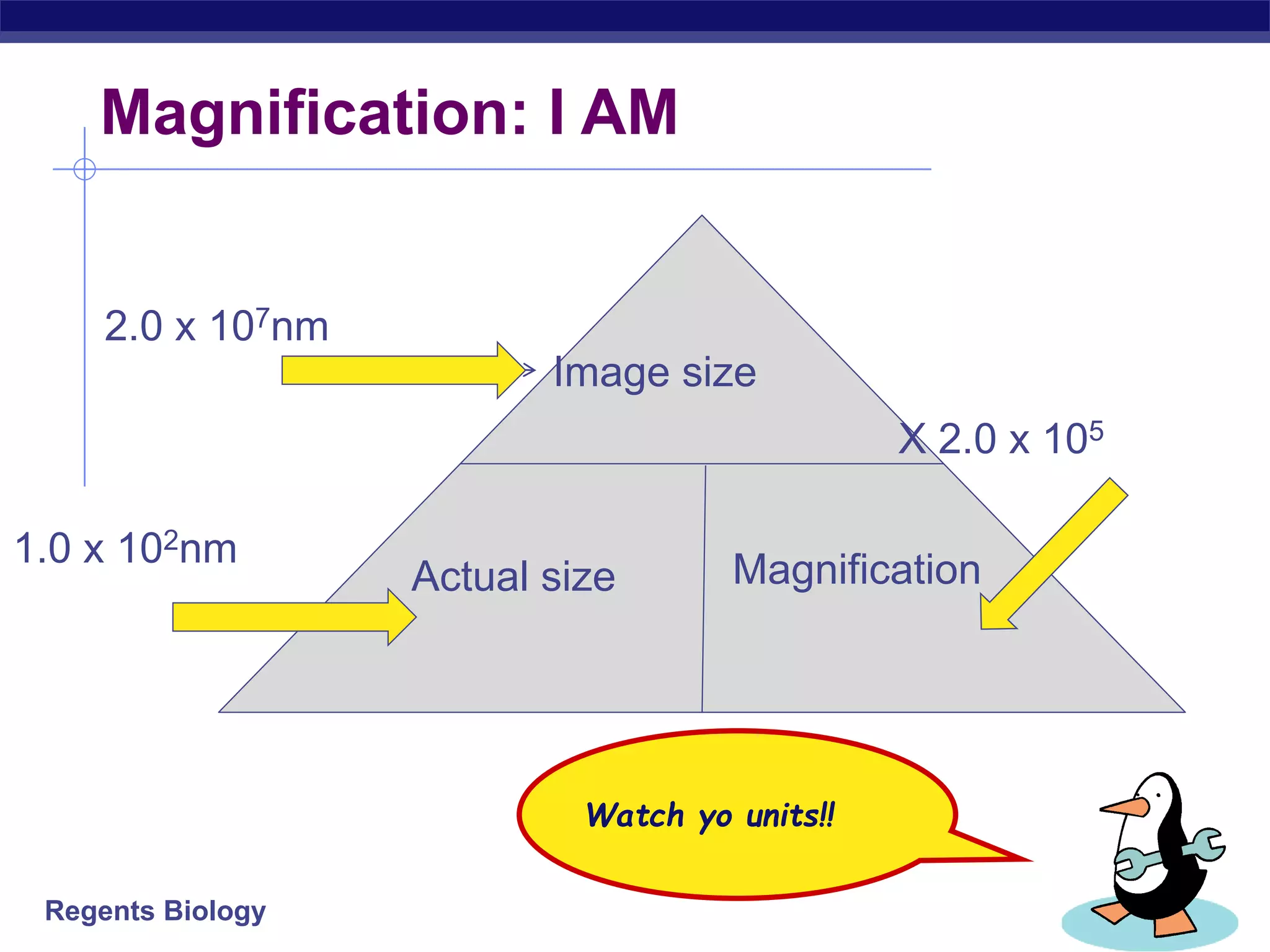
Magnification: IAM
Watch yo units!!
Image size
Actual size Magnification
2.0 x 10⁷
Need to figure this out
1.0 x 10²
#3 Technology and science are often used interchangably, but this requires some clarification.
Technology emerged before science. Materials were used to produce and used long before there was an understanding that different materials had different properties that could be used for different purposes.
Modern science often is the opposite. Scientific understanding is often the basis for technological development. These developments in technology in turn drive developments in science.
So technology applies the question: “does it work?” to its pursuit. Science is less bothered by the knowledge for the sake of knowledge
Desprite their mutual dependence, they are based on different values.
Science: evidence, rationality and the quest for deeper understanding
Tech: practical, appropriare and useful and an increasing emphasis on sustainability
#4 Technology and science are often used interchangably, but this requires some clarification.
Technology emerged before science. Materials were used to produce and used long before there was an understanding that different materials had different properties that could be used for different purposes.
Modern science often is the opposite. Scientific understanding is often the basis for technological development. These developments in technology in turn drive developments in science.
Both electron and light microscopes are technical devices which are used for visualizing structures that are too small to see with the unaided eye, and both types have relevant areas of applications in biology and the materials sciences. And this is pretty much it. The method of visualizing the structures is very different. Electron Microscopes use electrons and not photons (light rays) for visualization. The first electron microscope was constructed in 1931, compared to optical microscopes they are a very recent invention.
#12 Anything below 1 micron requires an electron microscope.
But there are STILL things we can’t see clearly with EM, like cell membranes!
#57 Probably seen under an electron microscope.
The virus surface is processed with metal like platinum, and the platinum is coated in a way that it coats the bumpy parts while misses the low parts. When the specimen is photographed under an electron microscope, the electrons are scattered by the metal atoms. In these areas, the electrons do not strike the photographic film, so they stay light. Theelectrons do strike the film in areas that are not coated by metal, so they appear dark.
#59 Calculation and drawing practice and homework
Lesson 7 Use microscopes to observe samples. Practical 1.


![[DSC Europe 25] Nikola Rajovic - Hardware Technologies Under the Hood: RISC-V...](https://cdn.slidesharecdn.com/ss_thumbnails/o2gptrmtoyqndgoshwgq-dsc2025-tenstorrent-rajovic-251205090438-814685f5-thumbnail.jpg?width=640&height=640&fit=bounds)


![[DSC Europe 25] Marko Krstic - Understanding the AI Threat Landscape - Risks,...](https://cdn.slidesharecdn.com/ss_thumbnails/tiyim1ins5jvbrvzpzla-2-251209104645-c69d3553-thumbnail.jpg?width=640&height=640&fit=bounds)

![[DSC Europe 25] Bogdan Daniel Maruneac - AI - It starts with you.pptx](https://cdn.slidesharecdn.com/ss_thumbnails/odov3snhrcqs9hx5ny2n-4-251205085715-f1daacfe-thumbnail.jpg?width=640&height=640&fit=bounds)
![[DSC Europe 25] Vid Stimac - Policy Parsimony: Between Oversimplifying and Ov...](https://cdn.slidesharecdn.com/ss_thumbnails/eqlepagzqp2rhg3gbluh-dsc-stimac-251120-251205090438-059e7f54-thumbnail.jpg?width=640&height=640&fit=bounds)
![[DSC Europe 25] Dragan Vucic - Building the Learning Organization - How AI Tr...](https://cdn.slidesharecdn.com/ss_thumbnails/8brigo2sbu6qur6gxrra-7-251205085715-6ae07d24-thumbnail.jpg?width=640&height=640&fit=bounds)
![[DSC Europe 25] Milan Zdravkovic - The road less traveled in District Heating...](https://cdn.slidesharecdn.com/ss_thumbnails/nfaboniqwsz4ucyctnmy-2-milan-zdravkovic-dsc2025-the-road-less-traveled-in-district-heating-operation-251208151905-f56388a5-thumbnail.jpg?width=640&height=640&fit=bounds)
![[DSC Europe 25] Vladimir Jelic - The AI-Driven Security Shift From Reactive D...](https://cdn.slidesharecdn.com/ss_thumbnails/6g5gj25mtjwayniqem1t-6-251209104645-7a5a5fc6-thumbnail.jpg?width=640&height=640&fit=bounds)


![[DSC Europe 25] Dobrica Cosic - Savings by the Second: How Dynamic Pricing an...](https://cdn.slidesharecdn.com/ss_thumbnails/znp09f3smtqz3w2sq6wn-1-dobrica-cosic-savings-by-the-second-how-dynamic-pricing-and-smart-data-are-bu-251208151905-26e6f41e-thumbnail.jpg?width=640&height=640&fit=bounds)

![[DSC Europe 25] Milan Sekuloski - Data, Defence, and Development: Cybersecuri...](https://cdn.slidesharecdn.com/ss_thumbnails/dfrkwwx4qly6atqpbl4z-4-251209104645-c3d4b0ca-thumbnail.jpg?width=640&height=640&fit=bounds)
![[DSC Europe 25] Sara Polak - The Ancient Operating System: What Archaeology T...](https://cdn.slidesharecdn.com/ss_thumbnails/3vch2p6tttdnwhsgazoz-3-sara-polak-smart-cities-251208152532-64404202-thumbnail.jpg?width=640&height=640&fit=bounds)
