Download to read offline
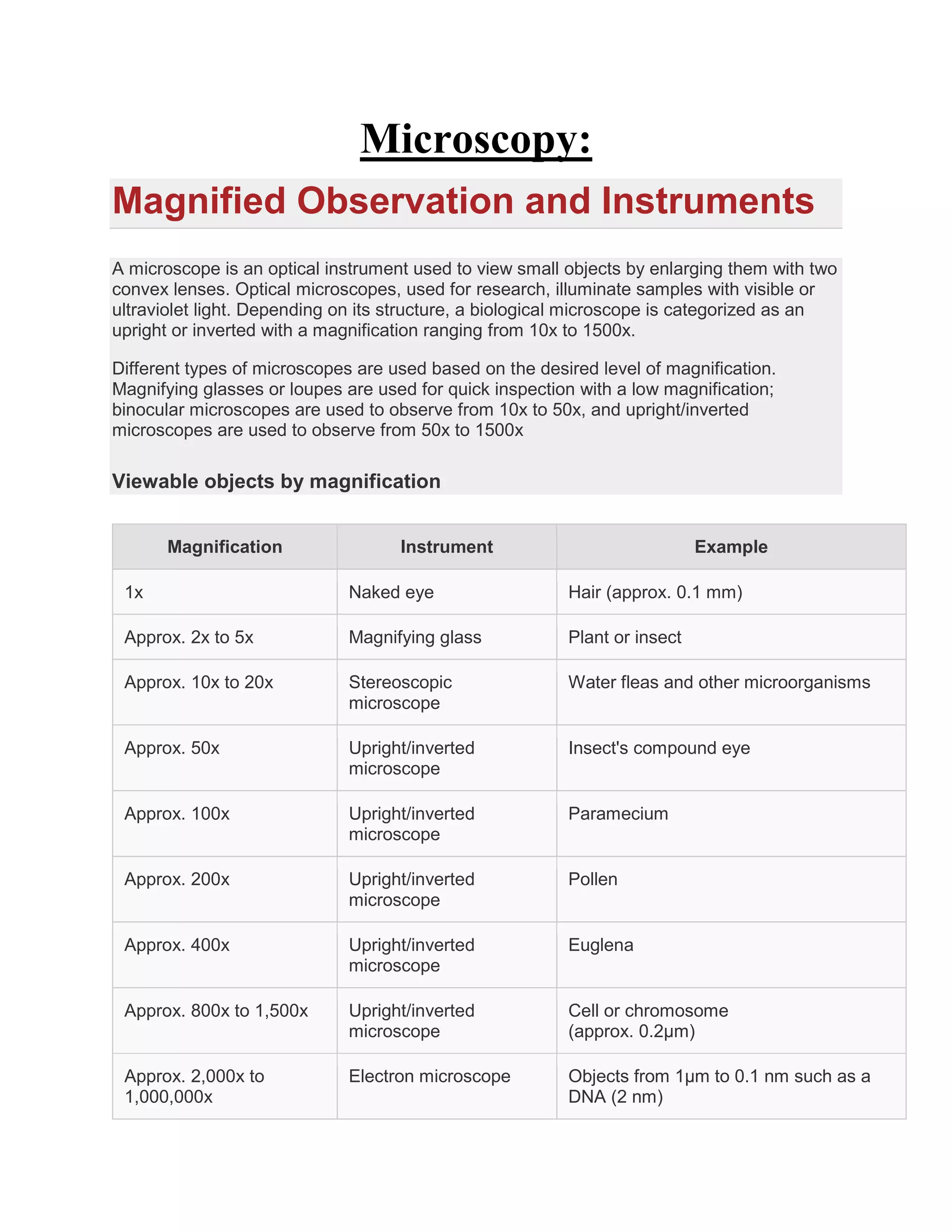
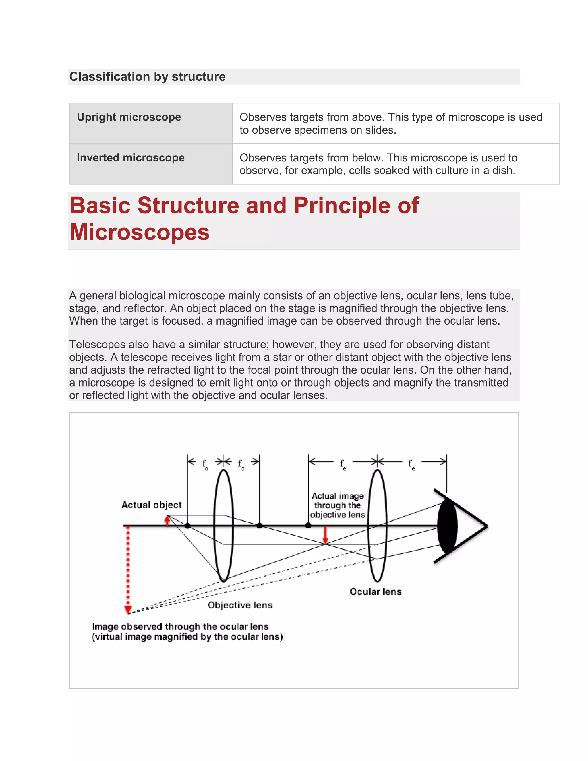

The document provides an overview of microscopy, detailing various types of microscopes, including optical, electron, and scanning probe microscopes, along with their specific applications and magnification ranges. It emphasizes the importance of magnification, resolution, and lens performance, including factors like numerical aperture and aberration. Additionally, it distinguishes between upright and inverted microscopes based on their structure and use in biological observations.


