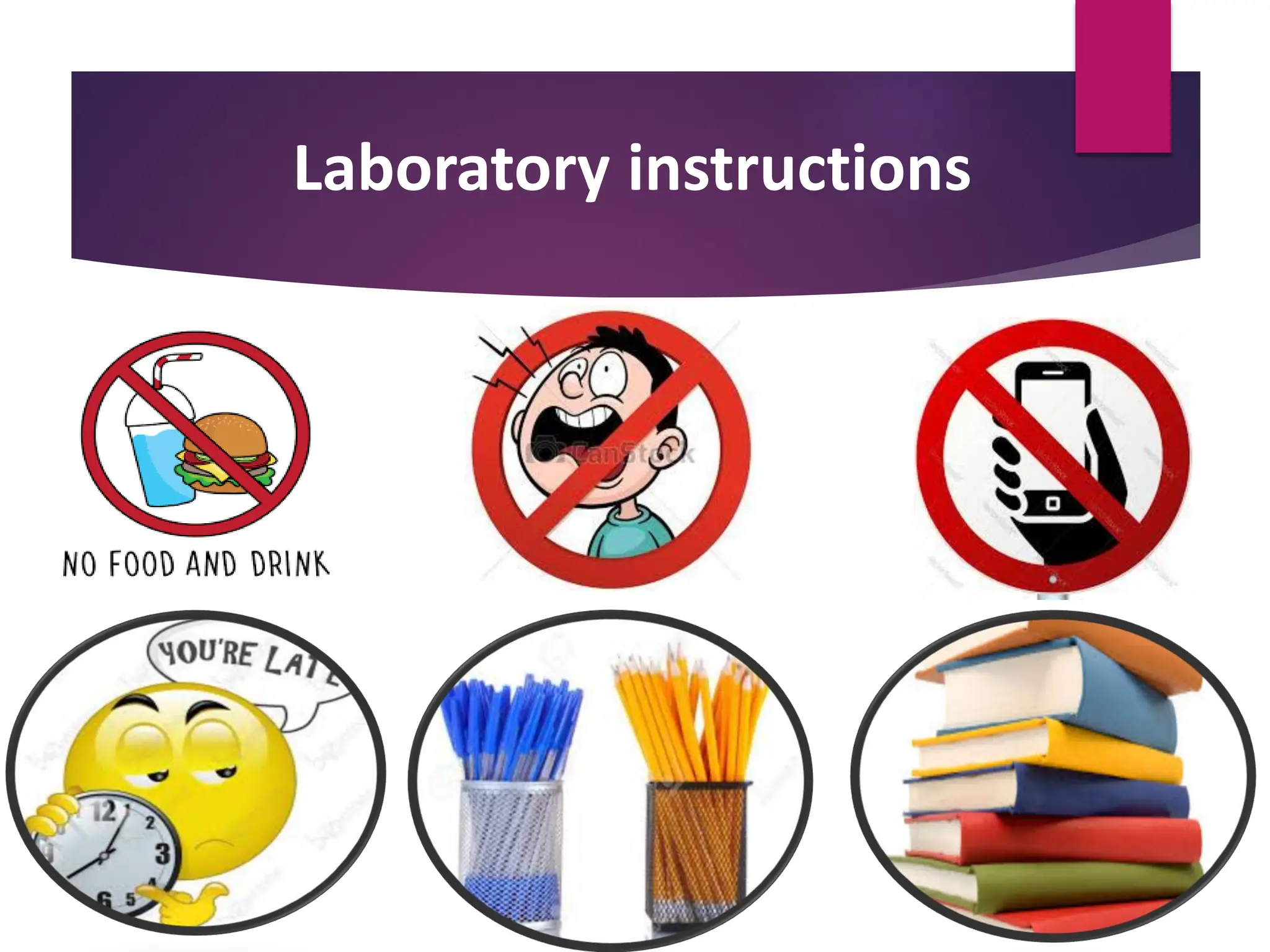
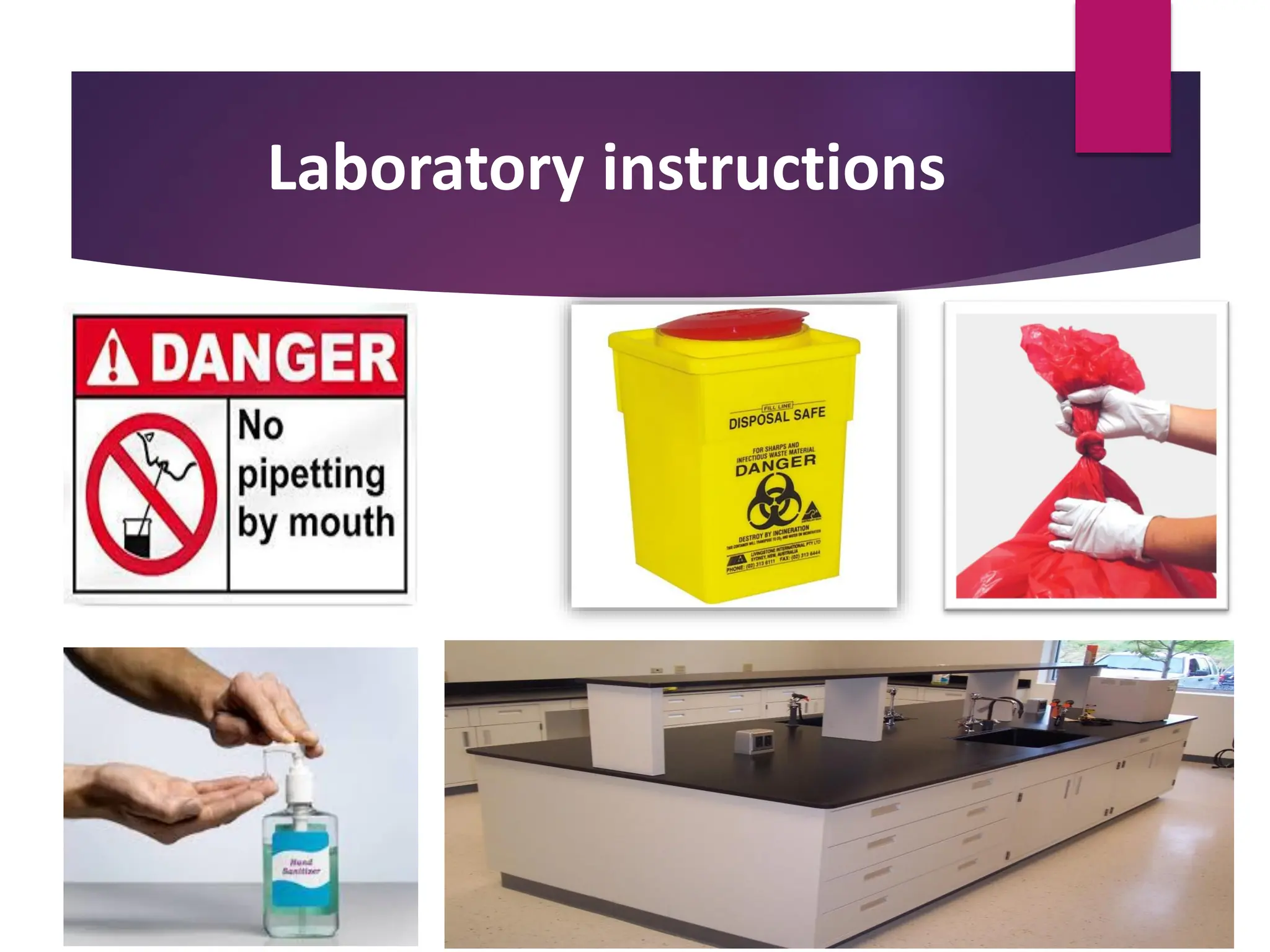
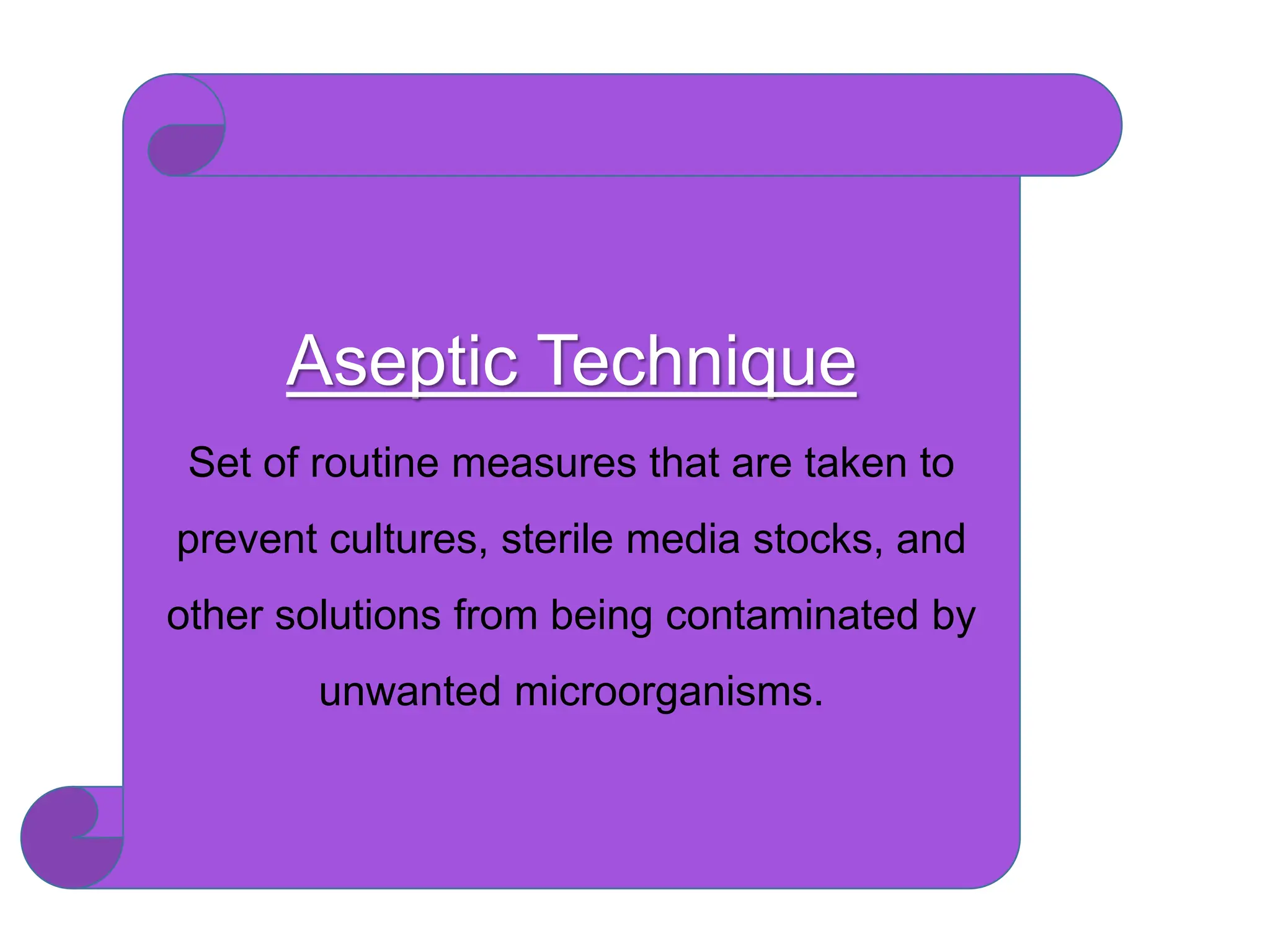
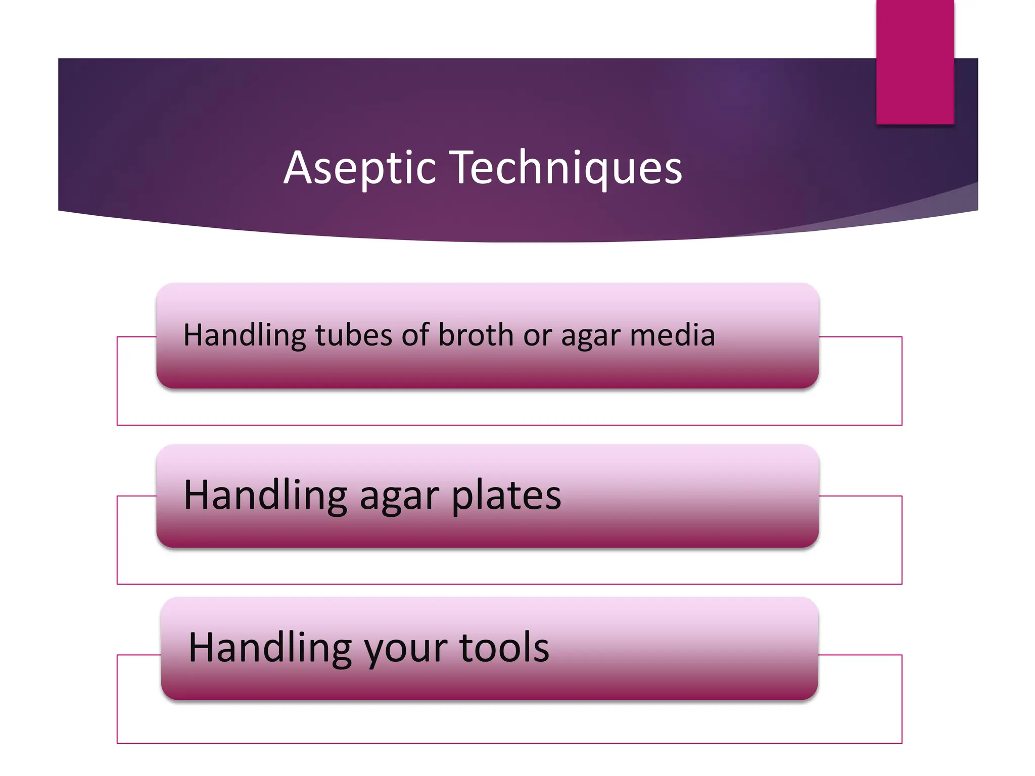
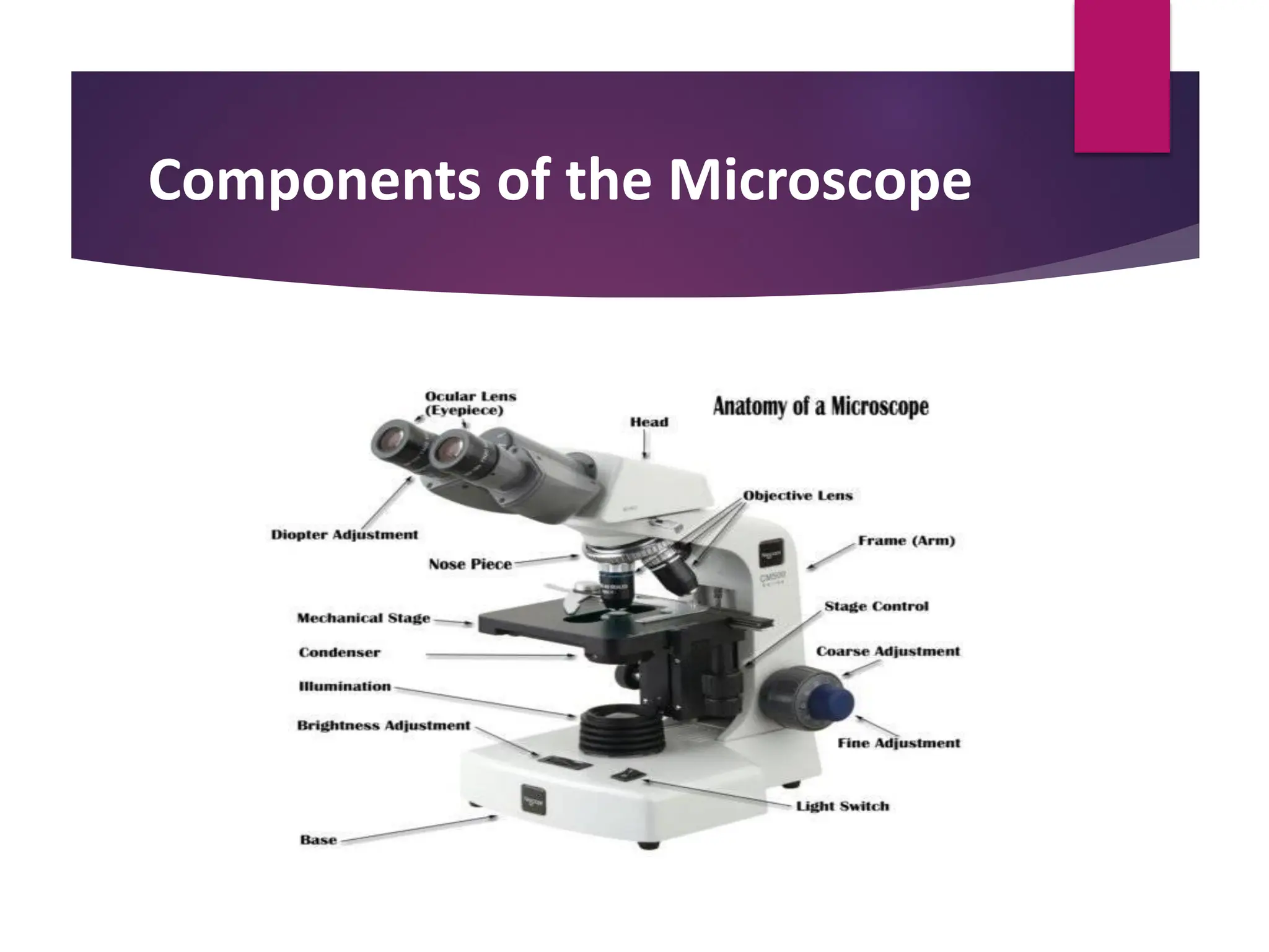
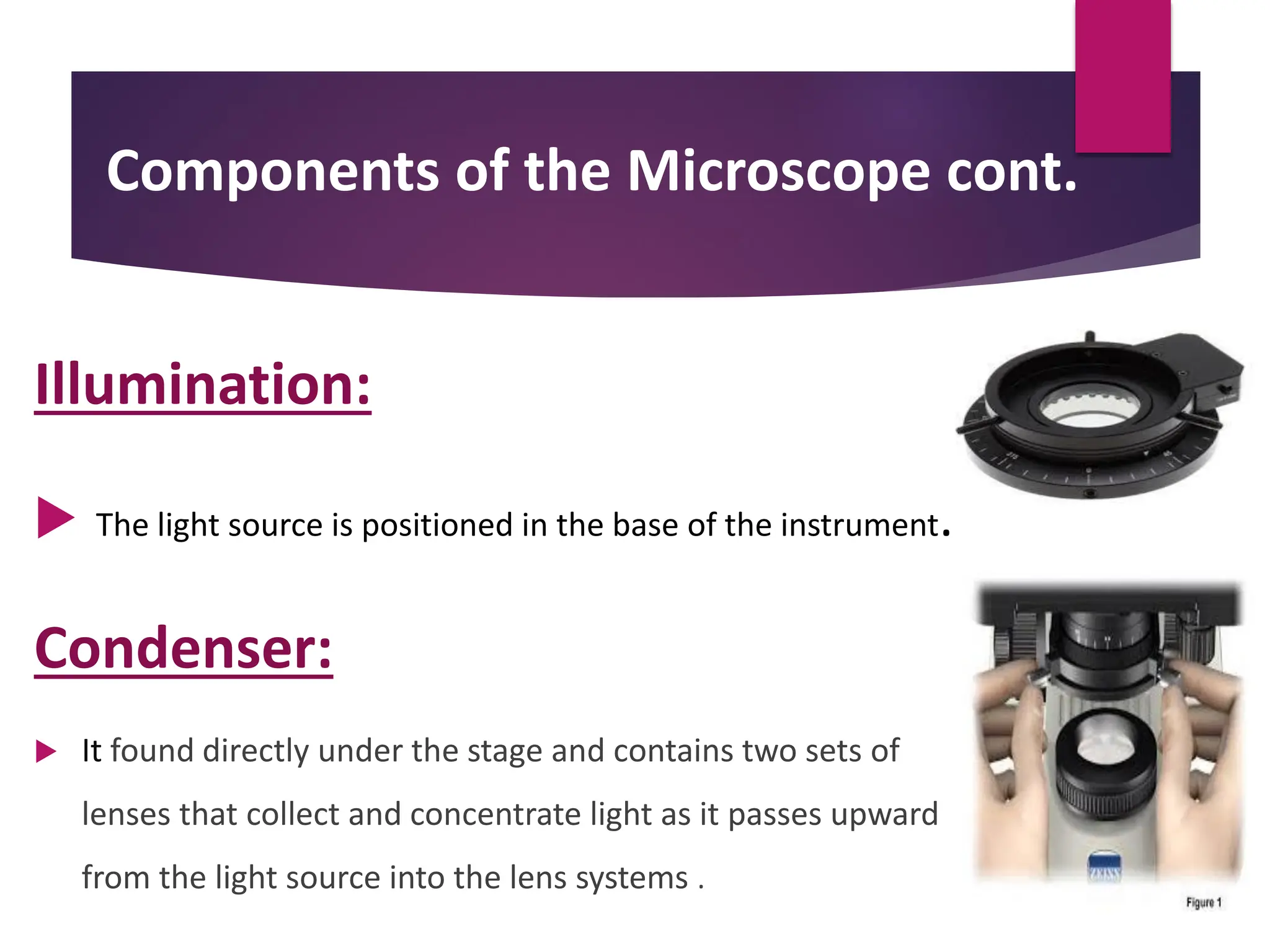
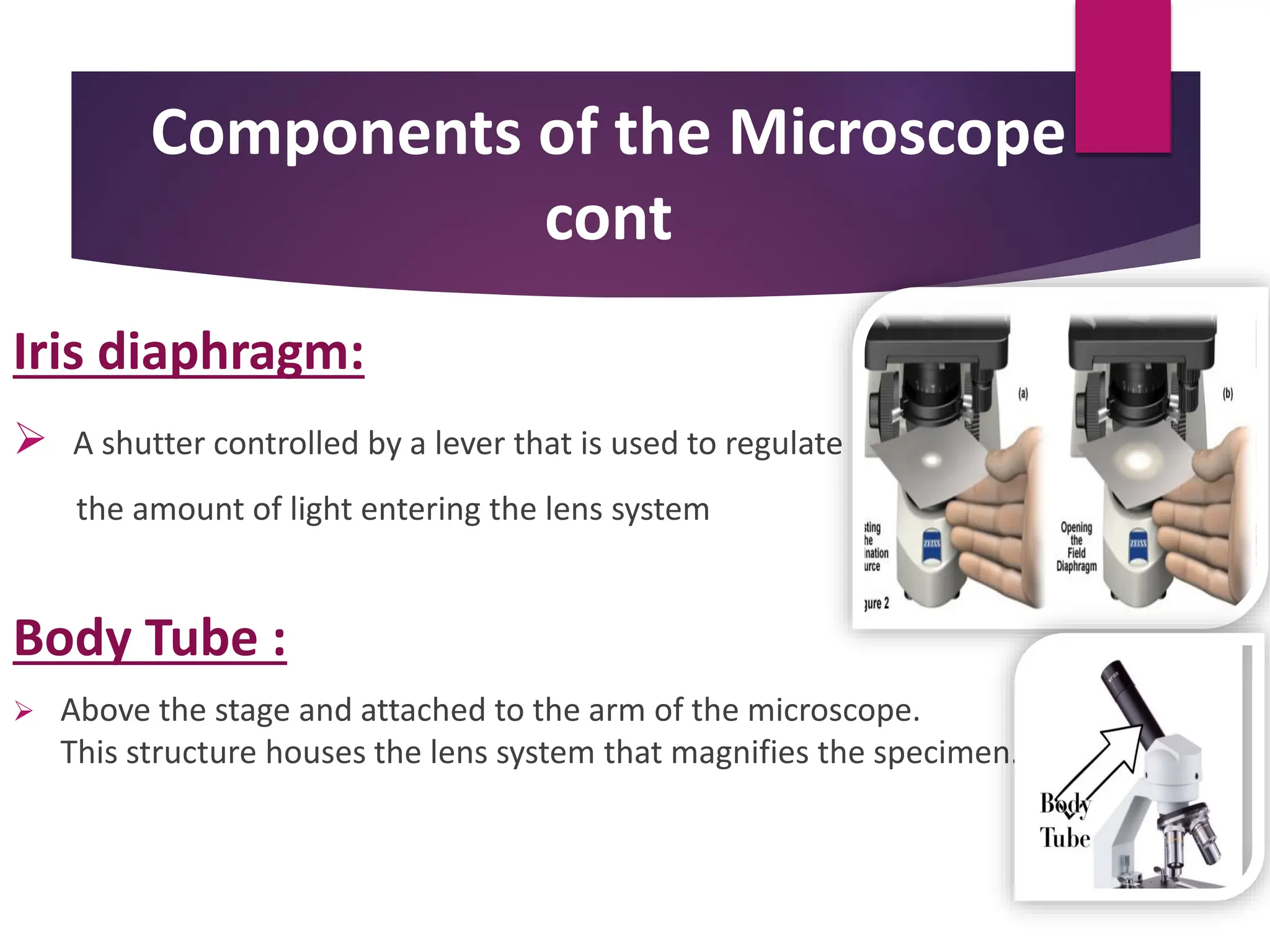
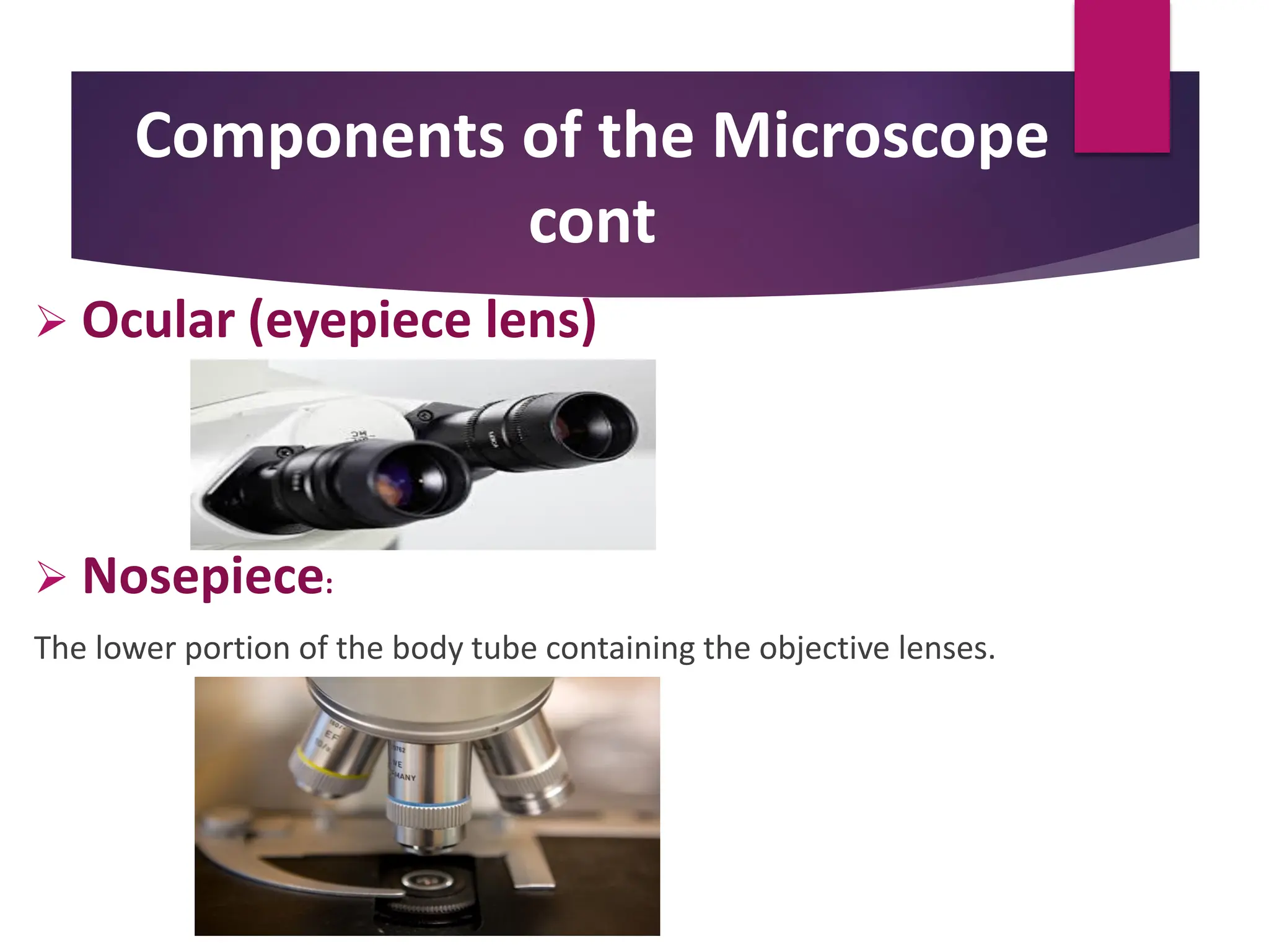
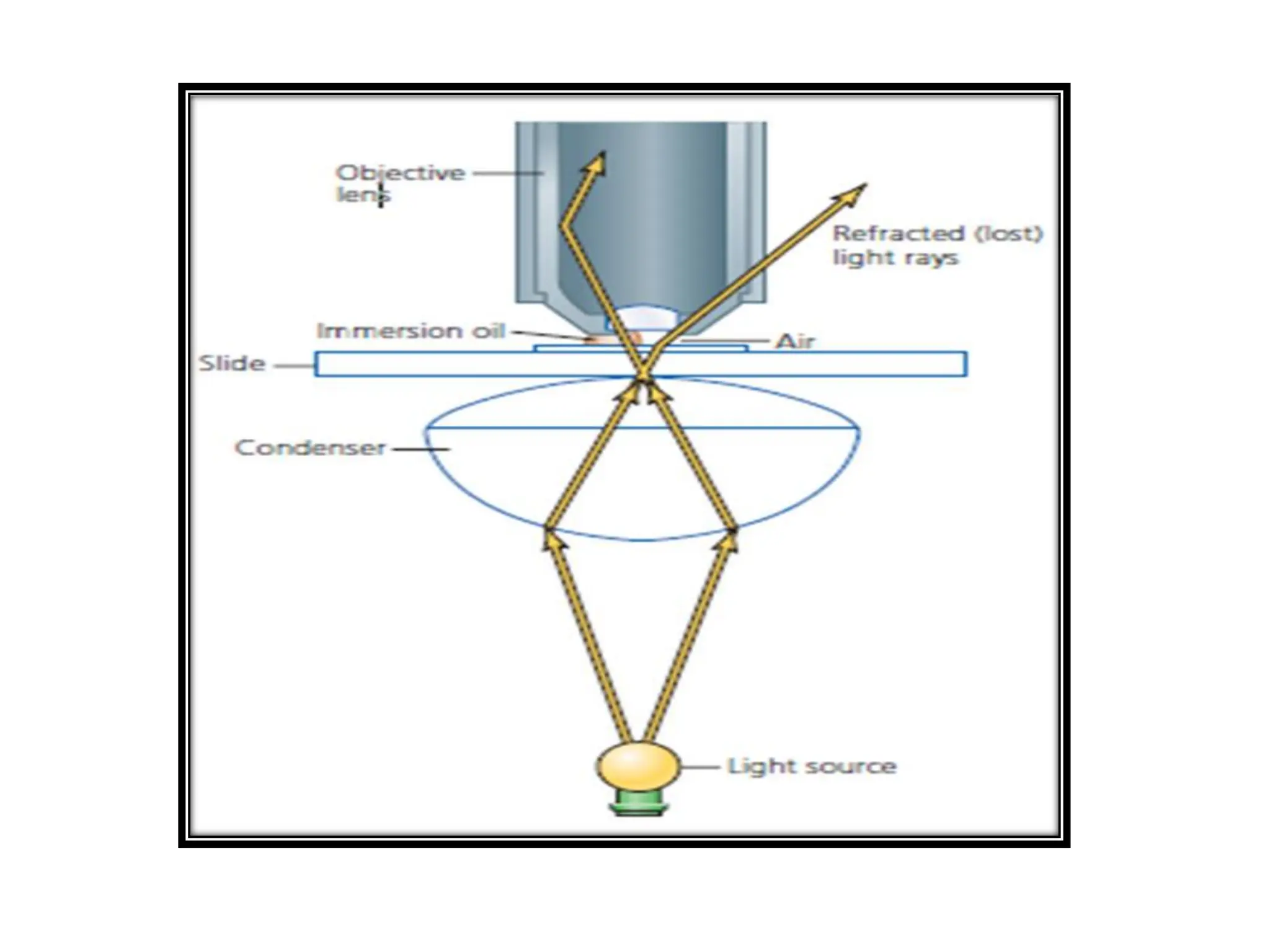
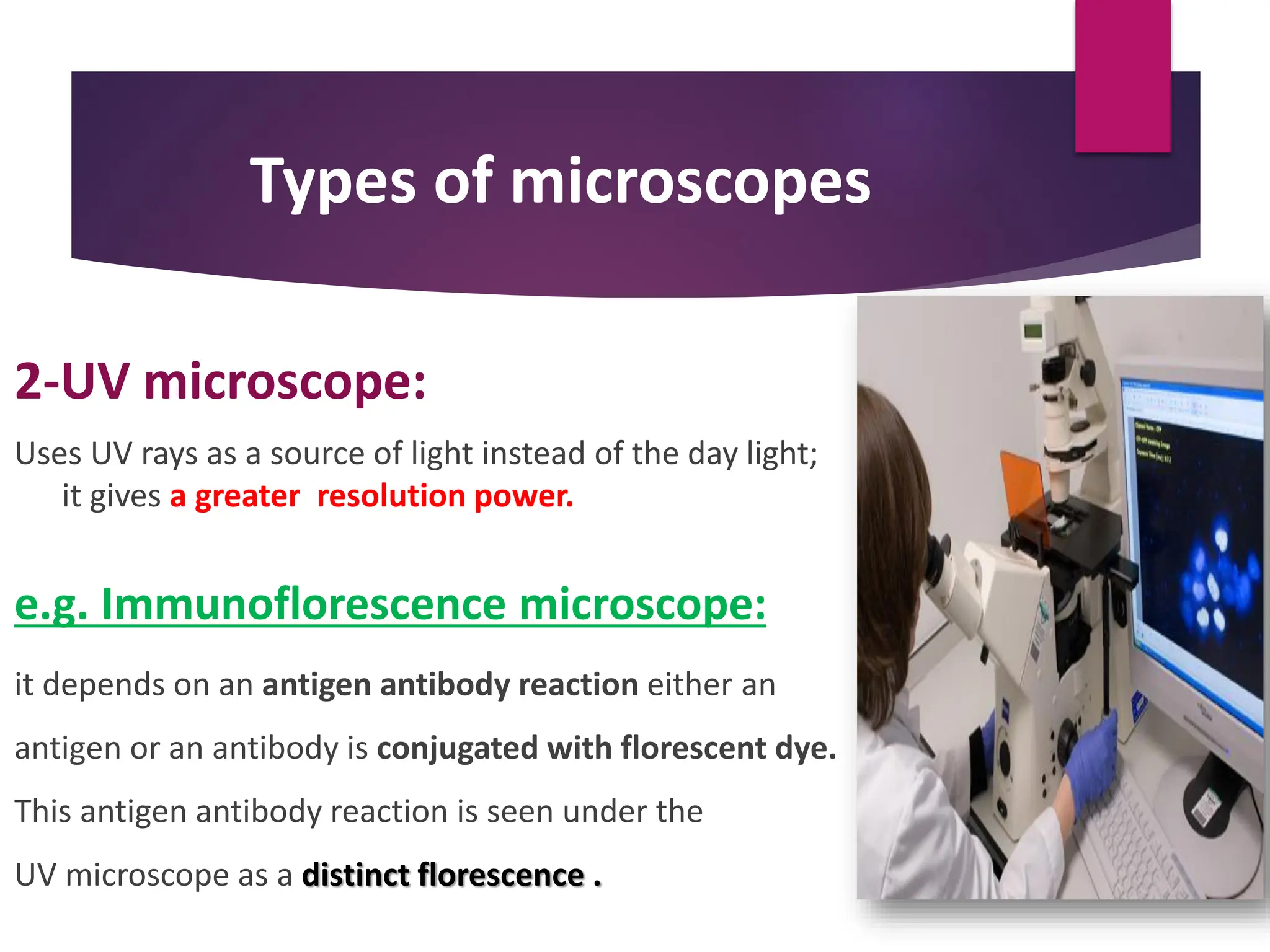
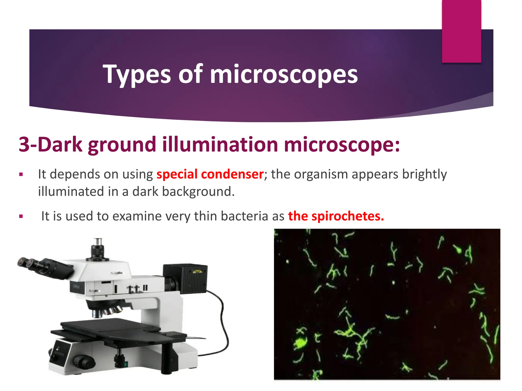
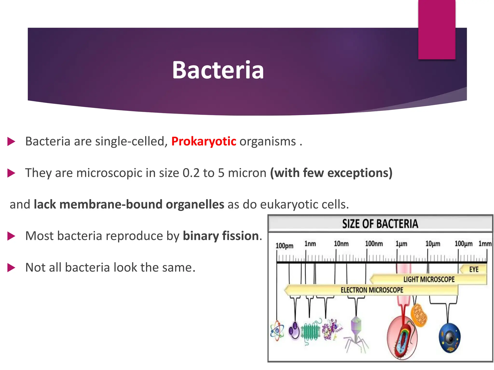
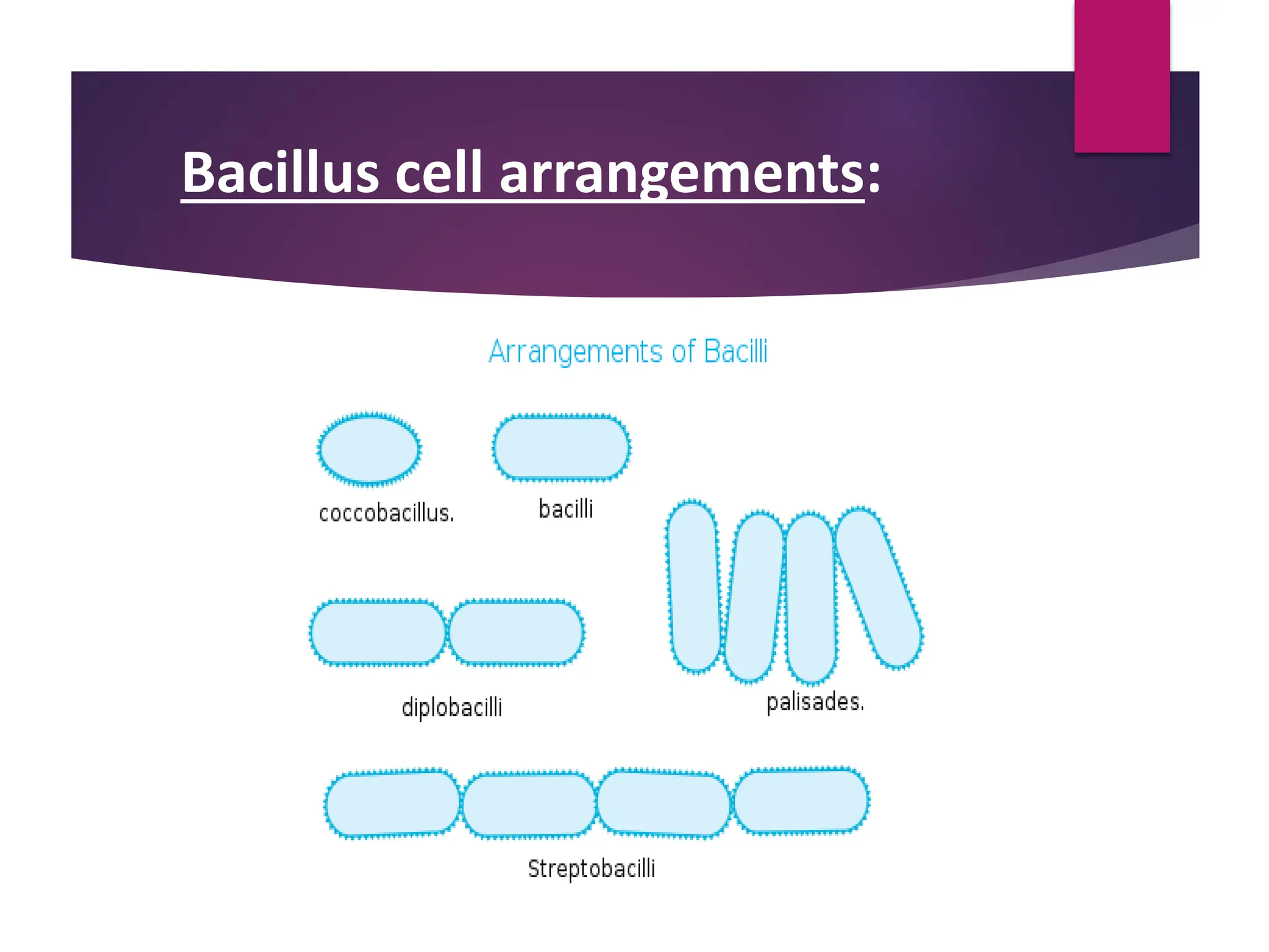
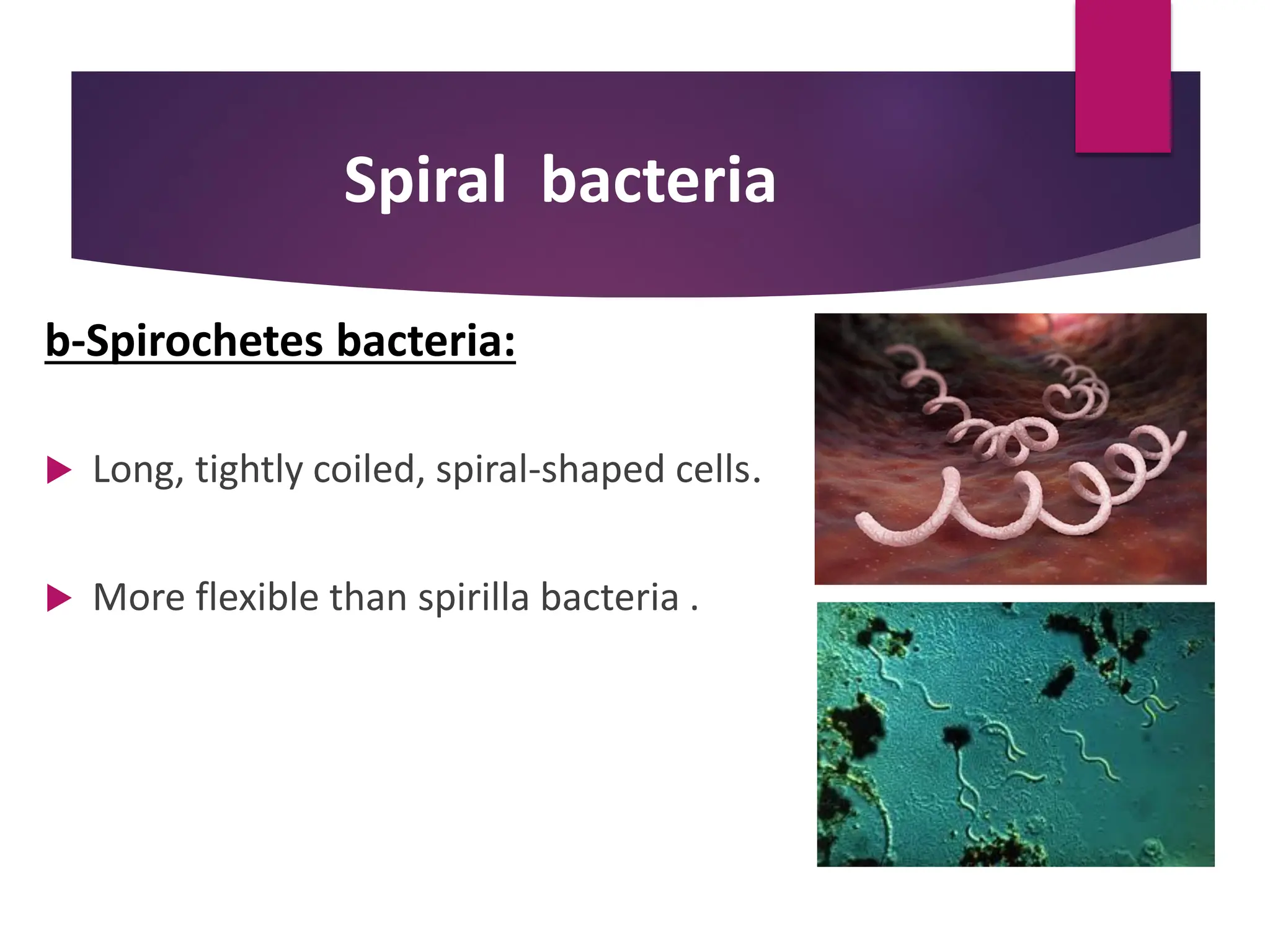
Laboratory instructions
Aseptic Technique
Set of routine measures that are taken to
prevent cultures, sterile media stocks, and
other solutions from being contaminated by
unwanted microorganisms.
Aseptic Techniques
Handling tubes of broth or agar media
Handling agar plates
Handling your tools
Handling tubes of broth or agar
media
Handling Agar plates
Handling your tools
Microscopy
•small
Micron
•To look at
Skopein
 It is an instrument for viewing objects that are too
small to be seen by the naked eye.
Components of the Microscope
Stage:
 A fixed platform with an opening in the center
allows the passage of light from an illuminating
source below to the lens system above the stage .
 provides a surface for the placement of a slide
with its specimen over the central opening.
Components of the Microscope cont.
Illumination:
 The light source is positioned in the base of the instrument.
Condenser:
 It found directly under the stage and contains two sets of
lenses that collect and concentrate light as it passes upward
from the light source into the lens systems .
Components of the Microscope
cont
Iris diaphragm:
 A shutter controlled by a lever that is used to regulate
the amount of light entering the lens system
Body Tube :
 Above the stage and attached to the arm of the microscope.
This structure houses the lens system that magnifies the specimen.
Components of the Microscope
cont
 Ocular (eyepiece lens)
 Nosepiece:
The lower portion of the body tube containing the objective lenses.
Components of the Microscope cont
 Coarse-adjustment and Fine-adjustment knobs:
 located above or below the stage .
 move the body tube up or down .
Components of the Microscope
cont
 Objective lenses :
 4x (scanning objective)
 10x (low power objective)
 40x (high power objective)
 100x (oil immersion lens)
Components of the Microscope
 Total Magnification power = magnification of ocular x
magnification of objective lens
Theoretical Principles of Microscopy
Magnification ??
 Unlimited enlargement is not possible by merely increasing the magnifying power of
the lenses or by using additional lenses, because lenses are limited by a property called
resolving power.
 Resolving Power or Resolution??:
Is how far apart two adjacent objects must be before a given lens shows them as
discrete entities. It depends on the wavelength of light and refractive index .
 When a lens cannot discriminate that is when the two objects appear as one, it has lost
resolution.
Oil immersion lens
 As light passes through various substances
(glass, air, specimens, etc.) it bends.
 This bending of light is called refraction.
 The refractive index of a substance is:
a measurement of the extent that the substance bends light.
Oil immersion lens
 Excessive refraction can cause distortion of the image.
 At magnification of less than 400x, the distortion is minimal. But at higher
magnifications, the distortion becomes so great that the image details are lost.
 An oil immersion lens helps to remedy this problem by eliminating the air gap
between the specimen and the objective lens.
 A drop of special immersion oil is placed on the microscope slide, and the oil
immersion objective lens is adjusted so that it is touching the oil.
 Immersion oil has the same refractive index as glass so that the light passes
through the slide, specimen, oil and objective lens as if they were a single piece of
glass.
Theoretical Principles of Microscopy
Illumination
 Effective illumination is required for efficient magnification and resolving power.
 Since the intensity of daylight is an uncontrolled variable, artificial light from a
tungsten lamp is the most commonly used light source in microscopy .
Types of microscopes
Optical microscope UV microscope
Electron
microscope
Dark ground
illumination
microscope
Types of microscopes
1-Optical microscope:
 Its based on visible wavelengths of light.
 It is the simplest and most widely used.
 The image seen with this type of microscope is two dimensional.
 Simple microscope: uses a single small lens and gives amazingly clear images.
 Compound microscope: uses a series of lenses and serves uses in many fields of
sciences .
Types of microscopes
2-UV microscope:
Uses UV rays as a source of light instead of the day light;
it gives a greater resolution power.
e.g. Immunoflorescence microscope:
it depends on an antigen antibody reaction either an
antigen or an antibody is conjugated with florescent dye.
This antigen antibody reaction is seen under the
UV microscope as a distinct florescence .
Types of microscopes
3-Dark ground illumination microscope:
 It depends on using special condenser; the organism appears brightly
illuminated in a dark background.
 It is used to examine very thin bacteria as the spirochetes.
Types of microscopes
4-Electron microscope:
 It uses beams of electrons instead of light, designed for very high
magnification usage.
 Electrons, which have a much smaller wavelength than visible light,
allow a much higher resolution.
 This electron beam must pass through a vacuum as air molecules
would otherwise scatter the beam ,The electrons are typically
detected to give the image.
 Scanning electron microscope (SEM): 3D image .
 Transmission electron microscope (TEM): This gives a 2D view.
Bacteria
 Bacteria are single-celled, Prokaryotic organisms .
 They are microscopic in size 0.2 to 5 micron (with few exceptions)
and lack membrane-bound organelles as do eukaryotic cells.
 Most bacteria reproduce by binary fission.
 Not all bacteria look the same.
Bacteria
Common shapes of bacteria
Coccus (spherical) Bacillus (rod)
Spiral
(curved or twisted)
 Though these are the most common shapes for bacteria ,some bacteria
have unusual and much less common forms. These bacteria have varying
shapes and are said to be pleomorphic .
 Other unusual bacteria forms include star-shapes, club-shapes, cube-
shapes, and filamentous branches.
 Bacteria can also have different arrangements of cells.
common shapes of bacteria
COCCI BACTERIA
 Coccus (cocci plural) bacteria are round, oval, or spherical in shape.
 These cell can exist in different arrangements.
Cocci cell arrangements
Common shapes of bacteria
Bacillus bacteria:
 Bacillus (bacilli plural) bacteria have rod-
shaped cells.
Bacillus cell arrangements:
Common shapes of bacteria
Spiral bacteria:
 Are twisted and commonly occur in three forms:
 Vibrio
 Spiruillum
 Spirochete
Spiral bacteria
a-Spirilla
 Elongated, spiral-shaped rigid cells.
 May have flagella , which are long protrusion used for movement .
Spiral bacteria
b-Spirochetes bacteria:
 Long, tightly coiled, spiral-shaped cells.
 More flexible than spirilla bacteria .
Spiral bacteria
C-Vibrio bacteria:
 Have a slight twist or curve and resemble the shape of a comma.
 They also have a flagellum , which is used for movement .
 A number of species of vibrio bacteria are pathogens and are
associated with food poisoning .
General Microbiology Section 1.pdf and bacteria

General Microbiology Section 1.pdf and bacteria

  • 3.
  • 4.
  • 5.
  • 6.
    Aseptic Technique Set ofroutine measures that are taken to prevent cultures, sterile media stocks, and other solutions from being contaminated by unwanted microorganisms.
  • 7.
    Aseptic Techniques Handling tubesof broth or agar media Handling agar plates Handling your tools
  • 8.
    Handling tubes ofbroth or agar media
  • 9.
  • 10.
  • 11.
    Microscopy •small Micron •To look at Skopein It is an instrument for viewing objects that are too small to be seen by the naked eye.
  • 12.
  • 13.
    Components of theMicroscope Stage:  A fixed platform with an opening in the center allows the passage of light from an illuminating source below to the lens system above the stage .  provides a surface for the placement of a slide with its specimen over the central opening.
  • 14.
    Components of theMicroscope cont. Illumination:  The light source is positioned in the base of the instrument. Condenser:  It found directly under the stage and contains two sets of lenses that collect and concentrate light as it passes upward from the light source into the lens systems .
  • 15.
    Components of theMicroscope cont Iris diaphragm:  A shutter controlled by a lever that is used to regulate the amount of light entering the lens system Body Tube :  Above the stage and attached to the arm of the microscope. This structure houses the lens system that magnifies the specimen.
  • 16.
    Components of theMicroscope cont  Ocular (eyepiece lens)  Nosepiece: The lower portion of the body tube containing the objective lenses.
  • 17.
    Components of theMicroscope cont  Coarse-adjustment and Fine-adjustment knobs:  located above or below the stage .  move the body tube up or down .
  • 18.
    Components of theMicroscope cont  Objective lenses :  4x (scanning objective)  10x (low power objective)  40x (high power objective)  100x (oil immersion lens)
  • 19.
    Components of theMicroscope  Total Magnification power = magnification of ocular x magnification of objective lens
  • 20.
    Theoretical Principles ofMicroscopy Magnification ??  Unlimited enlargement is not possible by merely increasing the magnifying power of the lenses or by using additional lenses, because lenses are limited by a property called resolving power.  Resolving Power or Resolution??: Is how far apart two adjacent objects must be before a given lens shows them as discrete entities. It depends on the wavelength of light and refractive index .  When a lens cannot discriminate that is when the two objects appear as one, it has lost resolution.
  • 21.
    Oil immersion lens As light passes through various substances (glass, air, specimens, etc.) it bends.  This bending of light is called refraction.  The refractive index of a substance is: a measurement of the extent that the substance bends light.
  • 22.
    Oil immersion lens Excessive refraction can cause distortion of the image.  At magnification of less than 400x, the distortion is minimal. But at higher magnifications, the distortion becomes so great that the image details are lost.  An oil immersion lens helps to remedy this problem by eliminating the air gap between the specimen and the objective lens.  A drop of special immersion oil is placed on the microscope slide, and the oil immersion objective lens is adjusted so that it is touching the oil.  Immersion oil has the same refractive index as glass so that the light passes through the slide, specimen, oil and objective lens as if they were a single piece of glass.
  • 24.
    Theoretical Principles ofMicroscopy Illumination  Effective illumination is required for efficient magnification and resolving power.  Since the intensity of daylight is an uncontrolled variable, artificial light from a tungsten lamp is the most commonly used light source in microscopy .
  • 25.
    Types of microscopes Opticalmicroscope UV microscope Electron microscope Dark ground illumination microscope
  • 26.
    Types of microscopes 1-Opticalmicroscope:  Its based on visible wavelengths of light.  It is the simplest and most widely used.  The image seen with this type of microscope is two dimensional.  Simple microscope: uses a single small lens and gives amazingly clear images.  Compound microscope: uses a series of lenses and serves uses in many fields of sciences .
  • 27.
    Types of microscopes 2-UVmicroscope: Uses UV rays as a source of light instead of the day light; it gives a greater resolution power. e.g. Immunoflorescence microscope: it depends on an antigen antibody reaction either an antigen or an antibody is conjugated with florescent dye. This antigen antibody reaction is seen under the UV microscope as a distinct florescence .
  • 28.
    Types of microscopes 3-Darkground illumination microscope:  It depends on using special condenser; the organism appears brightly illuminated in a dark background.  It is used to examine very thin bacteria as the spirochetes.
  • 29.
    Types of microscopes 4-Electronmicroscope:  It uses beams of electrons instead of light, designed for very high magnification usage.  Electrons, which have a much smaller wavelength than visible light, allow a much higher resolution.  This electron beam must pass through a vacuum as air molecules would otherwise scatter the beam ,The electrons are typically detected to give the image.  Scanning electron microscope (SEM): 3D image .  Transmission electron microscope (TEM): This gives a 2D view.
  • 30.
    Bacteria  Bacteria aresingle-celled, Prokaryotic organisms .  They are microscopic in size 0.2 to 5 micron (with few exceptions) and lack membrane-bound organelles as do eukaryotic cells.  Most bacteria reproduce by binary fission.  Not all bacteria look the same.
  • 31.
    Bacteria Common shapes ofbacteria Coccus (spherical) Bacillus (rod) Spiral (curved or twisted)
  • 32.
     Though theseare the most common shapes for bacteria ,some bacteria have unusual and much less common forms. These bacteria have varying shapes and are said to be pleomorphic .  Other unusual bacteria forms include star-shapes, club-shapes, cube- shapes, and filamentous branches.  Bacteria can also have different arrangements of cells. common shapes of bacteria
  • 33.
    COCCI BACTERIA  Coccus(cocci plural) bacteria are round, oval, or spherical in shape.  These cell can exist in different arrangements.
  • 34.
  • 35.
    Common shapes ofbacteria Bacillus bacteria:  Bacillus (bacilli plural) bacteria have rod- shaped cells.
  • 36.
  • 37.
    Common shapes ofbacteria Spiral bacteria:  Are twisted and commonly occur in three forms:  Vibrio  Spiruillum  Spirochete
  • 38.
    Spiral bacteria a-Spirilla  Elongated,spiral-shaped rigid cells.  May have flagella , which are long protrusion used for movement .
  • 39.
    Spiral bacteria b-Spirochetes bacteria: Long, tightly coiled, spiral-shaped cells.  More flexible than spirilla bacteria .
  • 40.
    Spiral bacteria C-Vibrio bacteria: Have a slight twist or curve and resemble the shape of a comma.  They also have a flagellum , which is used for movement .  A number of species of vibrio bacteria are pathogens and are associated with food poisoning .