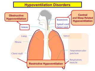
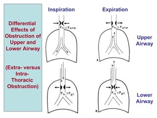
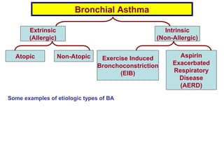
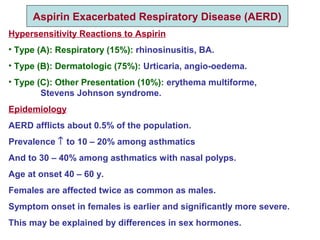
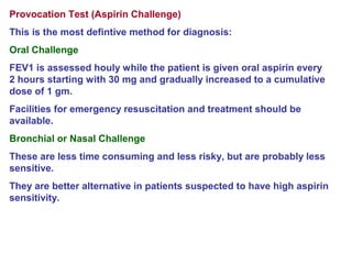
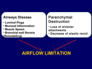
4. • Upper airway obstruction is usually more severe during inspiration.
Adventitious sound produced: Stridor.
• Lower airway obstruction is usually more severe during expiration.
Adventitious sound produced: Wheezes.
Wheezes Hyperinflation Atelectasis
Possible Consequences of Bronchial Obstruction
6. Tidal Volume X RR = Minute Ventilation (Minute Volume) (MV)
Forced Vital Capacity X RR = Maximum Breathing Capacity (MBC)
(FVC) (Maximum Voluntary Ventilation) (MVV)
….Multiplying by Respiratory Rate (RR):
7. (> 0.7)
(< 80%)
(< 80%) (< 80%)
FEV1
FVC
FEV1
FVC
Obstructive
(with Hyperinflation)
Restrictive
Two Main Patterns
of Hypoventilation Disorders
8. Obstructive Lung Diseases
Bronchial Asthma: a heterogeneous disease, usually characterized by
chronic airway inflammation. It is defined by the history of respiratory
symptoms such as wheezes, dyspnoea and cough that vary over time
together with variable expiratory airflow limitation (GINA 2015).
Chronic Bronchitis: chronic cough with expectoration for at least
6 months in a year or 3 months/Y for 2 successive years,
not due to lung disease.
Emphysema: persistent abnormal dilatation of the air spaces distal to
the terminal bronchioles (respiratory bronchioles, alveolar ducts,
alveoli), accompanied by destruction of the elastic tissues of the lungs.
Bronchiectasis: a chronic suppurative condition where the walls of the
bronchi are thickened from inflammation and infection.
Cystic Fibrosis: an inherited disease characterized by the build-up of
thick, sticky mucus that can damage many of the body's organs.
The thick mucus in the bronchial tree leads to variable degrees of
obstruction which become complicated by infection, lung damage and
ensuing respiratory failure.
13. – AtopyAtopy, the genetic predisposition for the development of an, the genetic predisposition for the development of an
immunoglobulin E (IgE)-mediated response to commonimmunoglobulin E (IgE)-mediated response to common
aeroallergens, is the strongest identifiable predisposingaeroallergens, is the strongest identifiable predisposing
factor for developing asthma.factor for developing asthma.
– However, not all people with allergies have asthma, and notHowever, not all people with allergies have asthma, and not
all cases of asthma can be explained by allergic response.all cases of asthma can be explained by allergic response.
– Viral respiratory infectionsViral respiratory infections are one of the most importantare one of the most important
causes of asthma exacerbation.causes of asthma exacerbation.
– In some patients, persistent changes in airway structure
occur, including sub-basement fibrosis, mucus
hypersecretion, injury to epithelial cells, smooth muscle
hypertrophy, and angiogenesis (remodeling)
14. Exercise Induced Bronchoconstriction (EIB)
Etiology
Osmotic Theory (most likely)
Hyperventilation during exercise → Water of airway surface liquid (ASL) is
evaporated faster than it is returned by condensation or from submucosa
→ ↑ osmotic pressure of ASL. Mast cells and basophils release mediators in
response to changes in osmolarity. Exercise itself is not needed to cause
bronchoconstriction, but just the creation of a hyperosmolar environment.
Moreover, dehydration of ASL ↓ its volume → ↓ mucocilliary clearance.
Thermal Theory (inadequate)
Exposure of airways to cool air → vasoconstriction of bronchial vasculature.
After cessation of exercise, ventilation ↓ and airways rewarm
→ reactive hyperaemia and oedema of airways.
Other Factors
• Poor air quality can be associated with a high prevalence of EIB in athletes.
• In swimmers, chloramines above the water can trigger EIB.
15. Occurrence
• EIB is reported frequently in asthmatics, but can also occur in
absence of chronic asthma.
• In asthmatics, the presence of EIB is a marker of poor control and
suggests the need to step up therapy.
• Commoner in athletes than in the general population.
Diagnosis
Self- reported symptoms are not reliable for diagnosis.
Formal diagnosis requires spirometry and
a bronchoprovocation challenge:
• Exercise: should be ramped up rapidly so that target heart rate is
achieved within 2 – 3 minutes and maintained for 6 minutes.
• Inhalation of dry mannitol powder.
16. Aspirin Exacerbated Respiratory Disease (AERD)
Hypersensitivity Reactions to Aspirin
• Type (A): Respiratory (15%): rhinosinusitis, BA.
• Type (B): Dermatologic (75%): Urticaria, angio-oedema.
• Type (C): Other Presentation (10%): erythema multiforme,
Stevens Johnson syndrome.
Epidemiology
AERD afflicts about 0.5% of the population.
Prevalence ↑ to 10 – 20% among asthmatics
And to 30 – 40% among asthmatics with nasal polyps.
Age at onset 40 – 60 y.
Females are affected twice as common as males.
Symptom onset in females is earlier and significantly more severe.
This may be explained by differences in sex hormones.
17. Pathogenesis
This is an extra- immunologic (pseudo- allergic) reaction which affects
both upper and lower airways.
Aspirin (and some other NSAIDs to variable extent) block
cyclooxygenase 1 (COX1) in mast cells, basophils and eosinophils →
dysregulation of arachidnonic acid (AA) metabolism with diversion
from COX1 to LOX5 (lipooxygenase 5) pathway →
↓ production of PGE1, ↑ production of CysLTs (LT C4, D4, E4).
CysLTs are highly effective proinflammatory mediators →
• Mucus hypersecretion.
• ↑ Vascular permeability.
• Bronchoconstriction.
• Eosinophilic infiltration.
19. Clinical Picture: Aspirin Triad: Samter`s Triad
Features may be seasonal or perennial.
Features may be triggered by stimuli other than aspirin.
Avoidance of aspirin and other NSAIDs does NOT lead to resolution of
BA or rhinosinusitis.
1) Aspirin Intolerance This is the main dysfunction:
Acute upper and lower respiratory tract reactions to aspirin ingestion
occurring 20 mins to 3 hours after its intake:
• Nasal congestion, oedema, eosinophilic infiltration
→ rhinorrhoea, anosmia, nasal obstruction.
• Post- nasal drip → dry irritant cough.
• Halitosis.
• Conjunctivitis → red eye, lacrymation.
• Laryngeal spasm.
20. 2) Bronchial Asthma (Aspirin Sensitive Asthma)
(Aspirin Intolerant Asthma)
Asthma is severe and refractory to treatment.
3) Chronic Rhinosinusitis with Nasal Polyps (CRSwNP)
The presence of nasal polyps signifies higher chronic nasal inflammation
with eosinophilc activation.
Polyps tend to recur rapidly after polypectomy.
The usual sequence of appearance of the triad components is:
Chronic rhinitis then BA then nasal polyposis
21. Provocation Test (Aspirin Challenge)
This is the most defintive method for diagnosis:
Oral Challenge
FEV1 is assessed houly while the patient is given oral aspirin every
2 hours starting with 30 mg and gradually increased to a cumulative
dose of 1 gm.
Facilities for emergency resuscitation and treatment should be
available.
Bronchial or Nasal Challenge
These are less time consuming and less risky, but are probably less
sensitive.
They are better alternative in patients suspected to have high aspirin
sensitivity.
22.
23.
24. Destruction of Alveolar Walls and Reduction of Alveolar Membrane Surface Area
In Emphysema
Assumed Naked
Eye Appearance
Histopathology
25. Burden of COPD
A leading cause of morbidity and mortality
worldwide.
Burden expected to increase due to
continued exposure to risk factors and the
aging of the world’s population.
associated with significant economic
burden.
31. Asthma – COPD Overlap Syndrome (ACOS)
Chronic
Bronchitis
Emphysema
COPD
Bronchial
Asthma
COPD: a common preventable and treatable disease characterized by
persistent airflow limitation that is usually progressive and
associated with enhanced chronic inflammatory responses in the
airways and lungs to noxious particles or gases. Exacerbations and
comorbidities contribute to overall severity (GOLD 2015).
ACOS: includes heterogeneous conditions characterized by
persistent airflow limitation with combined features of asthma and
COPD.
32. Asthma COPD Overlap Syndrome (ACOS)Asthma COPD Overlap Syndrome (ACOS)
Distinguishing asthma from COPD can be problematic, particularly in
smokers and older adults.
The distinction is relevant for treatment and prognosis.
LABA monotherapy should be avoided in BA and ACOS.
ICS monotherapy should be avoided in COPD.
BA has better prognosis than COPD. ACOS has the worst prognosis
compared to either disease entity alone:
• More frequent exacerbations.
• Faster decline of lung functions.
• Worse quality of life.
• Higher mortality.
33. Asthma COPD
Onset
Main Initial
Symptoms
Diurnal Variation
Smoking
Triggering by
allergens, drugs
Past History
Family History
< 20 Y
Wheezes, dyspnoea
Worse by night and early
morning
---
Marked
Asthma
Asthma or other allergic
conditions
> 40 Y
Cough, expectoration
Not evident
+++
Minimal or absent
COPD
Smoking
34. Asthma COPD
Course Marked variation in
symptoms, signs, airflow
limitation, occurring either
spontaneously or with
treatment. During
remissions, the patient
returns at or near baseline,
ie, only no or little
obstruction
Although remissions and
exacerbations are common,,
some degree of fixed airflow
limitation on a background
of chronic complications
and comorbidities persist
and ensue a slowly
progressive course so that a
baseline of normal
spirometric parameters is
not achievable.
Spirometry after
Bronchodilator
X-Ray
FEV1/FVC > 0.7
↑FEV1 > 12% or > 200 mL
Normal
FEV1/FVC < 0.7
↑FEV1 < 12% or < 200 mL
Hyperinflation
Editor's Notes
The requisites for normal ventilation are:
Patent airways.
Compliant lung and chest wall that can expand and recoil without undue resistance.
Functioning respiratory muscles, which depend on:
Central drive (respiratory center in brain stem).
Afferent and efferent respiratory center connections (spinal cord, nerve roots, peripheral nerves, chemoreceptors).
Muscles and motor end plates (neuromuscular junctions).
Accordingly, there are 3 major groups of hypoventilation disorders:
Obstructive Hypoventilation: due to obstruction of upper (extra-thoracic) or lower (intra-thoracic) airways.
Restrictive Hypoventilation: due to parenchymal (lung) or extra-parenchymal causes that restrict lung expansion
Central Hypoventilation and Sleep- Related Hypoventilation: due to congenital or acquired disorders
Central and sleep related hypoventilation are closely related.
Ventilation is normally reduced during sleep, more so in patients with central hypoventilation.
Therefore, when hypoventilation occurs only during sleep, this can be considered as an early phase of central hypoventilation.
Upper airway obstruction does not lead to air trapping, contrary to lower airway obstruction.
The consequences of bronchial obstruction depend on its degree:
Mild Obstruction (Bypass Valve): Air passes in and out with inspiration and expiration without air trapping but may produce wheezes.
Moderate Obstruction (Check or One Way Valve): Air enters with inspiration but is partially trapped during expiration hyperinflation
Severe (Complete) Obstruction (Stop Valve): Air does not pass in either direction.
Air previously trapped in the lung segment distal to obstruction becomes absorbed atelectasis
A brief introduction about lung function testing is mandatory for sound knowledge of hypoventilation disorders.
Tidal Volume (TV) is the volume of air expired or inspired during normal respiration.
Vital Capacity (VC) is the maximal volume of air that can be inspired after maximal expiration (or expired after maximal inspiration).
If vital capacity is intentionally exhaled as fast as possible, it is called forced vital capacity (FVC)
The volume of air expired during the first second during FVC maneuver is called Forced Expiratory Volume 1 (FEV1)
The maximal air flow rate that can be achieved during forcible expiration is called peak flow rate (PFR)
Pink Cylinder: Airway with obstructive lesion
Blue: vital capacity:
Green: restrictive lesion encroaching on the lung
In obstructive hypoventilation, the problem occurs mainly during expiration (can inspire an adequate amount but can not get it out rapidly).
In restrictive hypoventilation, the problem occurs mainly during inspiration (can get out rapidly what was inspired, but that was an already little amount)..
In obstructive pattern, FEV1/FVC &lt; 0.7 (absolute calculation, not % of predicted)
In obstructive ventilatory abnormality, low FEV1/FVC is the most important distinguishing feature. Low FEV1 is present in whatever ventilatory abnormality. FVC may be low or normal. If low, it does not distinguish between obstructive and restrictive.
In restrictive ventilatory abnormality, low FVC is the most important distinguishing feature. Low FEV1 is present in whatever ventilatory abnormality. FEV1/FVC may be normal or increased.
FEV1 ad FVC both decrease In obstructive, restrictive diseases, but In obstructive disease FEV1 decreases out of proportion to FVC so the most sensitive diagnostic parameter is the decrease of FEV1/FVC (measured, not % of predicted). In restrictive disease, FVC decreases in proportion to FEV1 so the ratio remains normal or may even increase
GINA: Global INitiative for Asthma
GOLD: Global initiative for chronic Obstructive Lung Disease
Atopic asthma is the most common form.
An atopic individual (having atopy) reacts with a broad range of allergens with formation of IgE (reaginic) antibodies, so he/she readily develops type I (immediate or anaphylactic) hypersensitivity reaction. Usually this person has a personal and family history of allergic problems such as allergic rhinitis, conjunctivitis, atopic dermatitis, drug reactions,… etc.
Hypersensitivity reaction is a harmful immunologic reaction.
Sensitization occurs with repeated exposures to the antigen.
The Ag is presented by Ag presenting cells (APCs) such as macrophages to CD4 T cells which in turn activate B cells to transfer into plasma cells that produce allergen specific IgE Ab which become fixed to mast cells and basophils.
Upon re-exposure to the Ag, it binds and cross-links 2 or more IgE Ab on mast cells leading to release of 3 categories of bioactive mediators.
Inflammation is the key underlying pathophysiologic mechanism
It produces airway obstruction either directly or by causing bronchial hyperresponsiveness
Increased microvascular permeability leads to mucosal oedema.
Inhibition of COX1 pathway leads to augmentation of LOX5 pathway.
There are 2 pathological types of emphysema:
Centrilobular (Cenracinar) Emphysema: Dilatation and destruction initially affect the respiratory bronchioles at the centre of the lung lobule (acinus).
This type commonly complicates chronic bronchitis
Panlobular (Panacinar) Emphysema: Dilatation and destruction initially affect the alveolar ducts and alveoli.
This type commonly complicates 1 antitrypsin deficiency.
The alveolar capillaries are compressed and attenuated, leading to pulmonary hypertension
Smoking is the main etiological factor for COPD
It causes airway inflammation thereby increasing production of neutrophil elastase
It also decreases hepatic production of alpha 1 antitrypsin
LABA: long acting beta agonists
ICS: inhaled corticosteroids