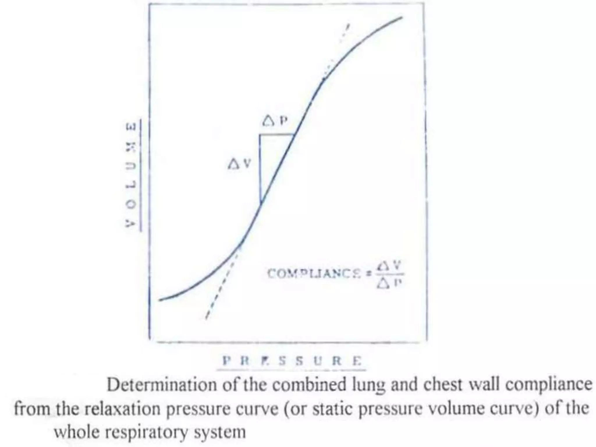
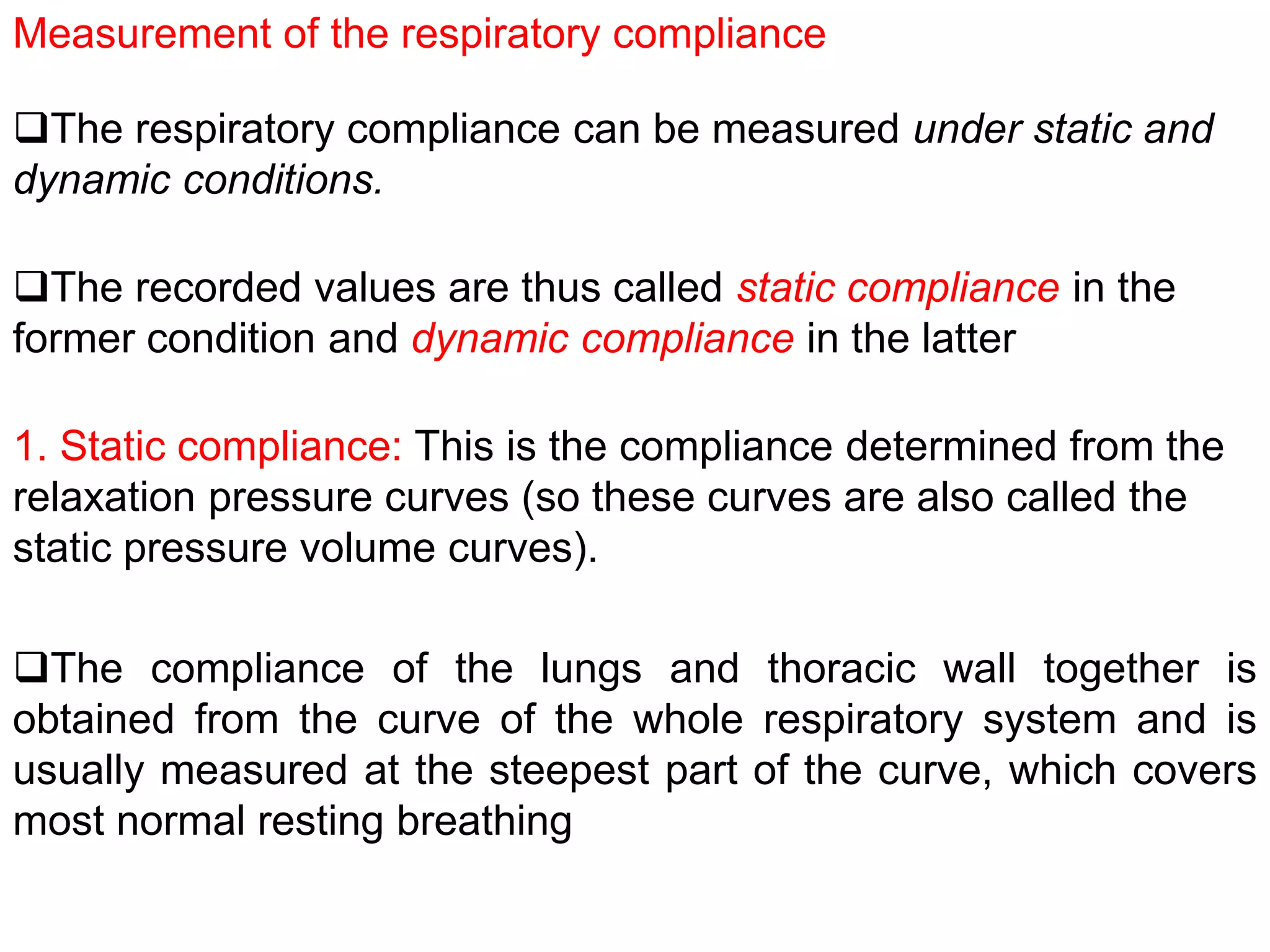
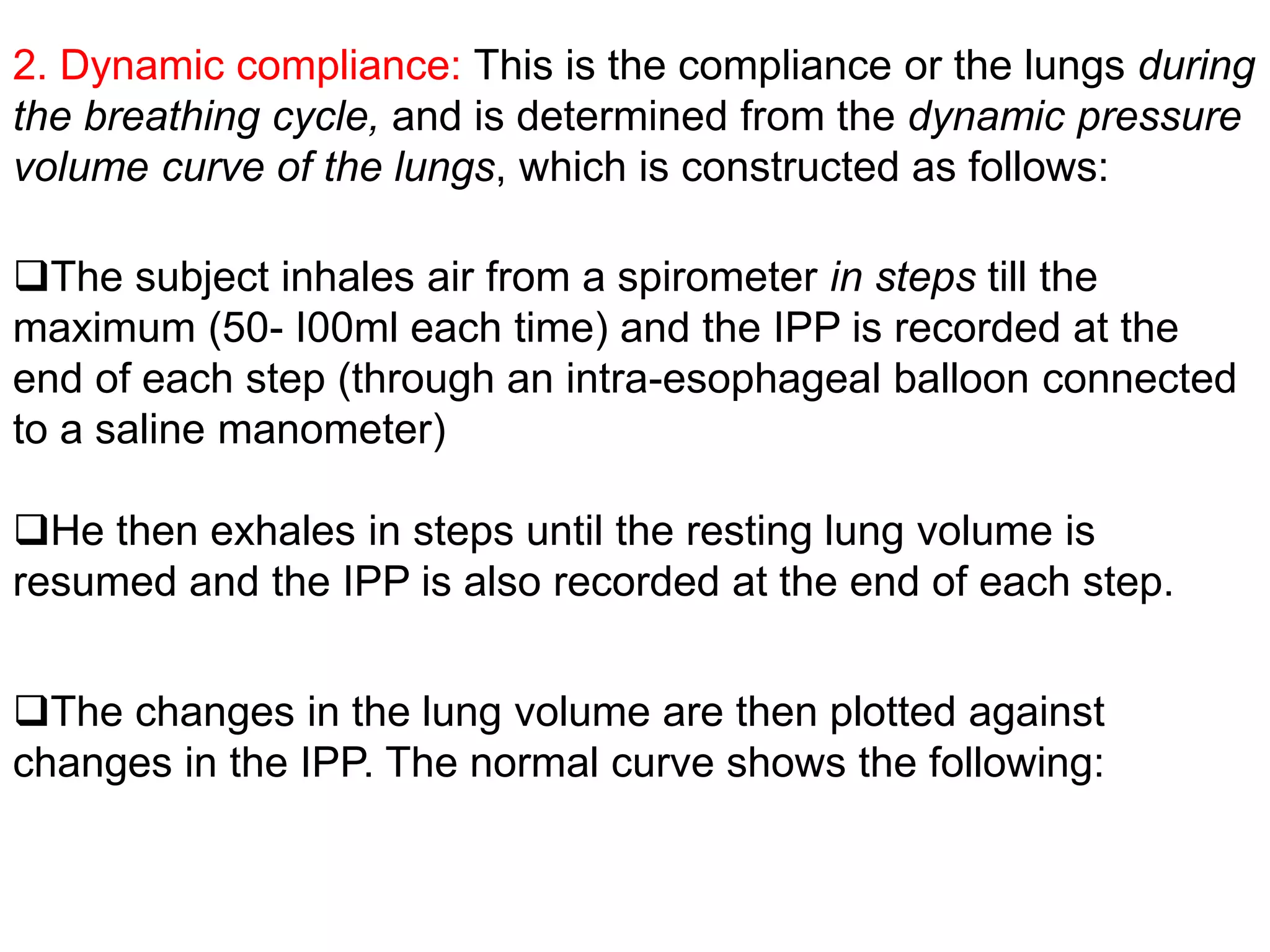
COMPLIANCE OF THE LUNGS AND
CHEST WALL
@
CBU SCHOOL OF MEDICINE
Compliance is a measurere for the distensibility (or expansibility
of elastic structures
lt is expressed as the increase in volume per unit increase
in the distending pressure
The compliance of the lungs alone (when removed outside the
body) is normally about 0.2 litre/cmH20 pressure while that of the
lungs and chest wall together is much less (about 0.1 litre/cmH20
pressure) because the lung's expansibility in the chest is limited
by the rigid thoracic cage.
Measurement of the respiratory compliance
The respiratory compliance can be measured under static and
dynamic conditions.
The recorded values are thus called static compliance in the
former condition and dynamic compliance in the latter
1. Static compliance: This is the compliance determined from the
relaxation pressure curves (so these curves are also called the
static pressure volume curves).
The compliance of the lungs and thoracic wall together is
obtained from the curve of the whole respiratory system and is
usually measured at the steepest part of the curve, which covers
most normal resting breathing
2. Dynamic compliance: This is the compliance or the lungs during
the breathing cycle, and is determined from the dynamic pressure
volume curve of the lungs, which is constructed as follows:
The subject inhales air from a spirometer in steps till the
maximum (50- I00ml each time) and the IPP is recorded at the
end of each step (through an intra-esophageal balloon connected
to a saline manometer)
He then exhales in steps until the resting lung volume is
resumed and the IPP is also recorded at the end of each step.
The changes in the lung volume are then plotted against
changes in the IPP. The normal curve shows the following:
1. It is in the form of a hyteresis loop i.e. the changes in the lung
volume during inflation and deflation are different (which is called
hysteresis) It is due to viscous properties (frictional resistance) in
the lungs.
2. The lung compliance is greater when measured during deflation
3. The dashed line (AYB) represents the average lung
compliance.
** The transpulmonarv (or transmural) pressure = Intrapulmonary
pressure - IPP. It is the pressure that actually affects lung inflation
and deflation. Therefore, it should be plotted against the changes
in the lung volume (and not the IPP)
However, since the intrapulmonary pressure at the end of
each inspiration or expiration is normally 0 mmHg, the IPP will he
equal to the transpulmonary pressure and thus, it can be used
instead.
**A newly born infant normally requires a transpulmonary pressure
of about+ 60 mmHg at the first breath to expand his lungs
Factors that affect lung compliance
1. The size of the lung and its initial volume
The lung compliance depends on its size, thus a lung of a
mouse has a smaller compliance than that of an elephant, and a
patient with one lung has nearly 1/2 normal compliance, and
children have 2-3 times smaller compliance than adults.
2. The surface tension of the alveolar fluid : This decreases the
compliance. Such effect is demonstrated by removing the lung of
a cat outside its body and inflating & deflating it, once with saline
and once with air while measuring the intrapulmonary pressure
Saline decreases the surface tension to nearly zero, and the
pressure-volume curve obtained with saline shows a higher
compliance and less hysteresis, compared with that obtained
when air was used for inflation and deflation of the lung
3. Diseases :The compliance is decreased in the RDS and
diseases that cause lung stiffness(e.g. pulmonary fibrosis, edema
& congestion). In such cases, the pressure-volume curve is
shifted downward & to the right.
**The thoracic compliance is decreased in cases of
(I) Deformities of the vertebral column e.g kyphosis(= antero-
posterior bending) and scoliosis ( lateral bending)
(II) Arthritis of the joints of the thoracic cage or the
vertebral column
(III) Skeletal muscle diseases
(IV)Obesity.
On the other hand, the curve is shifted upward & to the left in
conditions that cause increase of the lung compliance, e.g.
emphysema and old age.
THE WORK OF BREATHING
@
CBU SCHOOL OF MEDICINE
Almost all work of breathing is performed by the inspiratory
muscles because expiration is normally a passive process. lt is
normally 0.3-0.8 KgmeterI minute during eupnea, and its
components include the following :
2. Non-elastic (resistive) work : This is about 35% of the total work
and is used for resisting the frictional resistance to air movement
in the lungs It includes the following types of work:
1. Elastic (compliance) work: This is about 65% of the total work,
and is used for stretching the elastic tissues of the lungs and
chest wall (i.e for overcoming their elastic recoil forces)
a- Airway resistance work (28 %) : This is the work used to overcome 1he
resistance to air flow through the respiratory passages.
b- Tissue resistance work (7 %) : This is the work used to overcome
the viscous resistance in the lungs.
FACTORS THAT INCREASE THE WORK OF BREATHING
2. Reduction of the respiratory compliance (see causes above)
1. Hyperpnea : This may occur normally (e.g. during muscular
exercise) or abnormally (e.g. in cases of acidosis).
3. Deficient secretion of the surfactant (e.g in the RDS)
4. Diseases that increase the resistance to air outflow e,g. asthma
and emphysema (because the expiratory muscles also contract
actively).
EFFECTS OF GRAVITY ON THE
LUNGS
@
CBU SCHOOL OF MEDICINE
EFFECT OF GRAVITY ON VENTILATION
When such spring is freely suspended, it will be stretched
mainly at its upper part, and if pulled down. its length increases
mainly at its Iower part
In the upright position, the lungs are suspended in the thoracic
cavity like a slinky spring
Since the expanded parts are stiffer than the non-expanded
parts, further increases in volume I unit increase in IPP are
smaller in the lung apices than in the lung bases
The lungs are similarly more expanded in their upper pans
during standing. This is because the IPP is more -ve in the upper
parts (-10 cmH20) than in the lower parts (-2 5 cmH20) by the
effect of the lung weight
This causes regional differences in ventilation , which is greatest
in the lung bases and becomes progressively lesser towards their
apices
The IPP is small in the base (-2.5 cmH20), so this region has a
small resting volume (thus it expands markedly during inspiration
because it is situated at the steep part of the pressure volume
curve)
 The figure above shows the normal condition of the lungs
during standing in the midthoracic position (i.e. when they contain
the FRC and the intrapulmonary pressure is zero)
It is noted that after maximal expiration during standing, when
the lungs contain the RV, the IPP becomes- 4 cmH2O at the
apices and + 3. 5 cmH2O at the bases.This results in 2 effects
Conversely, the lPP is high in the apex (-10 cmH2O), so this
region has a large resting volume (thus it expands little during
inspiration).
Conversely, the lung apices are located at the steep part of
the pressure-volume curve, so they are better ventilated when
inspiration starts.
1- The lung bases are compressed and they are ventilated only
when the IPP falls below zero.
This indicates that the distribution of ventilation is reversed at
low lung volumes i.e. the lung apices are ventilated better than the
lung base.
2- Airway closure: The +ve lPP at the lung bases causes collapse
of the small airways in these parts (e.g. the respiratory
bronchioles) and trapping of a volume of air in the alveoli
The lung volume above the RV at which the small airways at the
lung bases begin to close is called the closing volume.
(2) EFFECT OF GRAVITY ON PULMONARY BLOOD FLOW
It is decreased in the lung apices to the extent that raises the
V/P ratio to 3.3. On the other hand, the increase in blood flow at
the lung bases reduces the V/P ratio there to 0.63.
Due to gravity, the pulmonary blood flow at the lung apices
during standing is much less than that at their bases.
PNEUMOTHORAX
@
CBU SCHOOL OF MEDICINE
The IPP increases, leading to collapse of the lung on the
affected side and shift of the mediastinum to the normal side.
This is entry of air inside the pleural cavity. This may occur due
to an external cause (e.g. injury of the thoracic wall) or an internal
cause (e.g.rupture of the alveoli into the pleural sac)
The condition is fatal if bilateral, and it is several types:
The shift of the mediastinum to the normal side increases the
resistance to air flow in the intact lung and leads to kinking of the
great vessels.
1. Closed pneumothorax: This is the mildest type, in which the
hole in the pleural sac seals off and the air that entered the pleural
cavity is gradually absorbed (so the collapsed lung will slowly re-
expand)
2. Open (sucking) pneumothorax: In this type, the pleural cavity
remains communicated to the external atmosphere, so the IPP
becomes atmospheric.
Respiratory distress is severe and respiration is stimulated by
(a) The developed hypoxia and hypercapnia
(b) Activation of the deflation receptors in the collapsed lung
Kinking of the great veins causes distension of the peripheral
veins and cyanosis. Shock may finally occur, which may lead to
death.
3. Tension (valvular) pneumothorax: In this type, there is a flap of
tissue over the hole in the pleural sac, which acts as a flutter valve
that permits air to enter during inspiration but prevents its escape
during expiration, so the IPP rises above atmospheric.
The hypoxia is more severe, and respiration is excessively
stimulated, which further increases the IPP (up to + 20 or + 30
mmHg)
However, this procedure is rarely performed now.
4. Artificial pneumothorax: This is a therapeutic procedure that is
performed to obliterate lung cavities that may form in many
diseases (specially tuberculosis).
Air is injected into the pleural sac in the region facing the cavity.
This leads to local collapse of the lung, which obliterates the
cavity and helps rapid healing.

Compliance of the lungs and chest wall

  • 1.
    COMPLIANCE OF THELUNGS AND CHEST WALL @ CBU SCHOOL OF MEDICINE
  • 2.
    Compliance is ameasurere for the distensibility (or expansibility of elastic structures lt is expressed as the increase in volume per unit increase in the distending pressure The compliance of the lungs alone (when removed outside the body) is normally about 0.2 litre/cmH20 pressure while that of the lungs and chest wall together is much less (about 0.1 litre/cmH20 pressure) because the lung's expansibility in the chest is limited by the rigid thoracic cage.
  • 4.
    Measurement of therespiratory compliance The respiratory compliance can be measured under static and dynamic conditions. The recorded values are thus called static compliance in the former condition and dynamic compliance in the latter 1. Static compliance: This is the compliance determined from the relaxation pressure curves (so these curves are also called the static pressure volume curves). The compliance of the lungs and thoracic wall together is obtained from the curve of the whole respiratory system and is usually measured at the steepest part of the curve, which covers most normal resting breathing
  • 5.
    2. Dynamic compliance:This is the compliance or the lungs during the breathing cycle, and is determined from the dynamic pressure volume curve of the lungs, which is constructed as follows: The subject inhales air from a spirometer in steps till the maximum (50- I00ml each time) and the IPP is recorded at the end of each step (through an intra-esophageal balloon connected to a saline manometer) He then exhales in steps until the resting lung volume is resumed and the IPP is also recorded at the end of each step. The changes in the lung volume are then plotted against changes in the IPP. The normal curve shows the following:
  • 7.
    1. It isin the form of a hyteresis loop i.e. the changes in the lung volume during inflation and deflation are different (which is called hysteresis) It is due to viscous properties (frictional resistance) in the lungs. 2. The lung compliance is greater when measured during deflation 3. The dashed line (AYB) represents the average lung compliance.
  • 8.
    ** The transpulmonarv(or transmural) pressure = Intrapulmonary pressure - IPP. It is the pressure that actually affects lung inflation and deflation. Therefore, it should be plotted against the changes in the lung volume (and not the IPP) However, since the intrapulmonary pressure at the end of each inspiration or expiration is normally 0 mmHg, the IPP will he equal to the transpulmonary pressure and thus, it can be used instead. **A newly born infant normally requires a transpulmonary pressure of about+ 60 mmHg at the first breath to expand his lungs
  • 9.
    Factors that affectlung compliance 1. The size of the lung and its initial volume The lung compliance depends on its size, thus a lung of a mouse has a smaller compliance than that of an elephant, and a patient with one lung has nearly 1/2 normal compliance, and children have 2-3 times smaller compliance than adults. 2. The surface tension of the alveolar fluid : This decreases the compliance. Such effect is demonstrated by removing the lung of a cat outside its body and inflating & deflating it, once with saline and once with air while measuring the intrapulmonary pressure Saline decreases the surface tension to nearly zero, and the pressure-volume curve obtained with saline shows a higher compliance and less hysteresis, compared with that obtained when air was used for inflation and deflation of the lung
  • 10.
    3. Diseases :Thecompliance is decreased in the RDS and diseases that cause lung stiffness(e.g. pulmonary fibrosis, edema & congestion). In such cases, the pressure-volume curve is shifted downward & to the right. **The thoracic compliance is decreased in cases of (I) Deformities of the vertebral column e.g kyphosis(= antero- posterior bending) and scoliosis ( lateral bending) (II) Arthritis of the joints of the thoracic cage or the vertebral column (III) Skeletal muscle diseases (IV)Obesity. On the other hand, the curve is shifted upward & to the left in conditions that cause increase of the lung compliance, e.g. emphysema and old age.
  • 11.
    THE WORK OFBREATHING @ CBU SCHOOL OF MEDICINE
  • 12.
    Almost all workof breathing is performed by the inspiratory muscles because expiration is normally a passive process. lt is normally 0.3-0.8 KgmeterI minute during eupnea, and its components include the following : 2. Non-elastic (resistive) work : This is about 35% of the total work and is used for resisting the frictional resistance to air movement in the lungs It includes the following types of work: 1. Elastic (compliance) work: This is about 65% of the total work, and is used for stretching the elastic tissues of the lungs and chest wall (i.e for overcoming their elastic recoil forces) a- Airway resistance work (28 %) : This is the work used to overcome 1he resistance to air flow through the respiratory passages. b- Tissue resistance work (7 %) : This is the work used to overcome the viscous resistance in the lungs.
  • 13.
    FACTORS THAT INCREASETHE WORK OF BREATHING 2. Reduction of the respiratory compliance (see causes above) 1. Hyperpnea : This may occur normally (e.g. during muscular exercise) or abnormally (e.g. in cases of acidosis). 3. Deficient secretion of the surfactant (e.g in the RDS) 4. Diseases that increase the resistance to air outflow e,g. asthma and emphysema (because the expiratory muscles also contract actively).
  • 14.
    EFFECTS OF GRAVITYON THE LUNGS @ CBU SCHOOL OF MEDICINE
  • 15.
    EFFECT OF GRAVITYON VENTILATION When such spring is freely suspended, it will be stretched mainly at its upper part, and if pulled down. its length increases mainly at its Iower part In the upright position, the lungs are suspended in the thoracic cavity like a slinky spring Since the expanded parts are stiffer than the non-expanded parts, further increases in volume I unit increase in IPP are smaller in the lung apices than in the lung bases The lungs are similarly more expanded in their upper pans during standing. This is because the IPP is more -ve in the upper parts (-10 cmH20) than in the lower parts (-2 5 cmH20) by the effect of the lung weight
  • 16.
    This causes regionaldifferences in ventilation , which is greatest in the lung bases and becomes progressively lesser towards their apices
  • 17.
    The IPP issmall in the base (-2.5 cmH20), so this region has a small resting volume (thus it expands markedly during inspiration because it is situated at the steep part of the pressure volume curve)  The figure above shows the normal condition of the lungs during standing in the midthoracic position (i.e. when they contain the FRC and the intrapulmonary pressure is zero) It is noted that after maximal expiration during standing, when the lungs contain the RV, the IPP becomes- 4 cmH2O at the apices and + 3. 5 cmH2O at the bases.This results in 2 effects Conversely, the lPP is high in the apex (-10 cmH2O), so this region has a large resting volume (thus it expands little during inspiration).
  • 18.
    Conversely, the lungapices are located at the steep part of the pressure-volume curve, so they are better ventilated when inspiration starts. 1- The lung bases are compressed and they are ventilated only when the IPP falls below zero. This indicates that the distribution of ventilation is reversed at low lung volumes i.e. the lung apices are ventilated better than the lung base.
  • 19.
    2- Airway closure:The +ve lPP at the lung bases causes collapse of the small airways in these parts (e.g. the respiratory bronchioles) and trapping of a volume of air in the alveoli The lung volume above the RV at which the small airways at the lung bases begin to close is called the closing volume.
  • 20.
    (2) EFFECT OFGRAVITY ON PULMONARY BLOOD FLOW It is decreased in the lung apices to the extent that raises the V/P ratio to 3.3. On the other hand, the increase in blood flow at the lung bases reduces the V/P ratio there to 0.63. Due to gravity, the pulmonary blood flow at the lung apices during standing is much less than that at their bases.
  • 21.
  • 22.
    The IPP increases,leading to collapse of the lung on the affected side and shift of the mediastinum to the normal side. This is entry of air inside the pleural cavity. This may occur due to an external cause (e.g. injury of the thoracic wall) or an internal cause (e.g.rupture of the alveoli into the pleural sac) The condition is fatal if bilateral, and it is several types:
  • 23.
    The shift ofthe mediastinum to the normal side increases the resistance to air flow in the intact lung and leads to kinking of the great vessels. 1. Closed pneumothorax: This is the mildest type, in which the hole in the pleural sac seals off and the air that entered the pleural cavity is gradually absorbed (so the collapsed lung will slowly re- expand) 2. Open (sucking) pneumothorax: In this type, the pleural cavity remains communicated to the external atmosphere, so the IPP becomes atmospheric. Respiratory distress is severe and respiration is stimulated by (a) The developed hypoxia and hypercapnia (b) Activation of the deflation receptors in the collapsed lung
  • 24.
    Kinking of thegreat veins causes distension of the peripheral veins and cyanosis. Shock may finally occur, which may lead to death. 3. Tension (valvular) pneumothorax: In this type, there is a flap of tissue over the hole in the pleural sac, which acts as a flutter valve that permits air to enter during inspiration but prevents its escape during expiration, so the IPP rises above atmospheric. The hypoxia is more severe, and respiration is excessively stimulated, which further increases the IPP (up to + 20 or + 30 mmHg)
  • 25.
    However, this procedureis rarely performed now. 4. Artificial pneumothorax: This is a therapeutic procedure that is performed to obliterate lung cavities that may form in many diseases (specially tuberculosis). Air is injected into the pleural sac in the region facing the cavity. This leads to local collapse of the lung, which obliterates the cavity and helps rapid healing.