Downloaded 830 times


This document provides an overview of the anatomy of the lumbosacral plexus, which is formed from the combination of the lumbar and sacral plexuses. It describes the roots, branches, divisions, and terminal branches that form the various nerves. These include the femoral, obturator, superior gluteal, inferior gluteal, and sciatic nerves. It also outlines the motor and sensory distributions of the nerves of the lumbosacral plexus to the lower limbs and related structures.
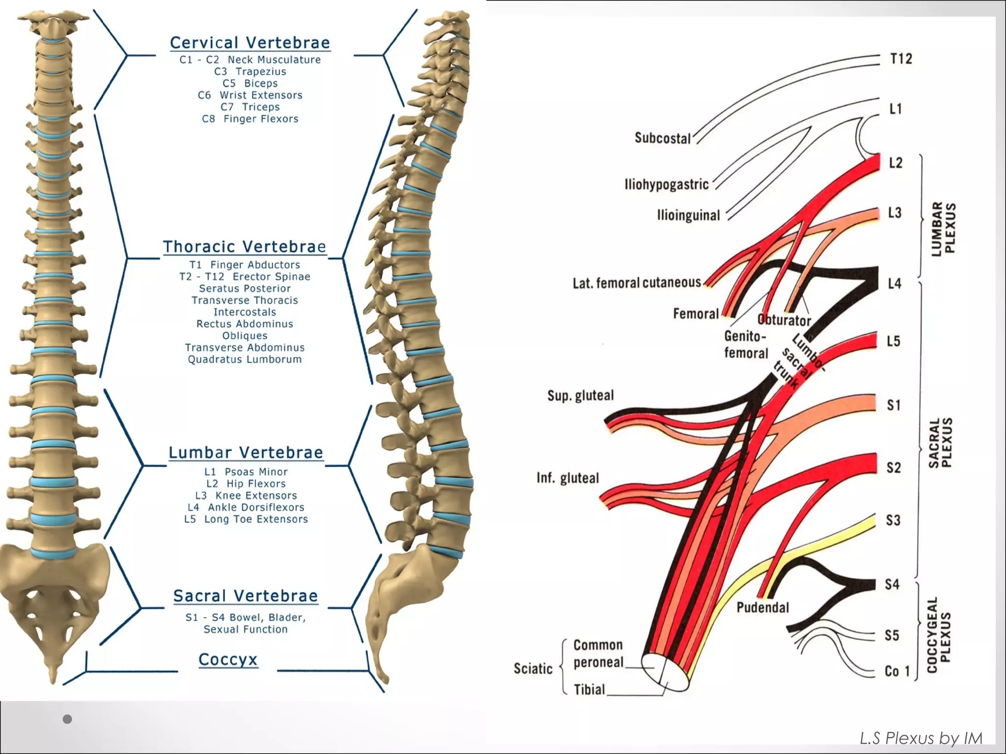
Introduces the topic by outlining spinal cord anatomy and components of the lumbar and sacral plexuses.
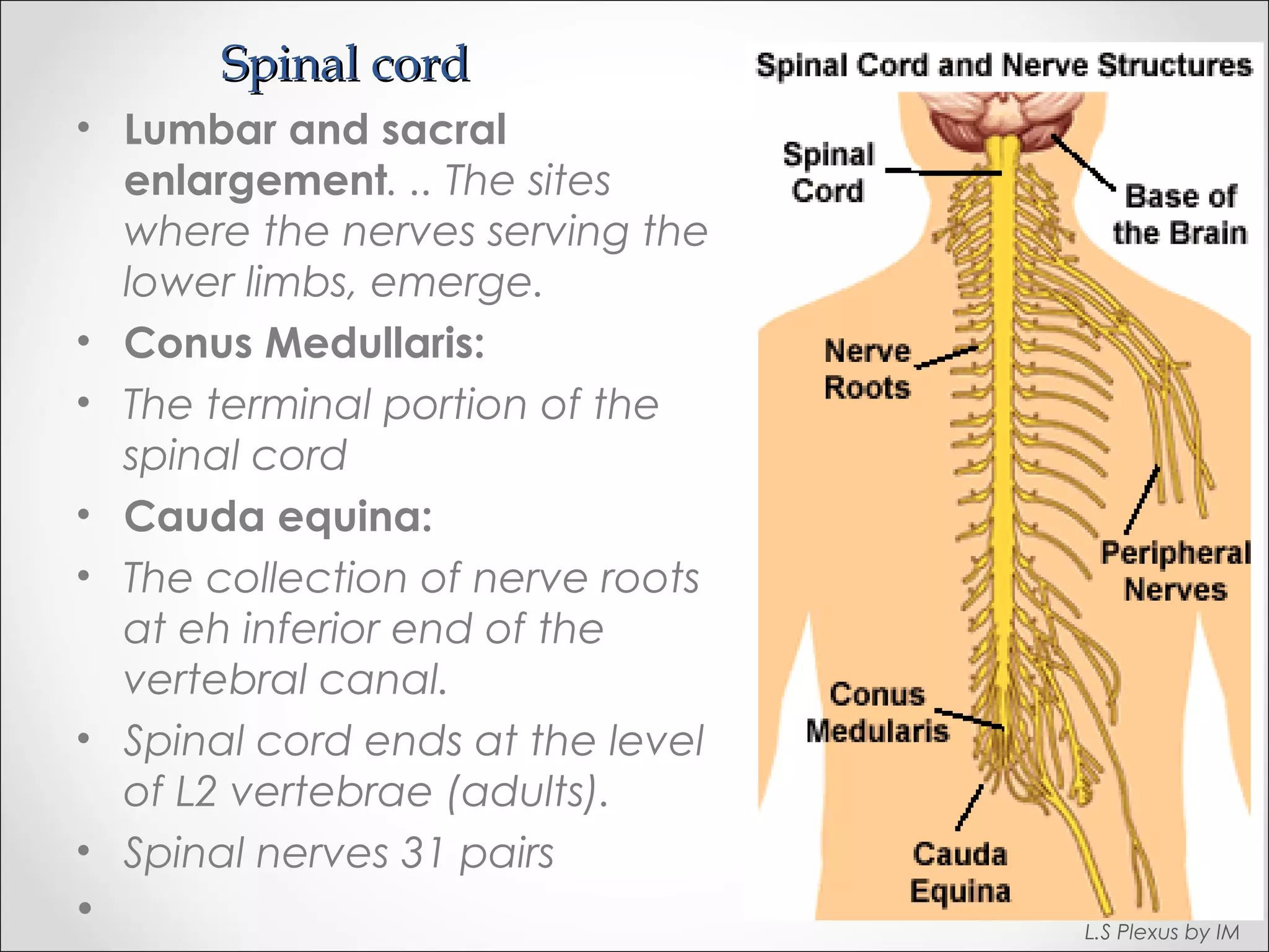
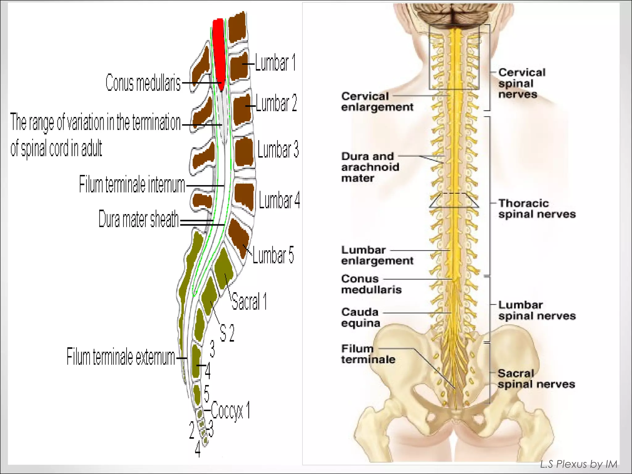
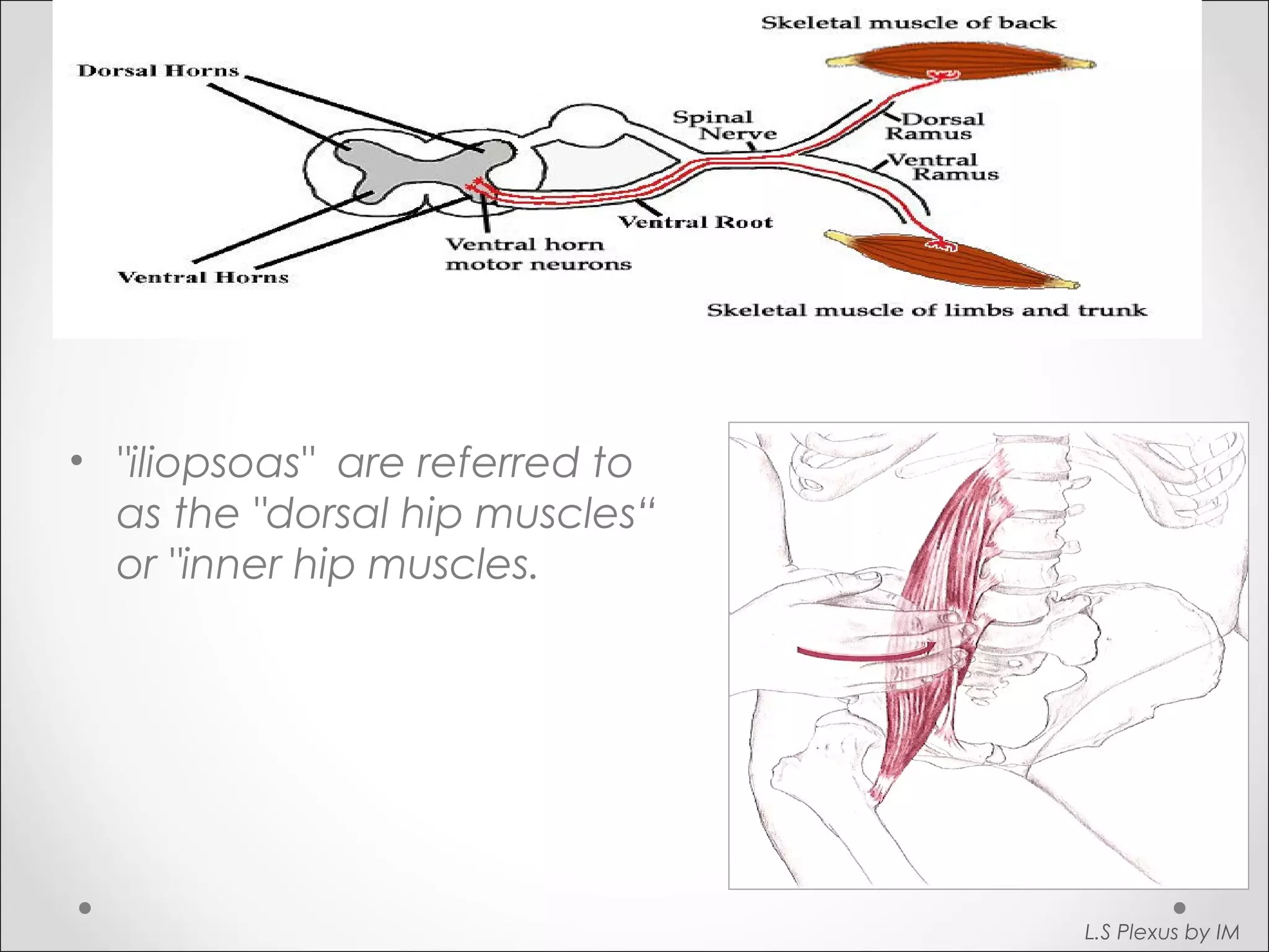
Details the lumbar and sacral enlargements, conus medullaris, and cauda equina in the spinal cord.
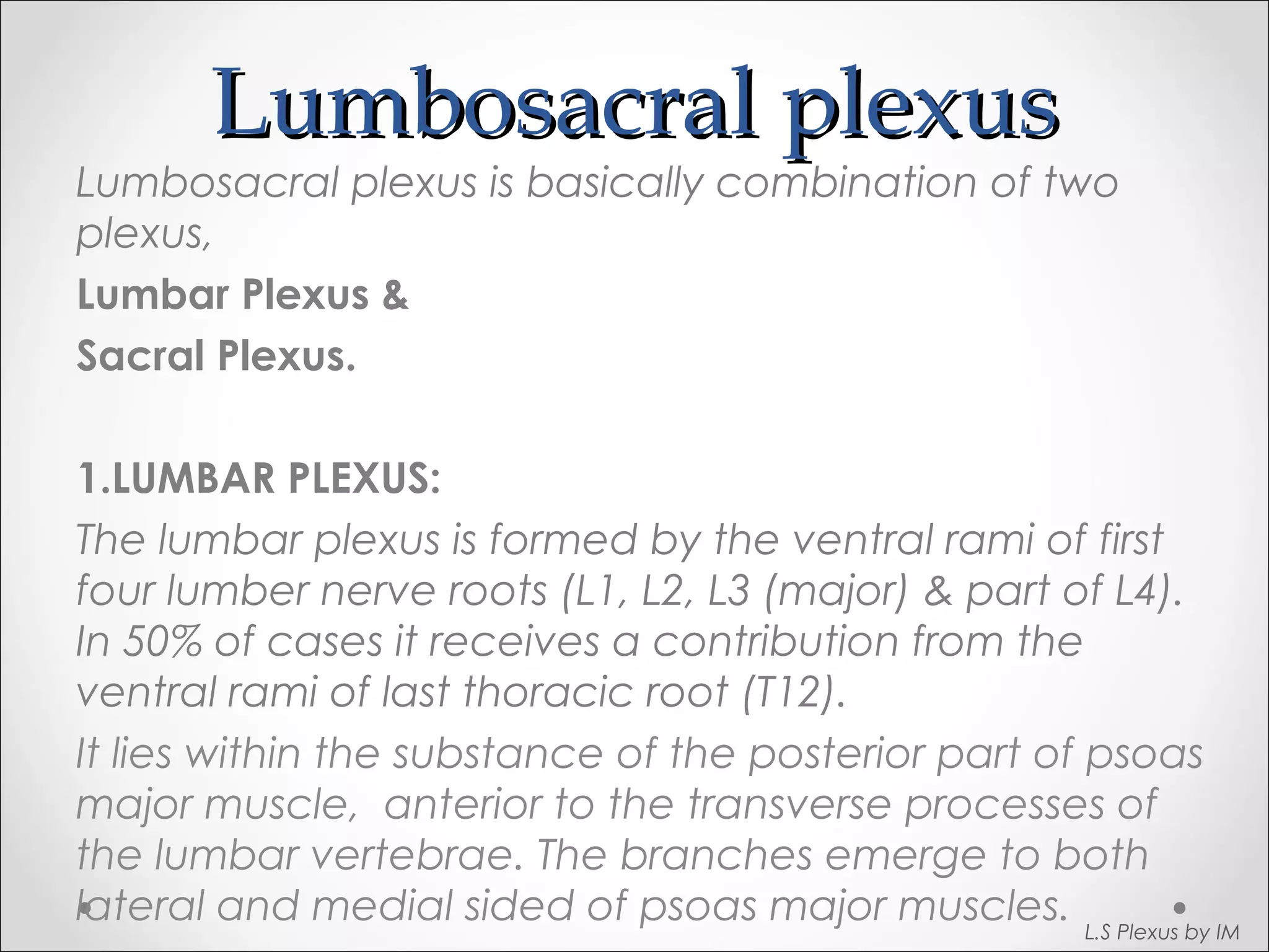
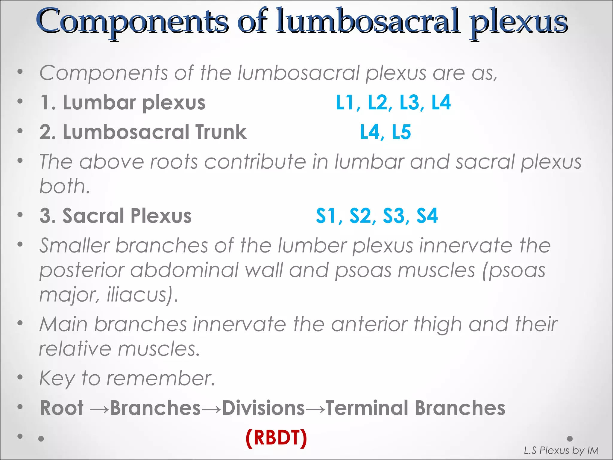
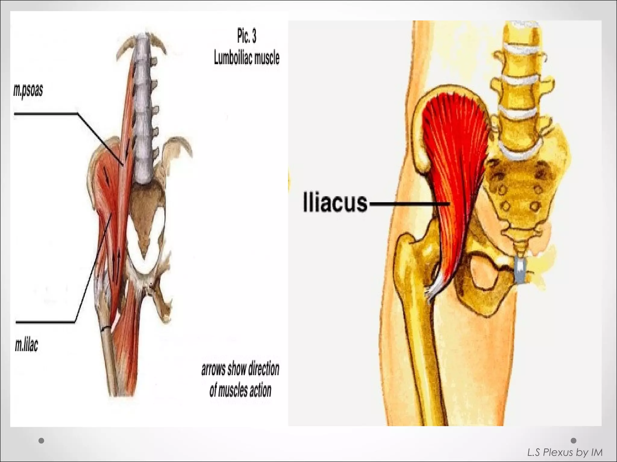
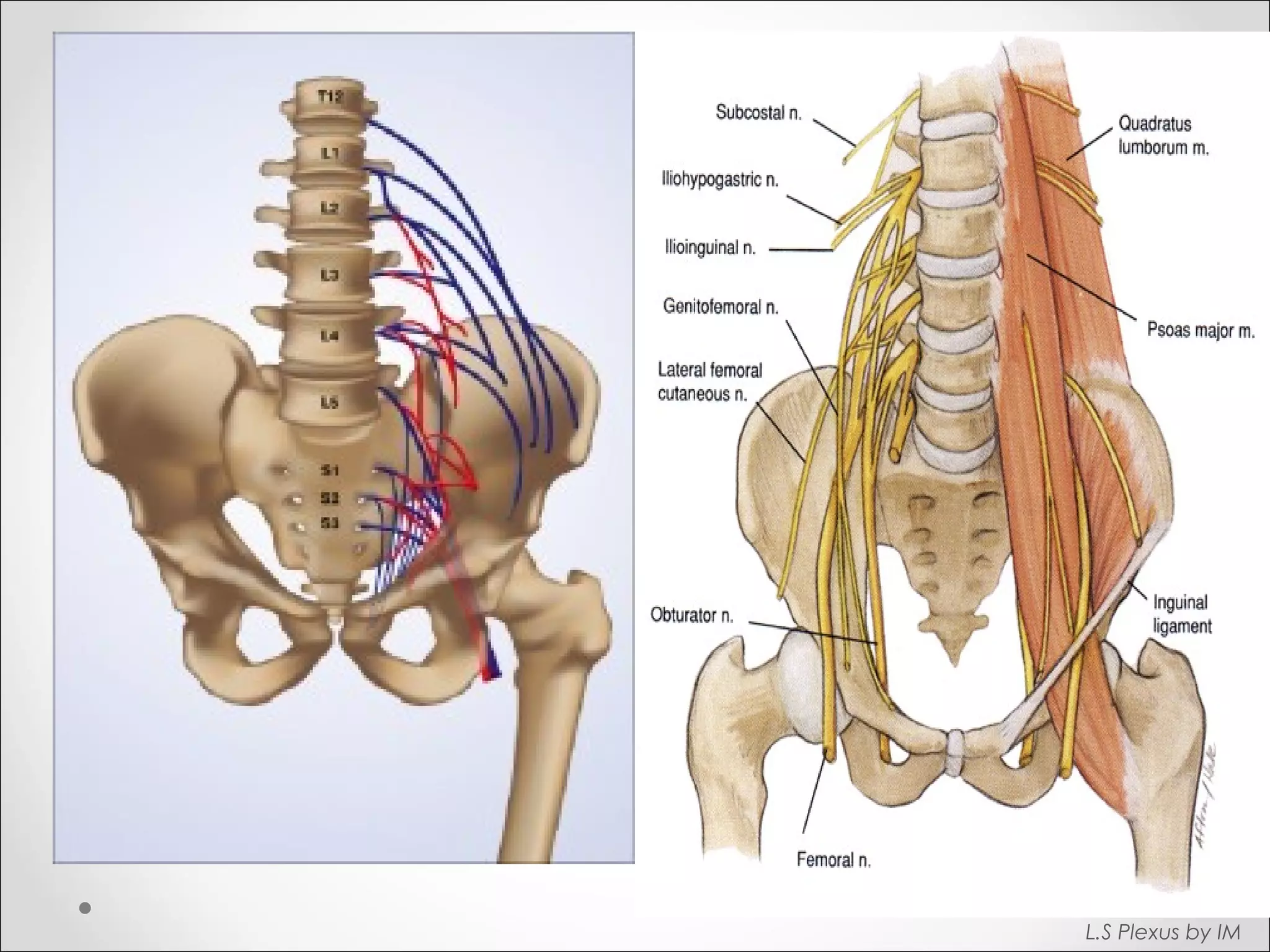
Describes the lumbosacral plexus, its combined structure of lumbar and sacral plexus, and key components.
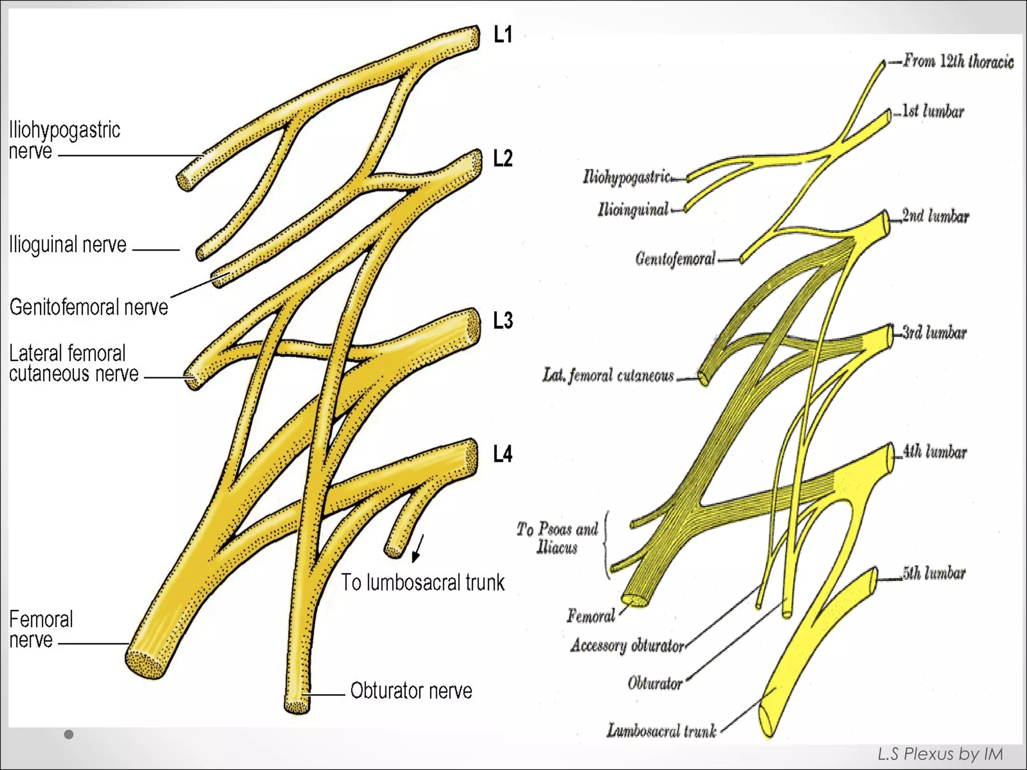
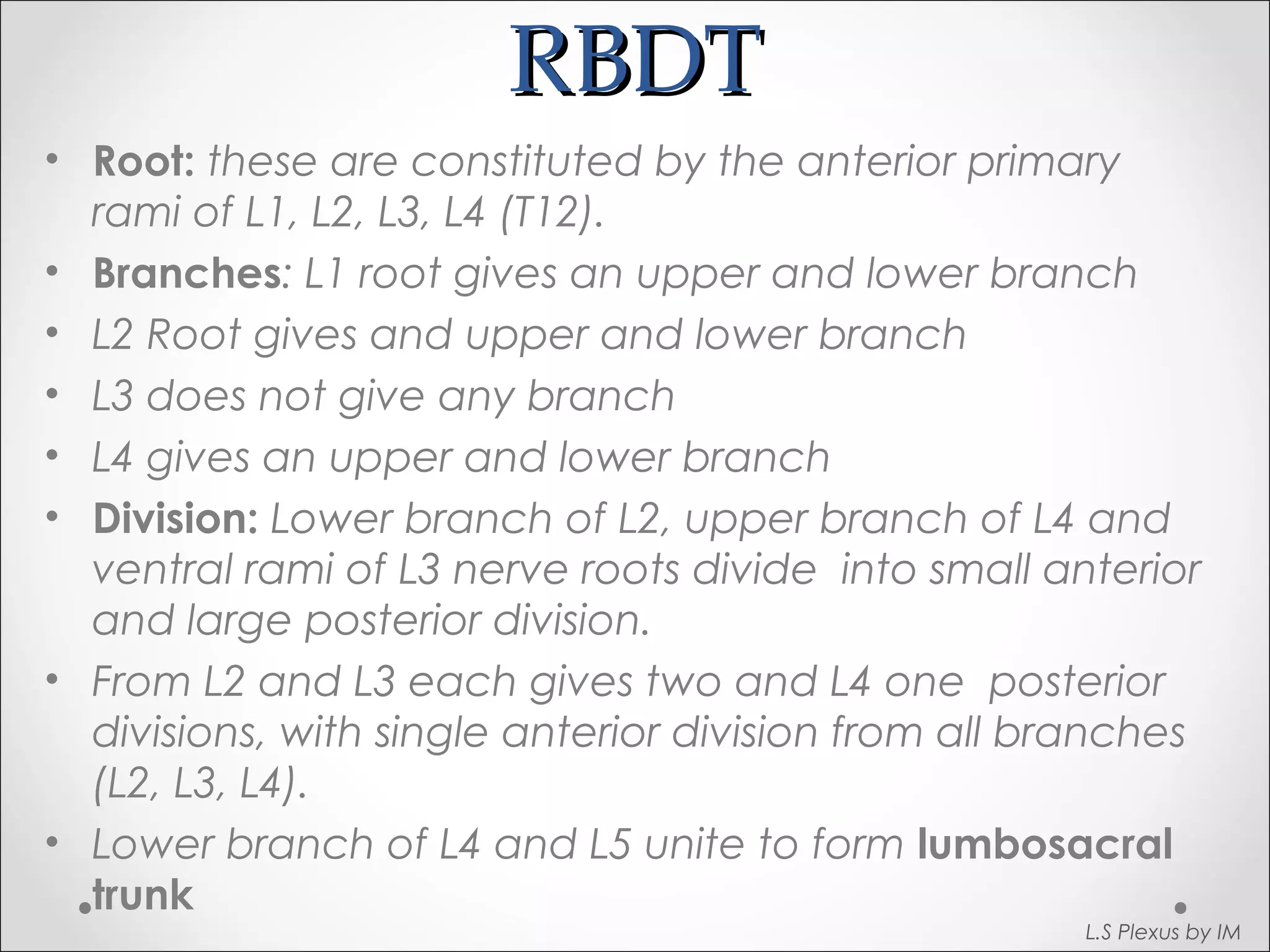
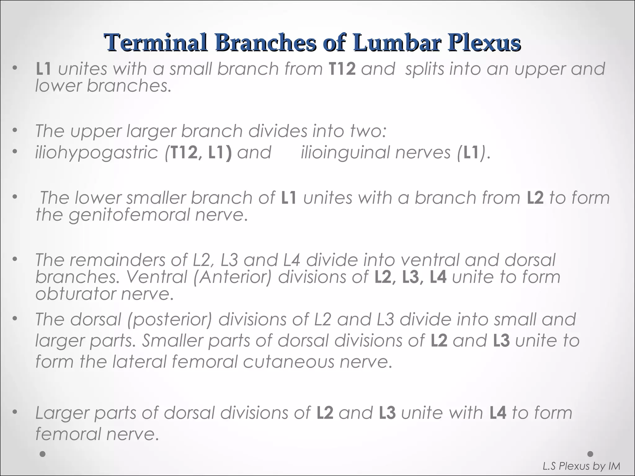
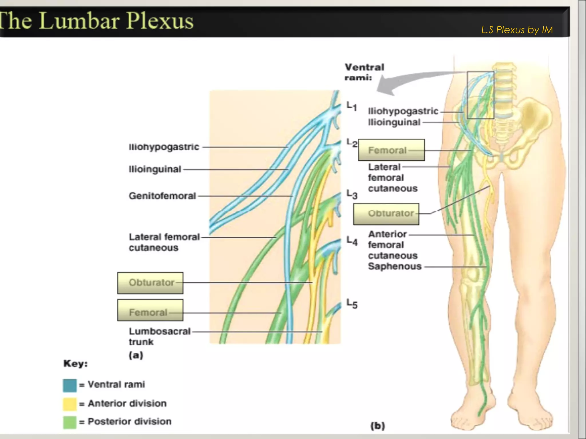
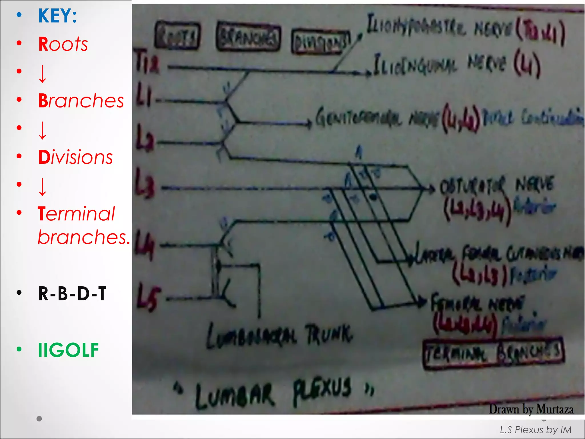
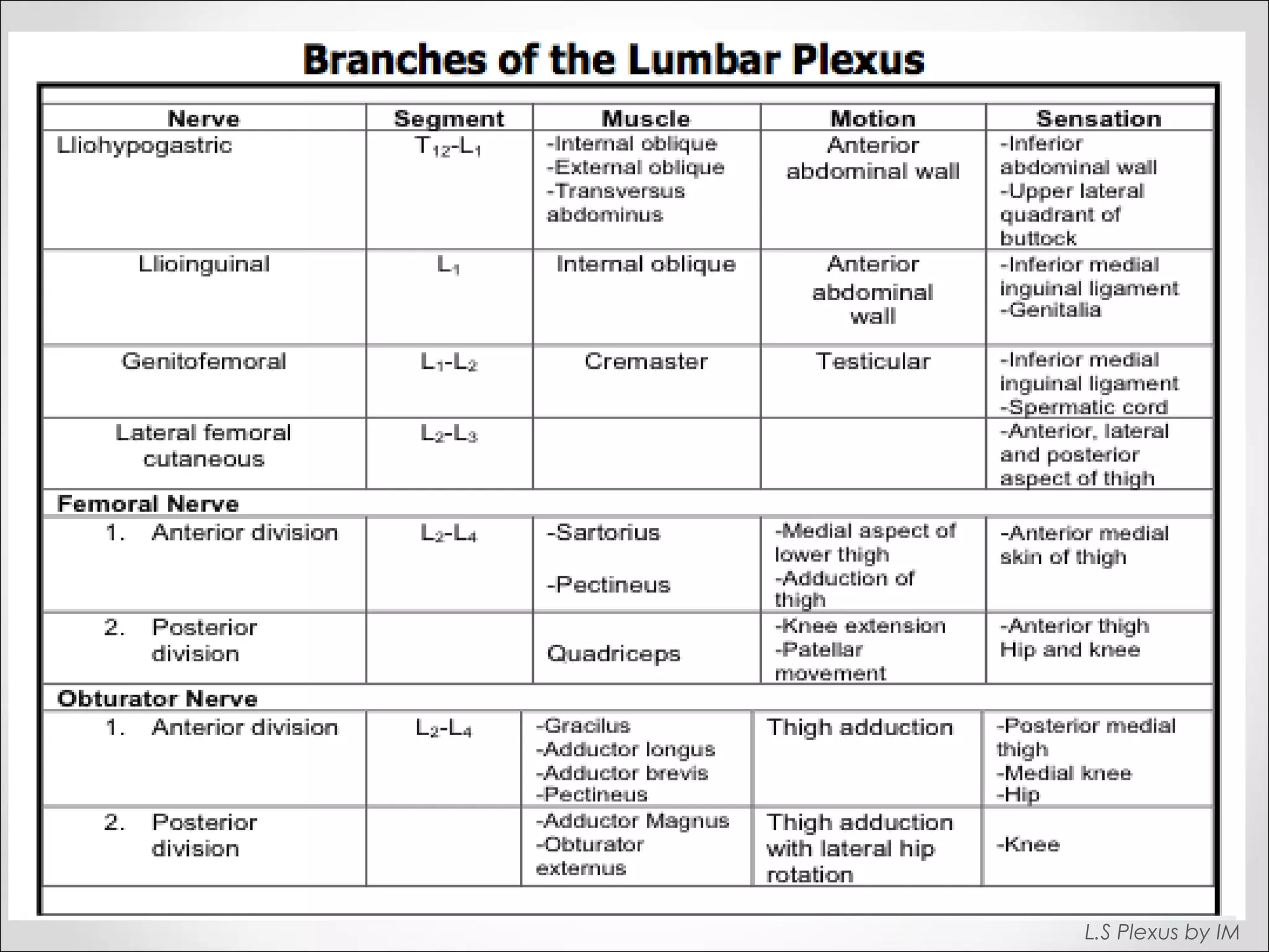
Explains root, branches, divisions, and terminal branches (R-B-D-T) of lumbar plexus with a focus on major nerves.
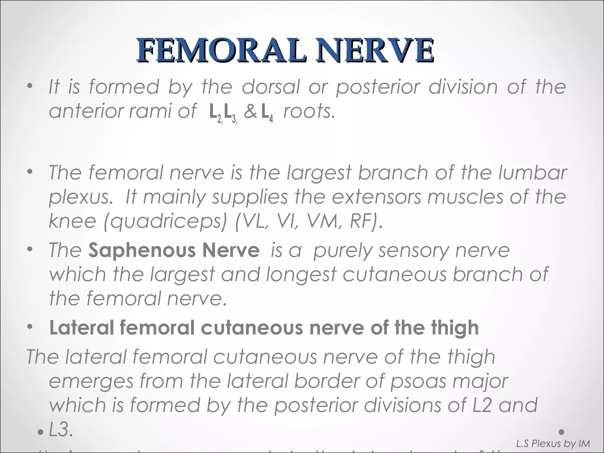
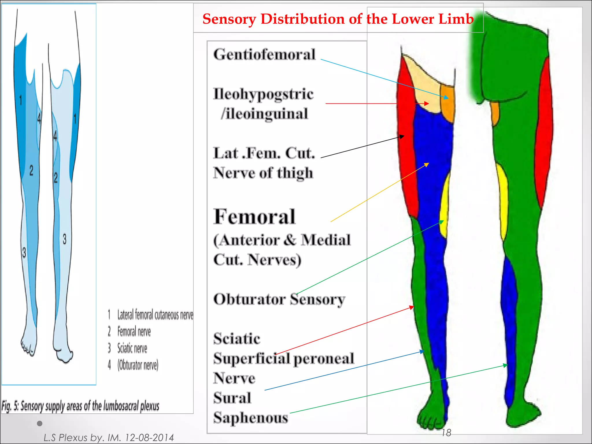
Details the formation and function of the femoral nerve and its sensory branches like the saphenous nerve.
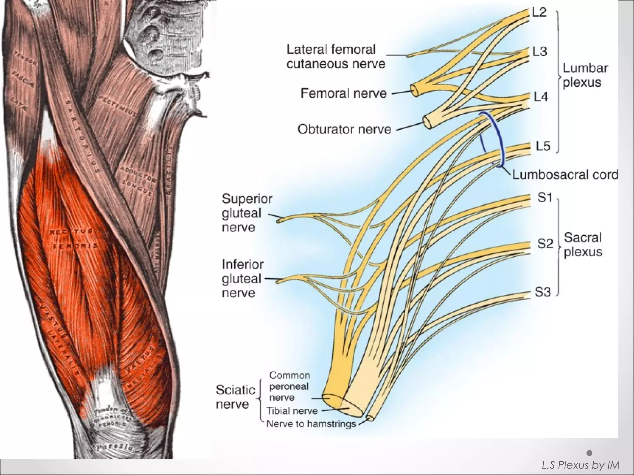
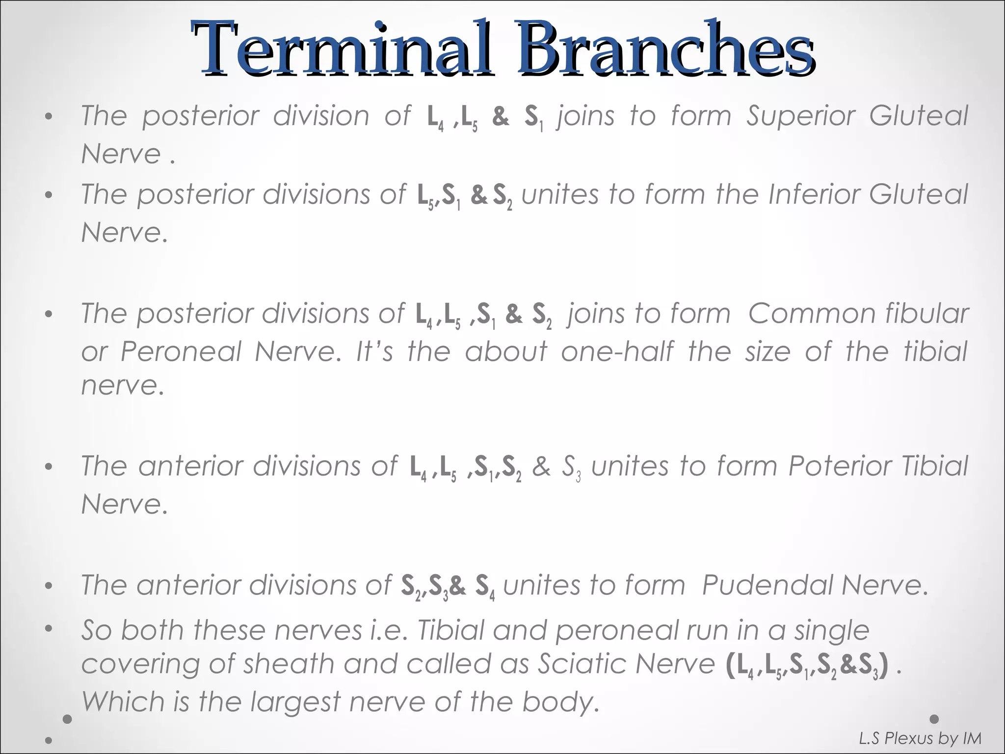
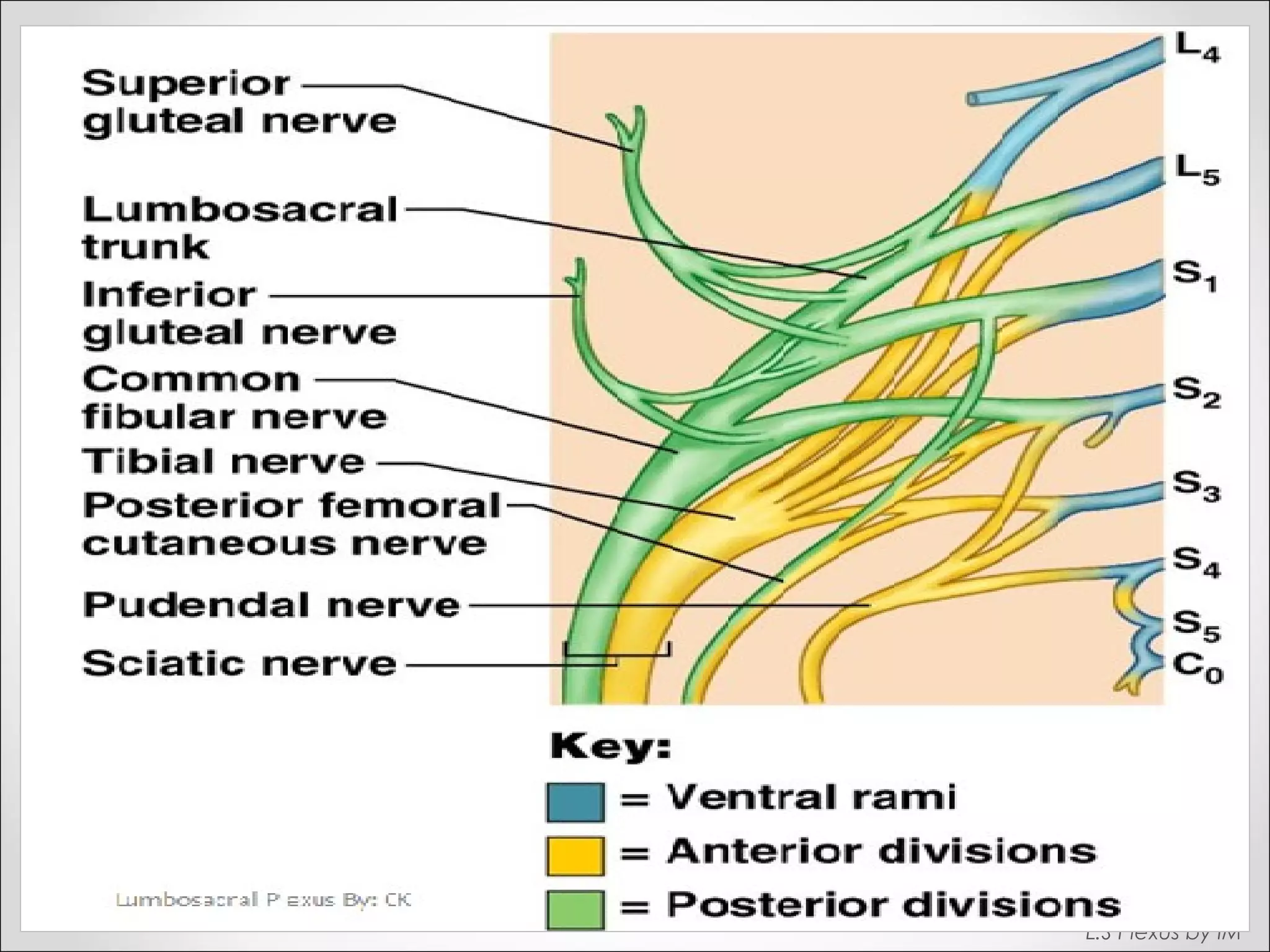
Explains formation of the sacral plexus and its terminal branches, including gluteal and sciatic nerves.
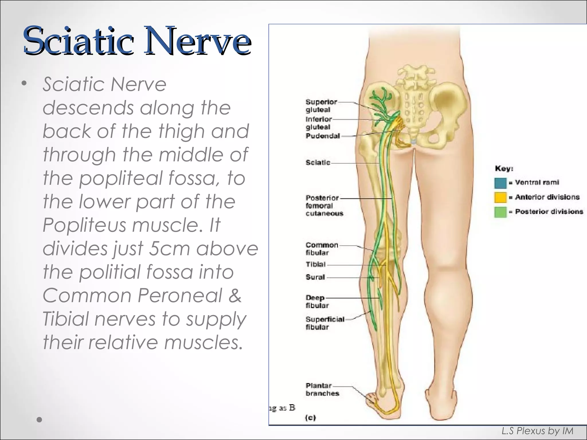
Describes the pathway of the sciatic nerve and its division into common peroneal and tibial nerves.
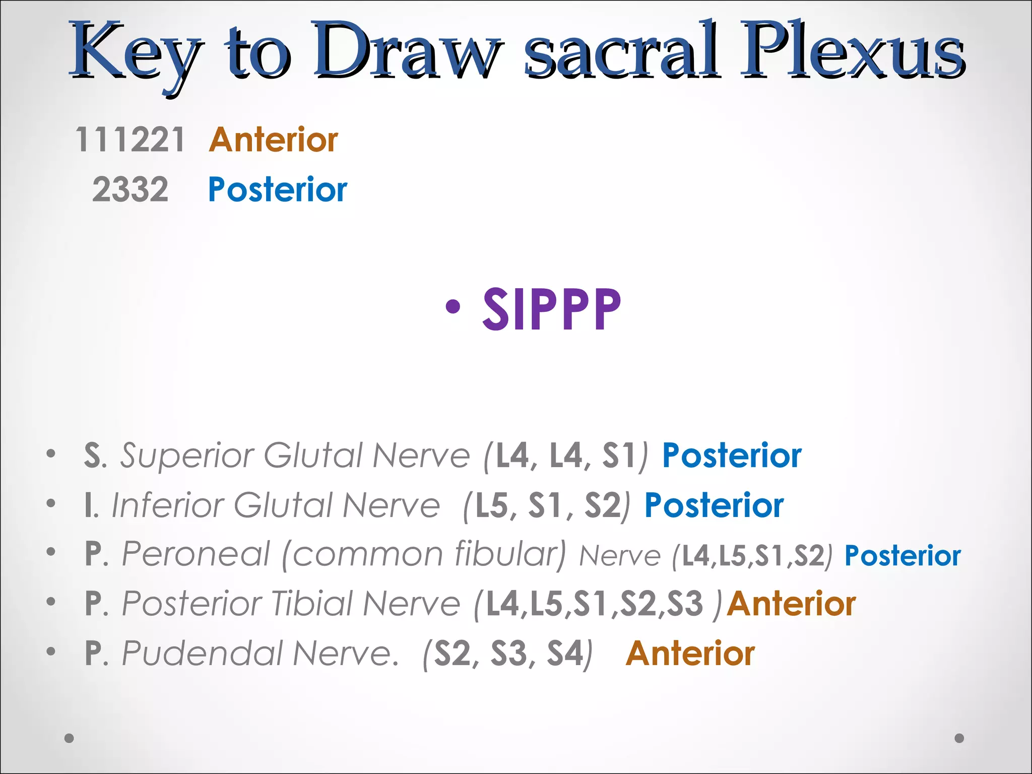
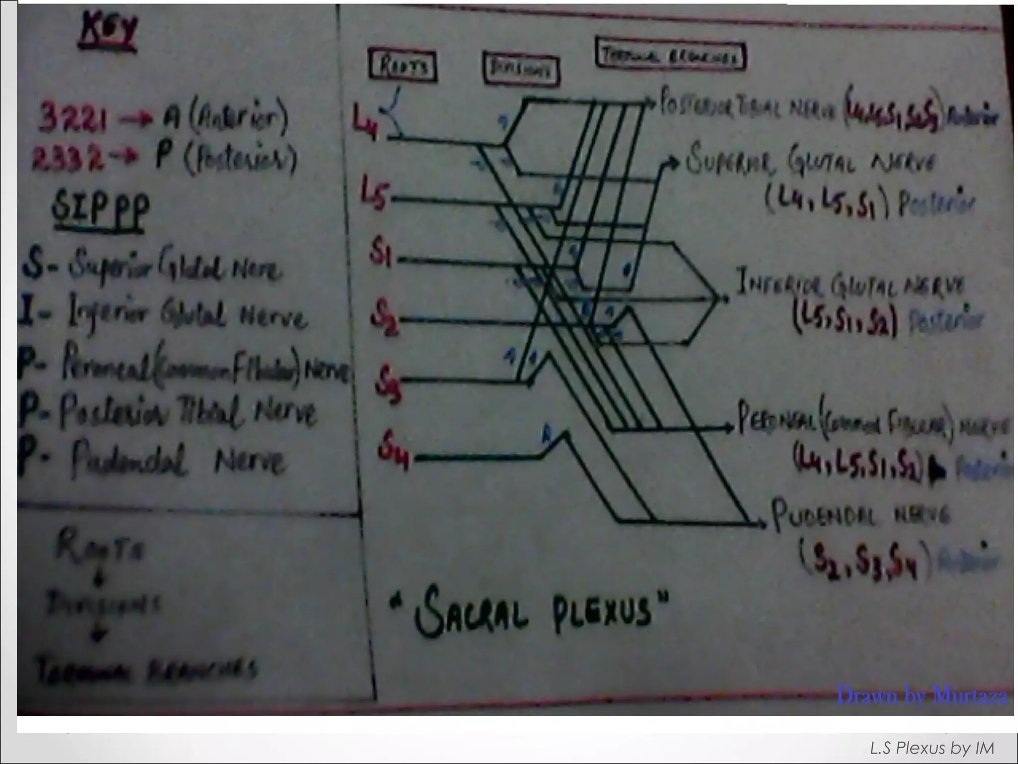
Provides mnemonics for identifying the key terminal branches of the sacral plexus for better understanding.
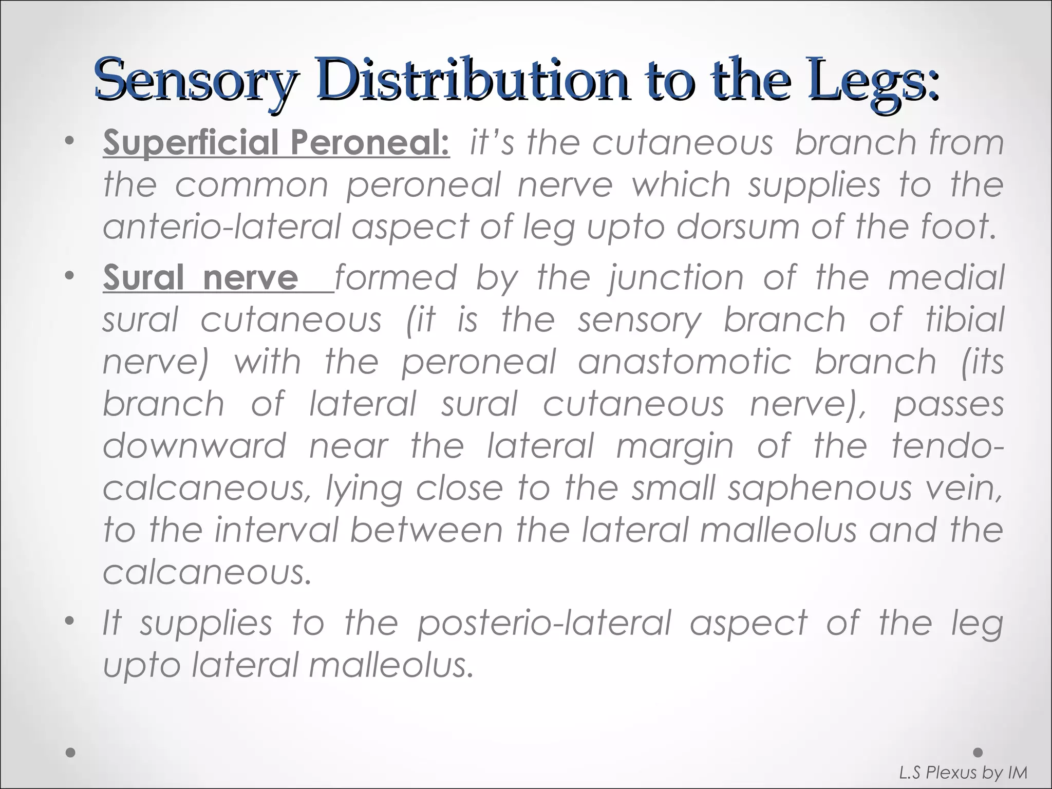
Explains the cutaneous branches from nerves supplying sensory innervation to the posterior and lateral aspects of the legs.
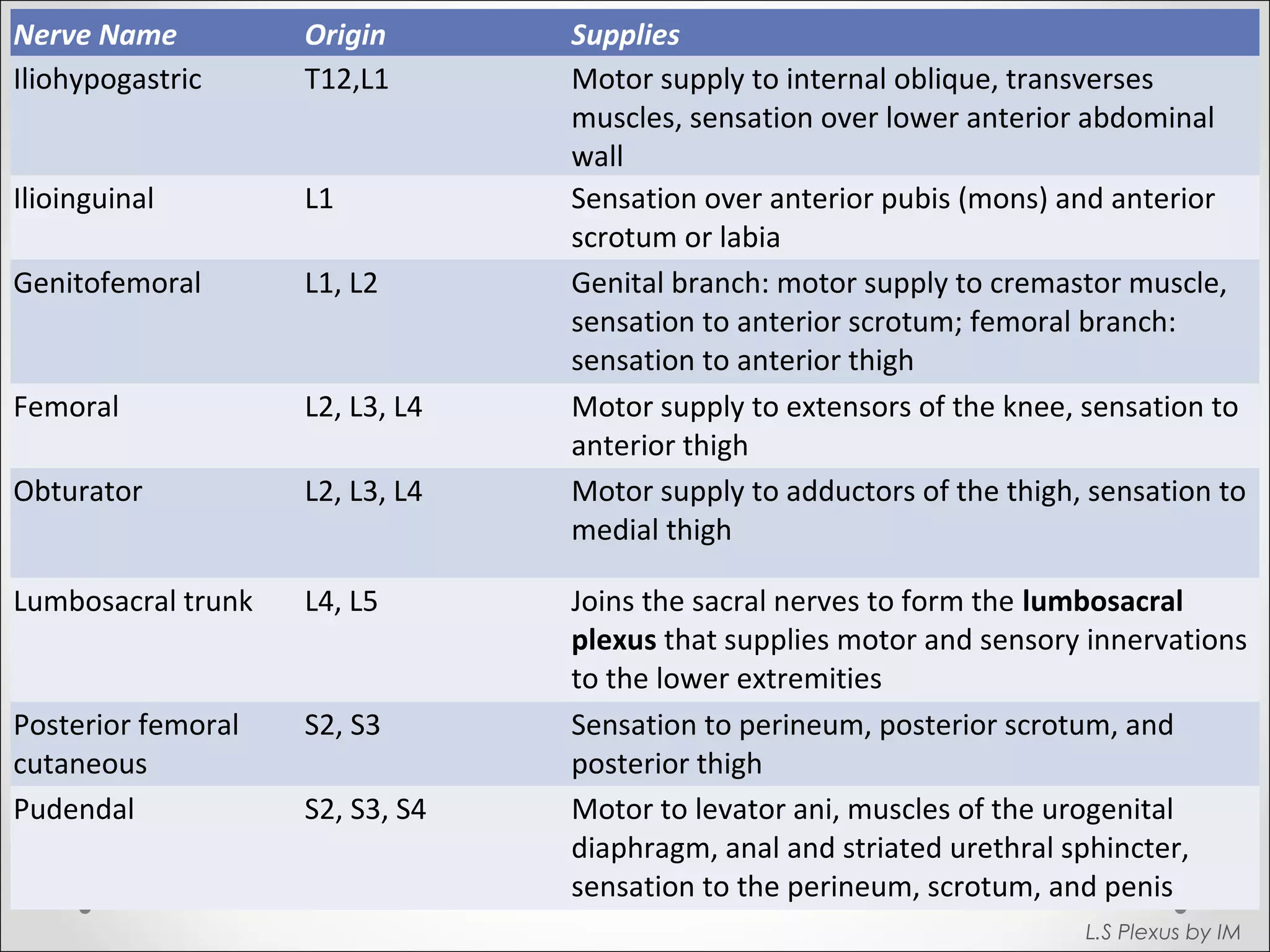
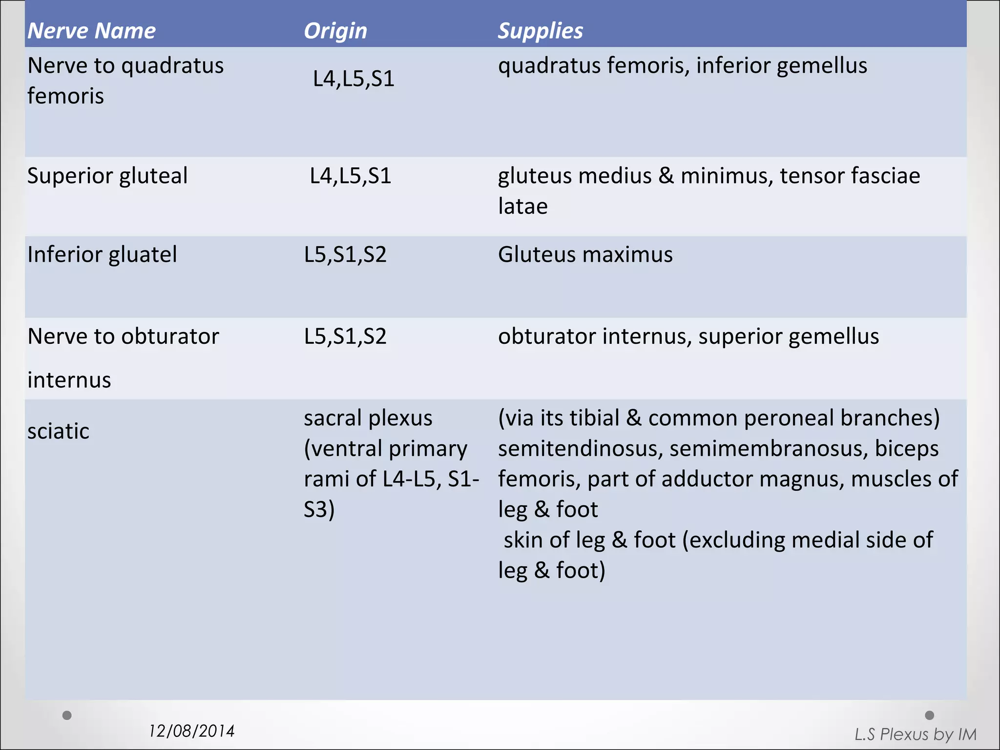
Lists various nerves, their origins, and the muscles or areas they supply, providing a comprehensive overview of innervations.
Concludes the discussion with acknowledgment of key references and thanks for the audience's patience.


![1. brachial plexus & its applied anatomy[1]](https://cdn.slidesharecdn.com/ss_thumbnails/1-brachialplexusitsappliedanatomy1-100602035429-phpapp01-thumbnail.jpg?width=640&height=640&fit=bounds)


