The document summarizes the major nerves of the lower limb, including the femoral nerve, obturator nerve, sciatic nerve, tibial nerve, and common fibular nerve. It describes the formation, course, and branches of each nerve as well as the muscles and skin areas they innervate. The tibial and common fibular nerves are terminal branches of the sciatic nerve. In the foot, the tibial nerve bifurcates into the medial and lateral plantar nerves, which supply intrinsic foot muscles and skin.
A. Femoral nerve
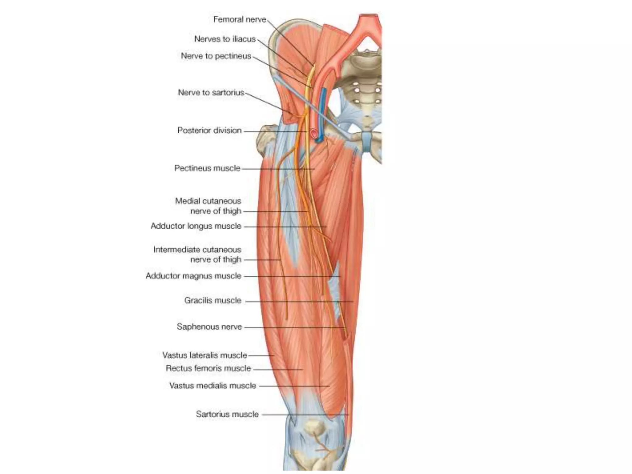
•Formed by branches of lumbar plexues
• ( L2,3,4)
• it is largest branch of the lumbar plexus
3.
• passes deepto ingiunal ligament and enters the
femoral triangle, lateral to the femoral vessels.
• Before entering the thigh, the femoral nerve
supplies branches to the iliacus and pectineus
muscles
• After entering the triangle, the femoral nerve
divides into several branches to the anterior thigh
muscles.
4.
• Immediately afterpassing under the inguinal
ligament, the femoral nerve divides into anterior
and posterior divisions,
• which supply muscles of the anterior compartment
of thigh and
• skin on the anterior and medial aspects of the
thigh and on the medial sides of the leg and foot.
5.
• Branches ofthe femoral nerve include
• anterior cutaneous branches, (ant. Division)
• numerous motor nerves, (post. division)
• one long cutaneous nerve, the saphenous nerve,
6.
• anterior cutaneousbranches
• penetrate deep fascia to supply skin on the front of the
thigh and knee
• numerous motor nerves
• supply the quadriceps femoris muscles (rectus femoris,
vastus lateralis, vastus intermedius, and vastus medialis
muscles) and the sartorius muscle
7.
• the saphenousnerve,
• supplies skin as far distally as the medial side of the foot.
9.
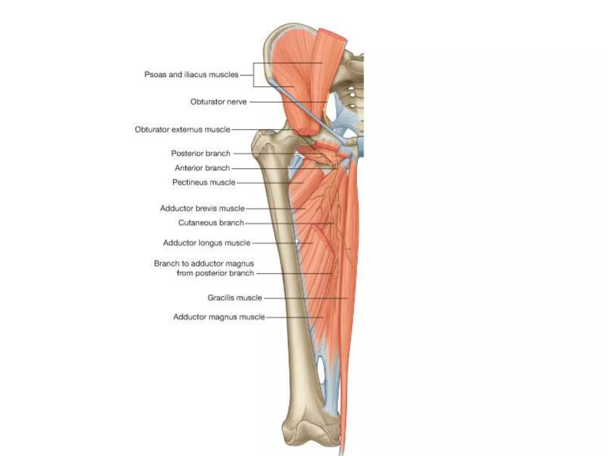
• B. Theobturator nerve(L3-4)
• The obturator nerve is a branch of the lumbar plexus on
the posterior abdominal wall.
• passes out of the medial margin of the psoas muscle to
enter the pelvis
• It supplies most of the adductor muscles and skin on the
medial aspect of the thigh
11.
• As theobturator nerve enters the thigh, it divides
into two branches, an anterior branch and a
posterior branch, which are separated by the
adductor brevis muscle
• the posterior branch
• supplies the obturator externus and adductor brevis
muscles and the part of adductor magnus that attaches
to the linea aspera;
12.
• the anteriorbranch
• it supplies branches to the adductor longus, gracilis, and
adductor brevis muscles, and the pectineus muscle, and
cutaneous branches innervate the skin on the medial
side of the thigh.
13.
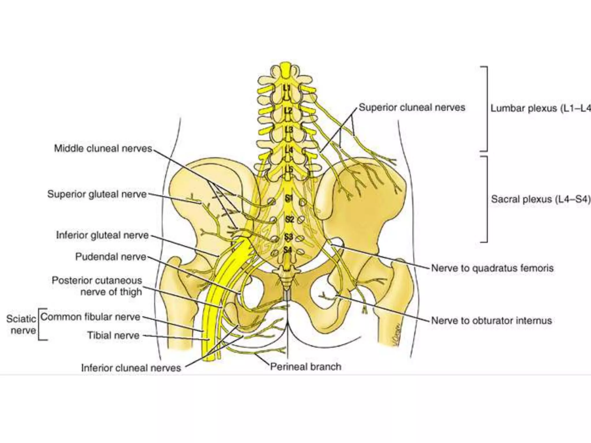
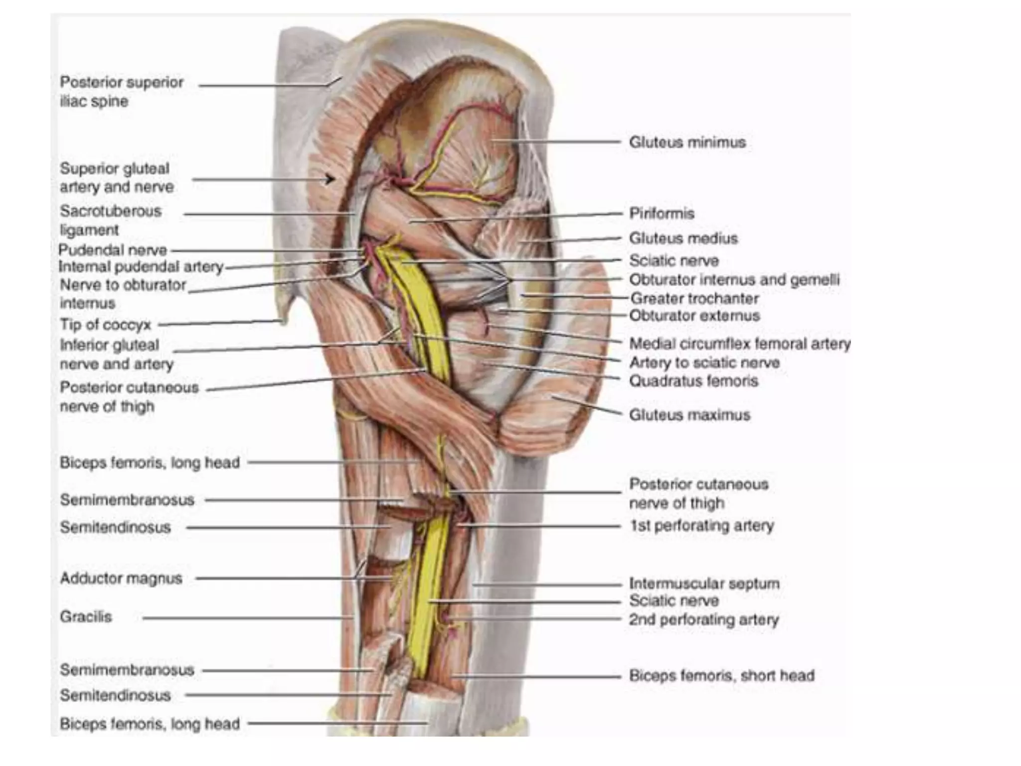
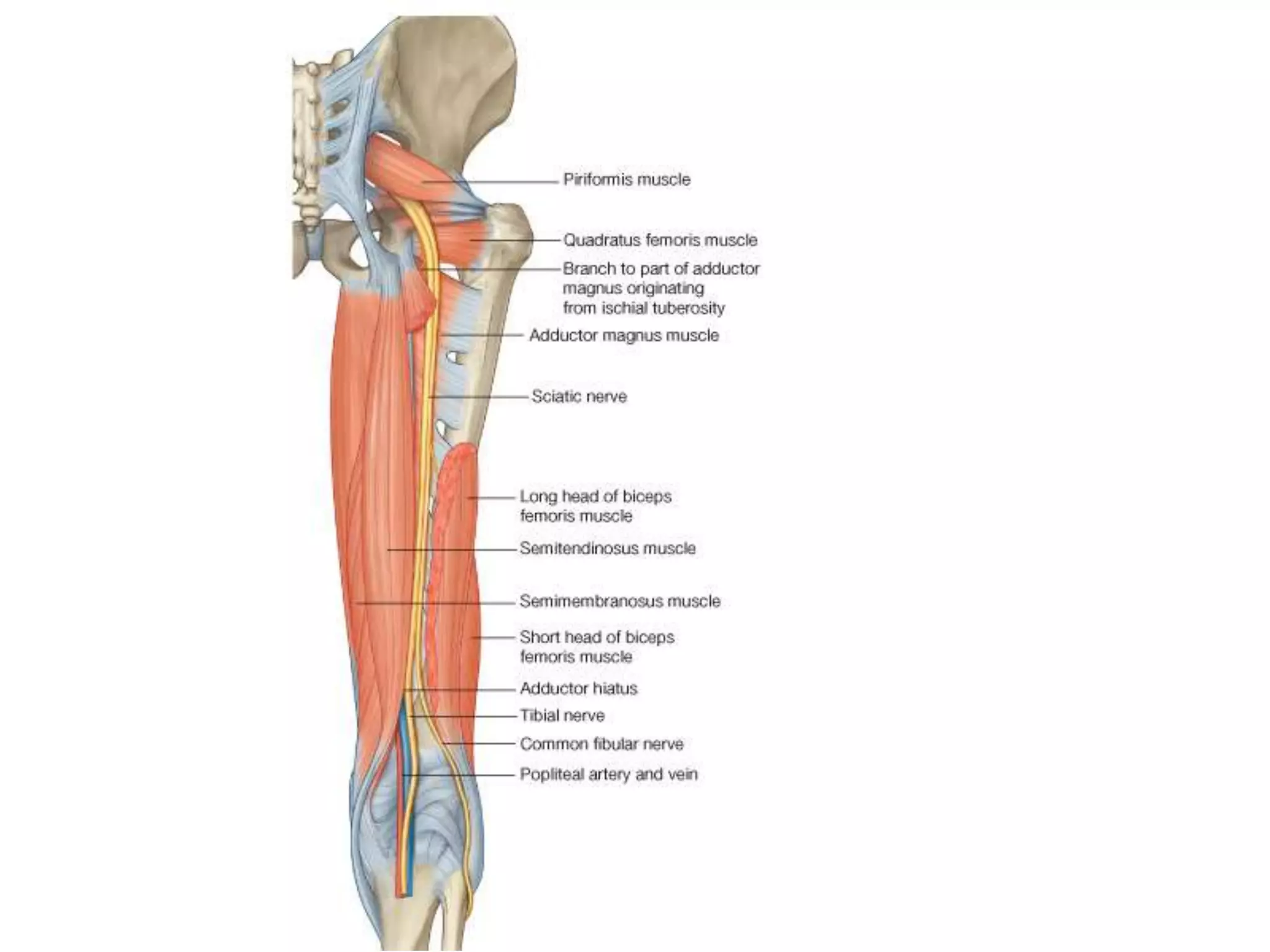
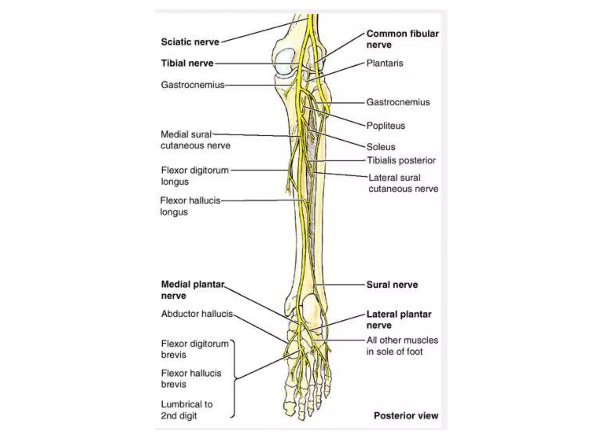
• C. Sciaticnerve (L4-S3)
• The sciatic nerve descends into the posterior
compartment of thigh from the gluteal region
• It innervates all muscles in the posterior compartment
of thigh and then its branches continue into the leg and
foot.
16.
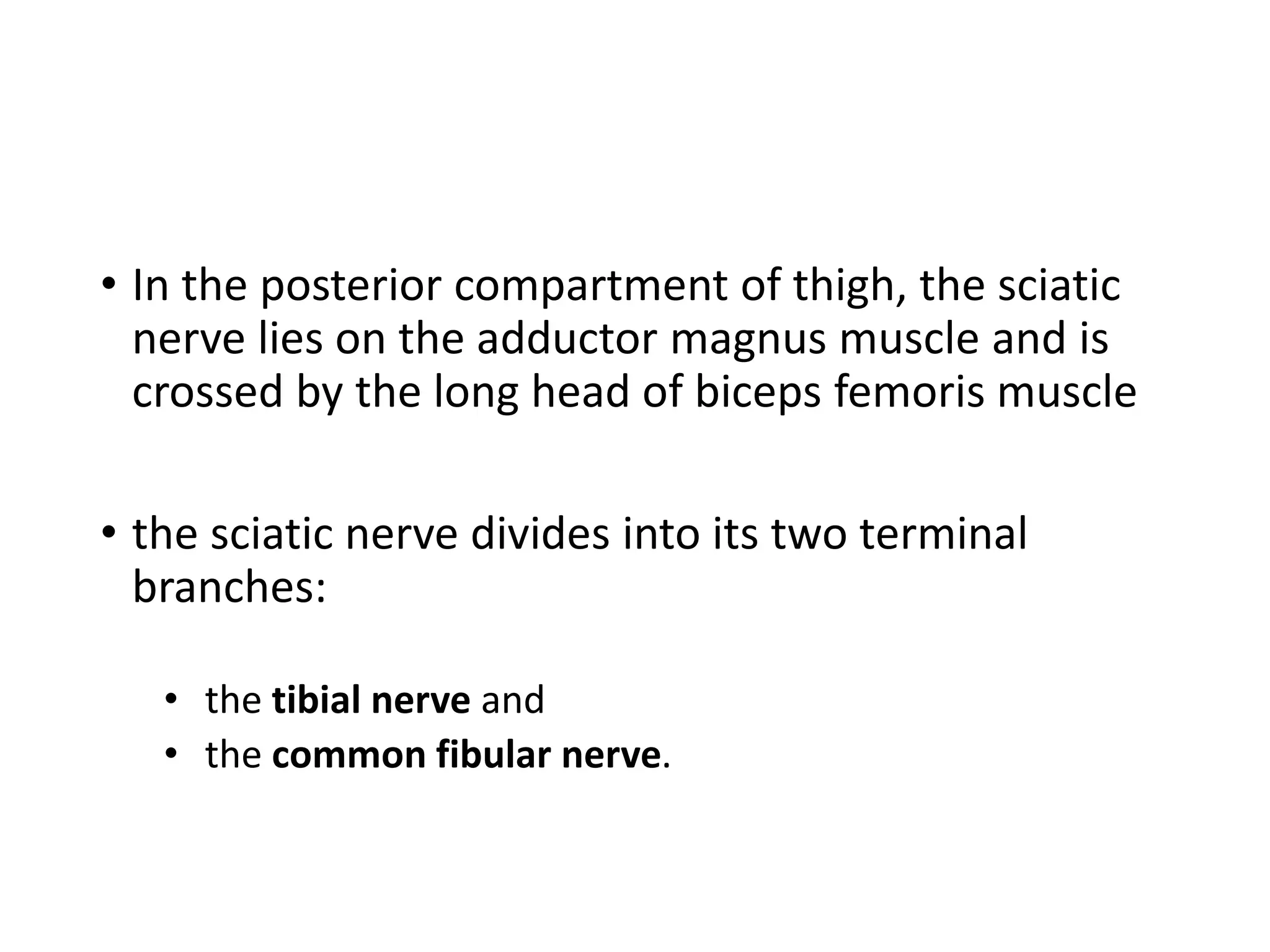
• In theposterior compartment of thigh, the sciatic
nerve lies on the adductor magnus muscle and is
crossed by the long head of biceps femoris muscle
• the sciatic nerve divides into its two terminal
branches:
• the tibial nerve and
• the common fibular nerve.
18.
• Tibial nerve
•The tibial part of the sciatic nerve, either before or after
its separation from the common fibular nerve
• supplies branches to all muscles in the posterior
compartment of thigh (long head of biceps femoris,
semimembranosus, semitendinosus)
• except the short head of biceps femoris, which is
innervated by the common fibular part
19.
• The tibialnerve descends through the popliteal
fossa, enters the posterior compartment of leg and
continues into the sole of the foot
20.
• The tibialnerve innervates:
• all muscles in the posterior compartment of leg;
• all intrinsic muscles in the sole of the foot except for the
first two dorsal interossei muscles, which are innervated
by the deep fibular nerve;
• skin on the posterolateral side of the lower half of the
leg and medial side of the ankle, foot, and little toe, and
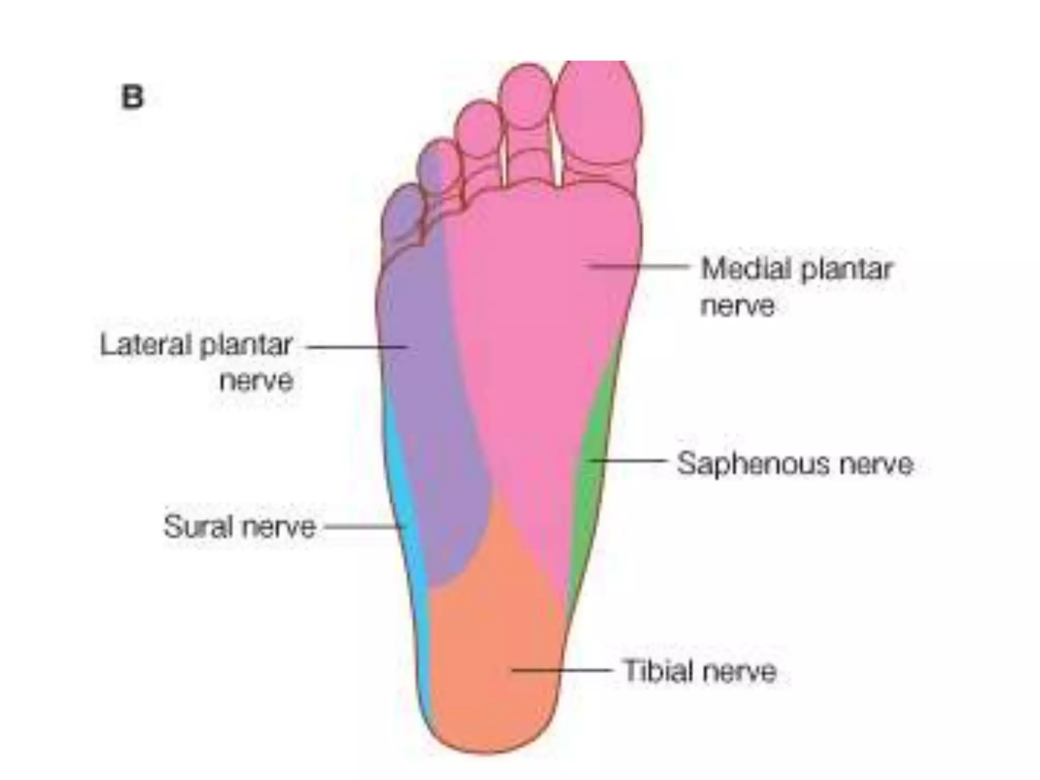
skin on the sole of the foot and toes.
22.
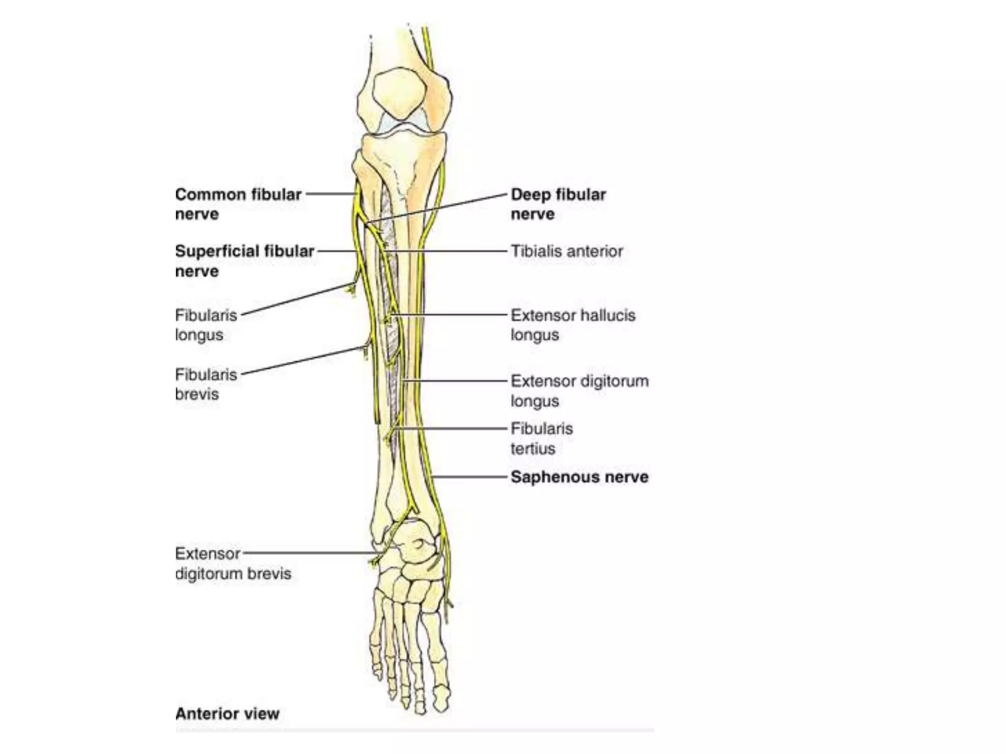
• Common fibularnerve
• The common fibular part of the sciatic nerve innervates
the short head of biceps femoris in the posterior
compartment of thigh and
• continues into the lateral and anterior compartments of
leg and onto the foot
23.
• The commonfibular nerve innervates:
• muscles in the anterior and lateral compartments
of leg;
• one muscle (extensor digitorum brevis) on the
dorsal aspect of the foot;
• the first two dorsal interossei muscles in the sole of
the foot;
• skin over the lateral aspect of the leg, and ankle,
and over the dorsal aspect of the foot and toes
25.
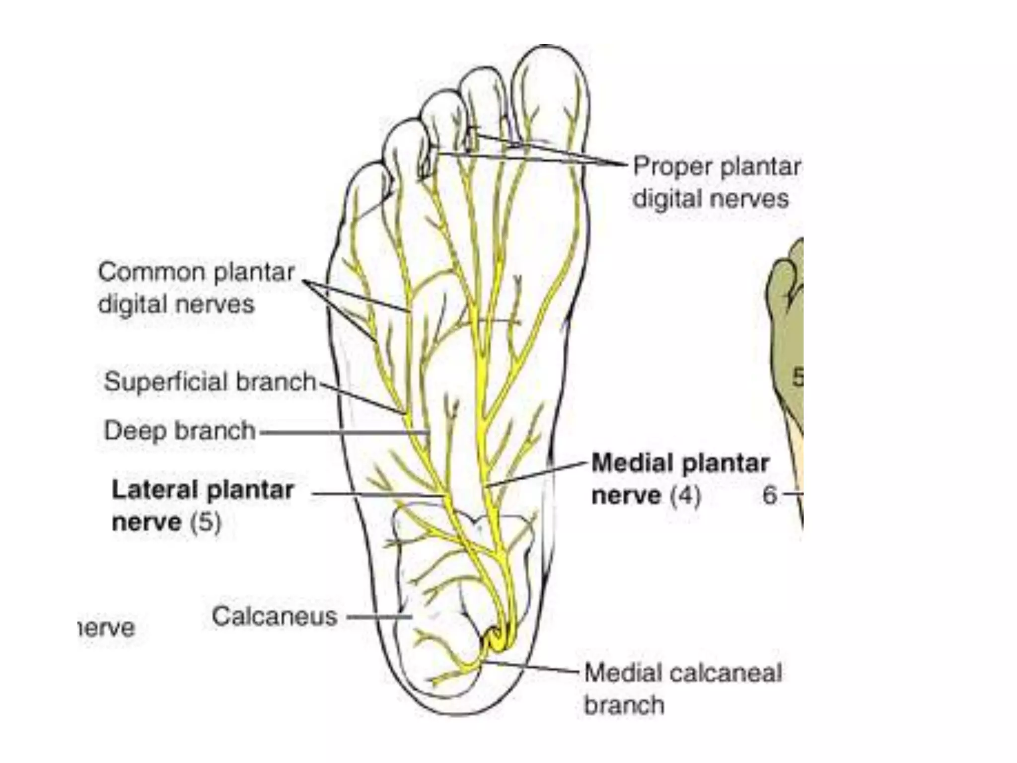
• The tibialnerve enters the foot through the tarsal
tunnel posterior to the medial malleolus
• gives origin to medial calcaneal branches, which
penetrate the flexor retinaculum to supply the heel
26.
• the tibialnerve bifurcates with the posterior tibial
artery into:
• a large medial plantar nerve;
• a smaller lateral plantar nerve
27.
• The medialplantar nerve
• is the major sensory nerve in the sole of the foot
• It innervates skin on most of the anterior two-thirds of
the sole and adjacent surfaces of the medial three and
one-half toes, which includes the great toe
• In addition to this large area of plantar skin, the nerve
also innervates four intrinsic muscles-abductor hallucis,
flexor digitorum brevis, flexor hallucis brevis, and the
first lumbrical.
28.
• Lateral plantarnerve
• an important motor nerve in the foot because it
innervates all intrinsic muscles in the sole
• except for the muscles supplied by the medial plantar
nerve