Downloaded 124 times
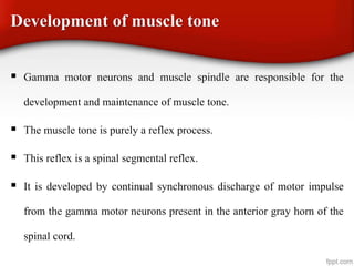
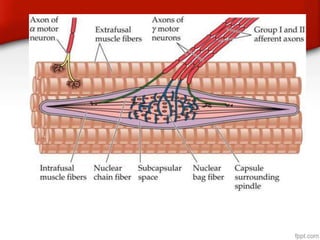
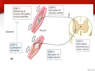


1) The document discusses posture and equilibrium, defining posture as the subconscious adjustment of muscle tone to maintain balance and the body's relationship to gravity. 2) It describes how muscle tone, regulated by gamma motor neurons, and stretch reflexes originating from muscle spindles work together to maintain posture through continuous low-level muscle contraction. 3) Various static and statokinetic reflexes involving the limbs, eyes, and whole body work in coordination to automatically right the body and keep it balanced during both motion and rest.


