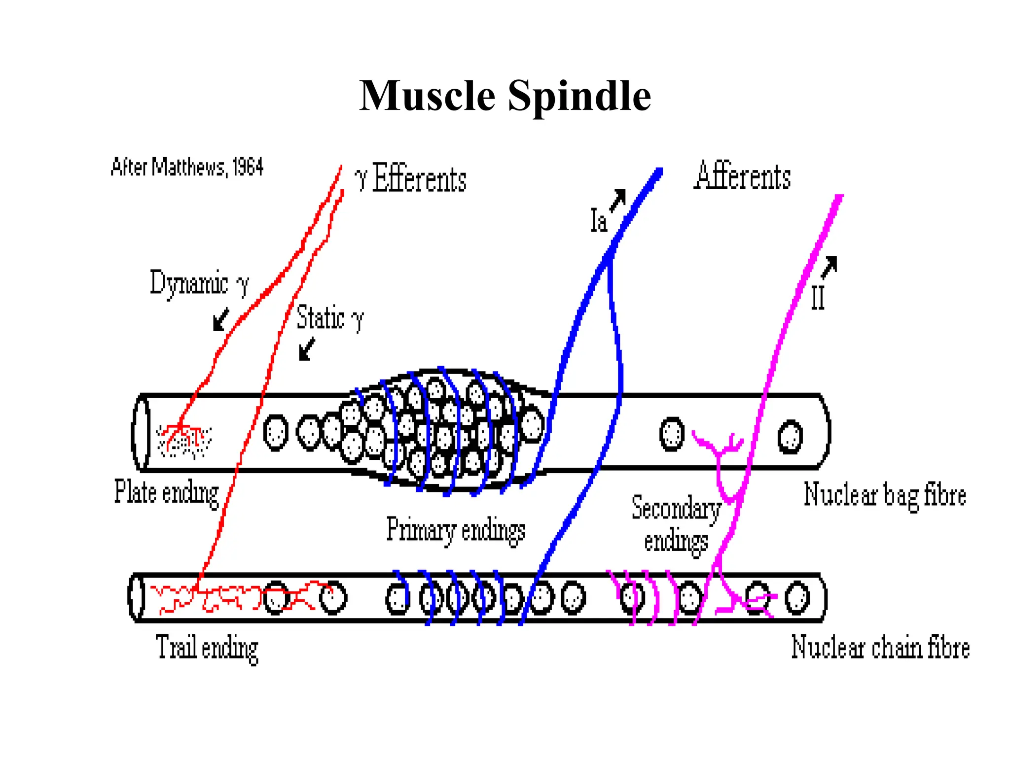
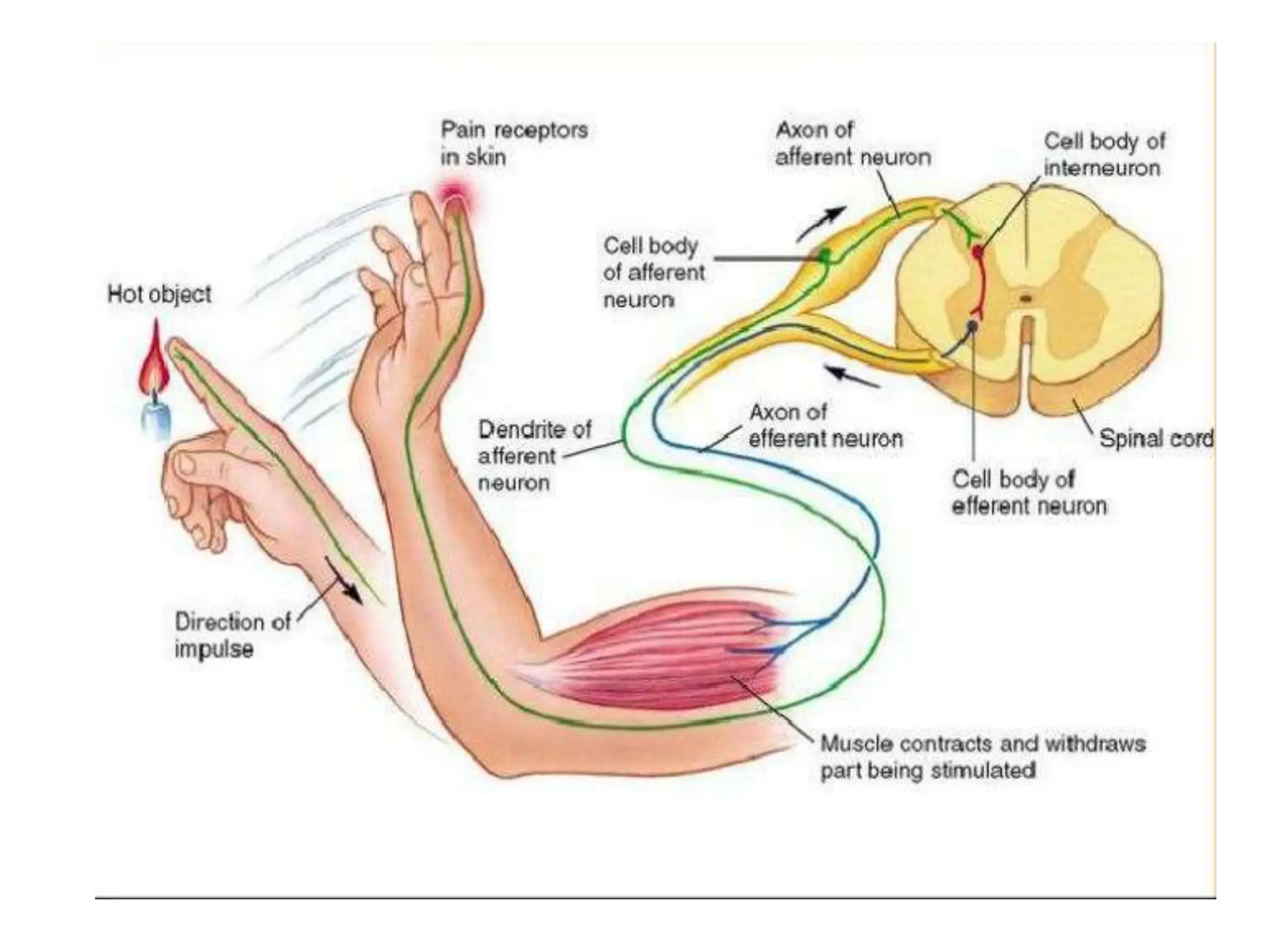
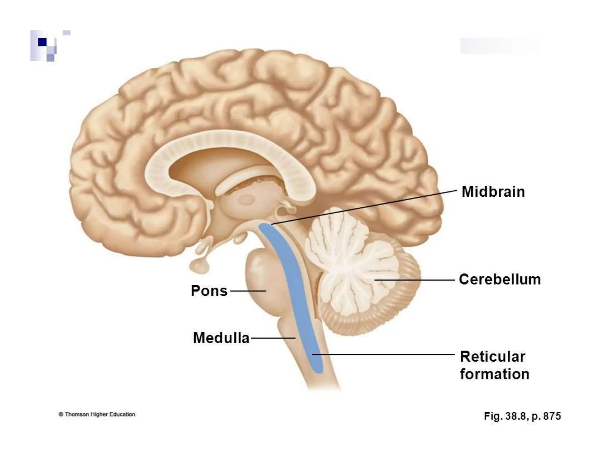
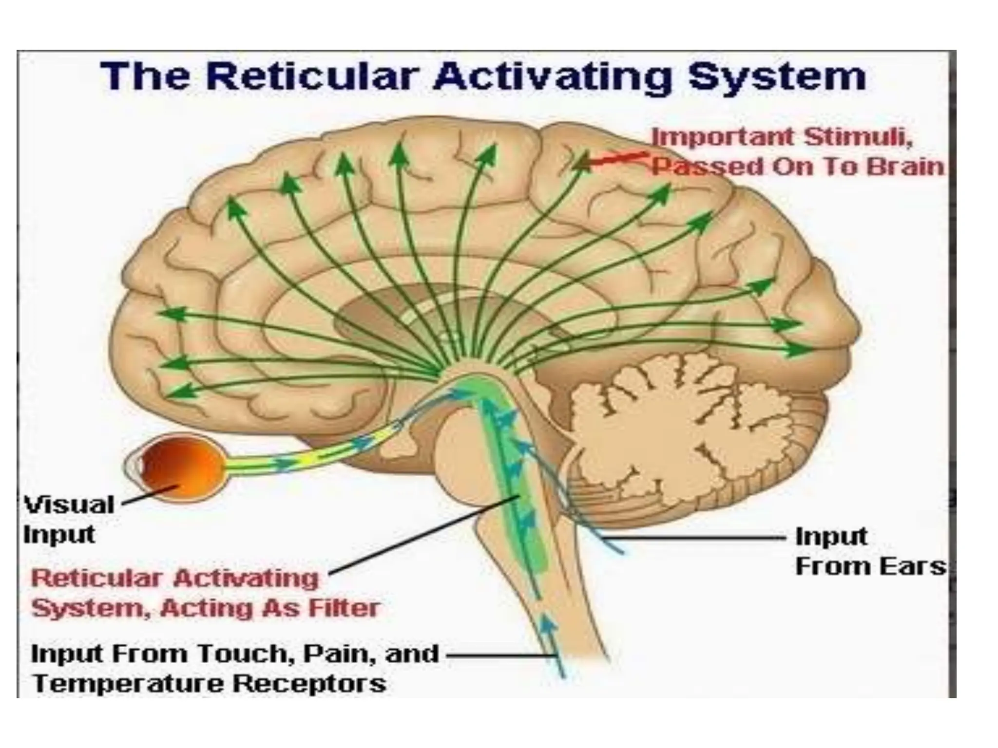
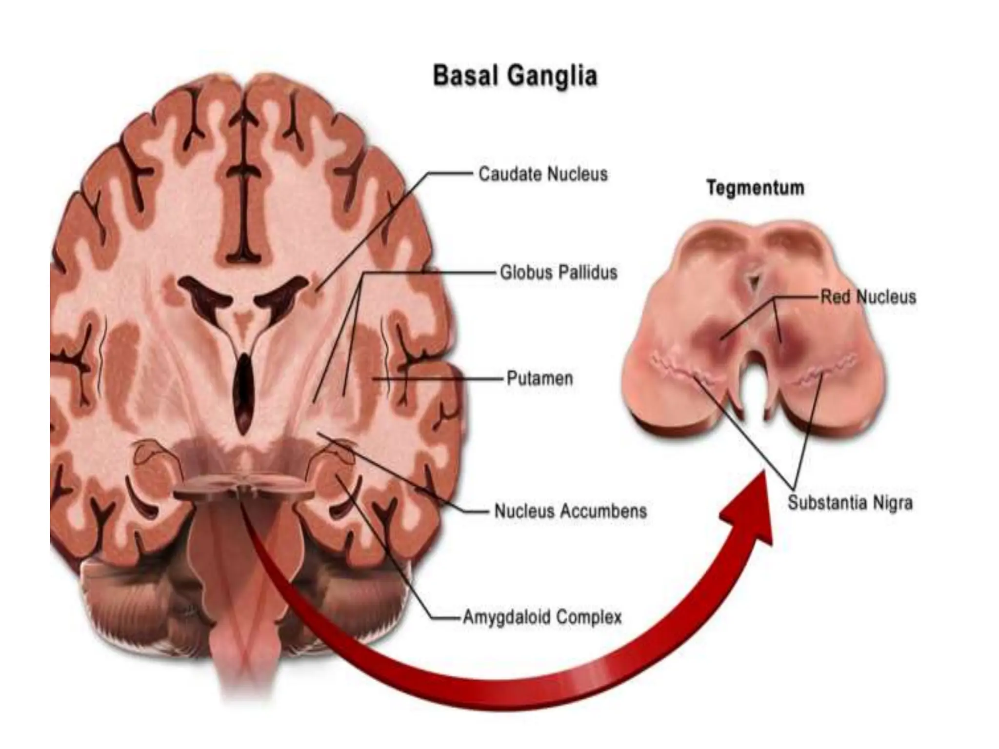
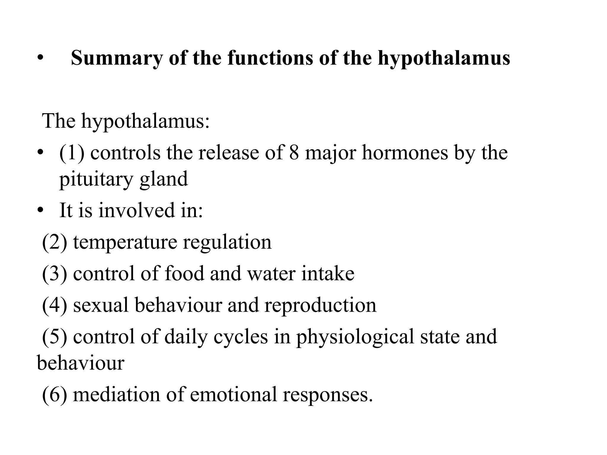
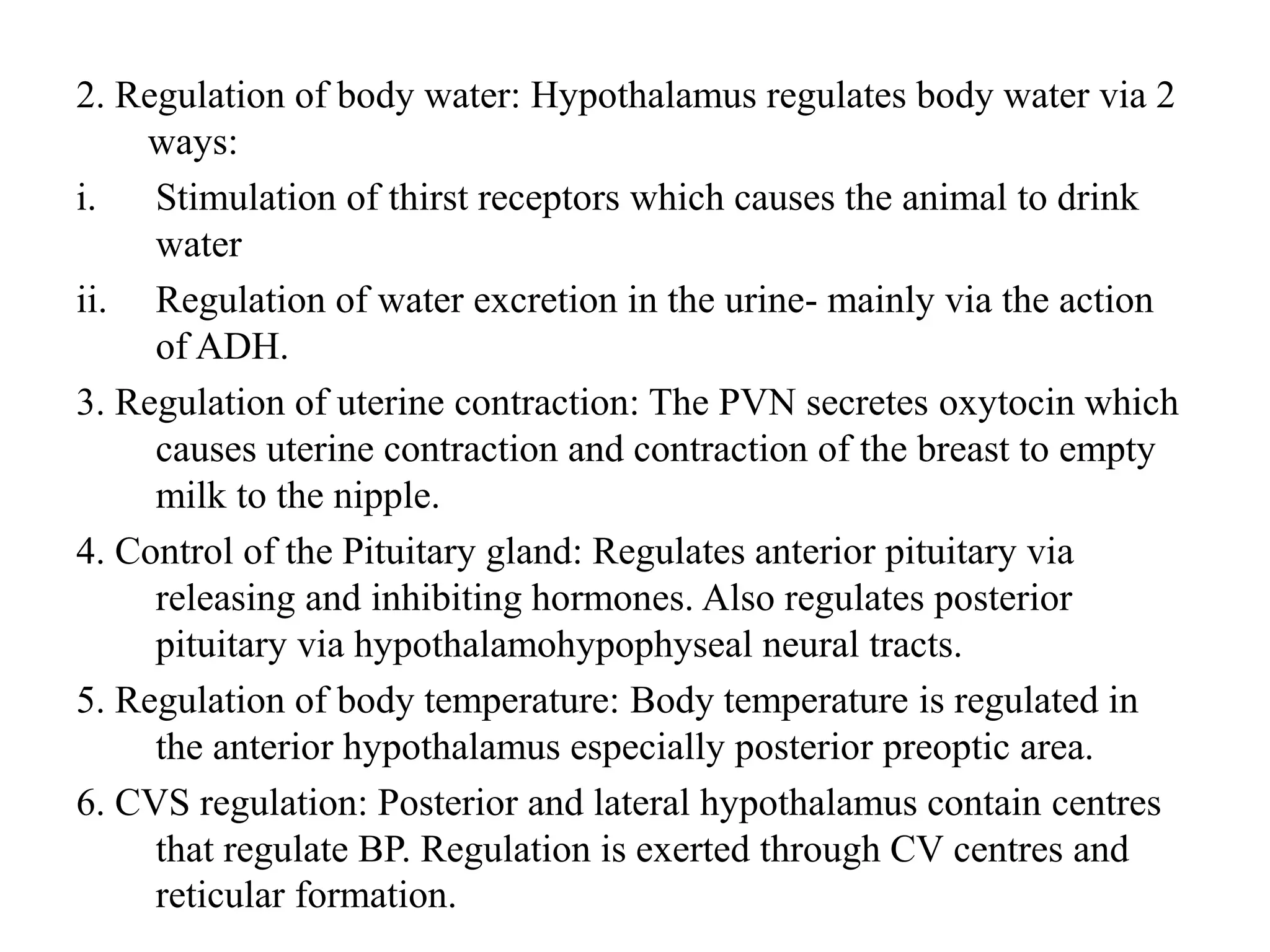
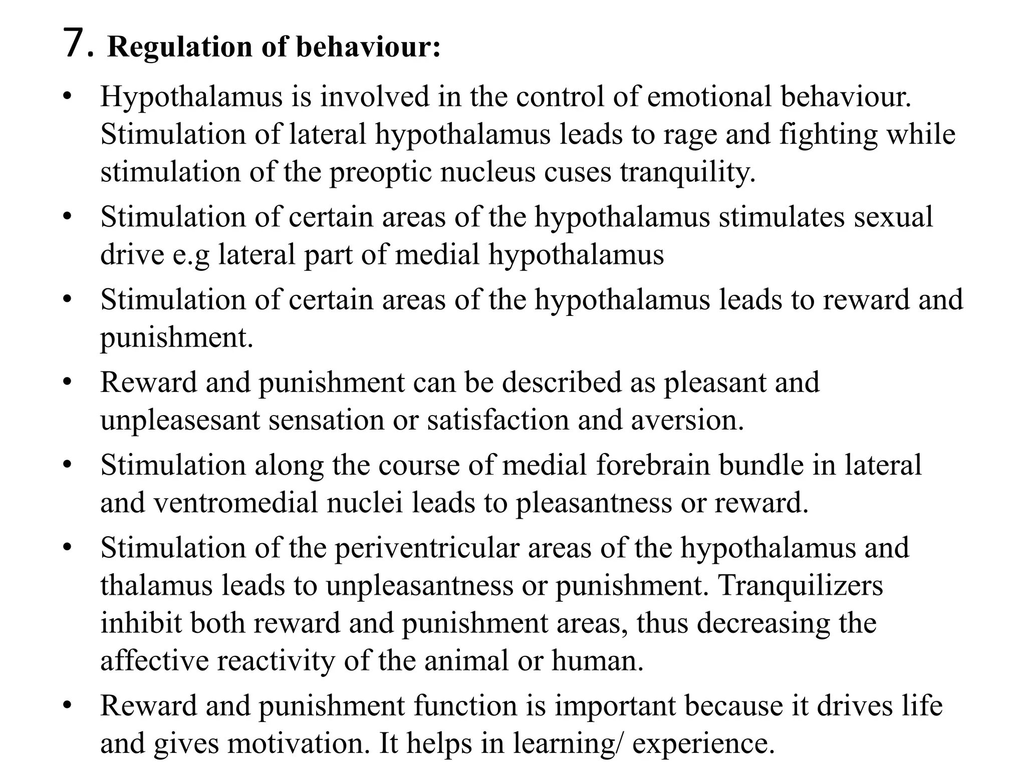
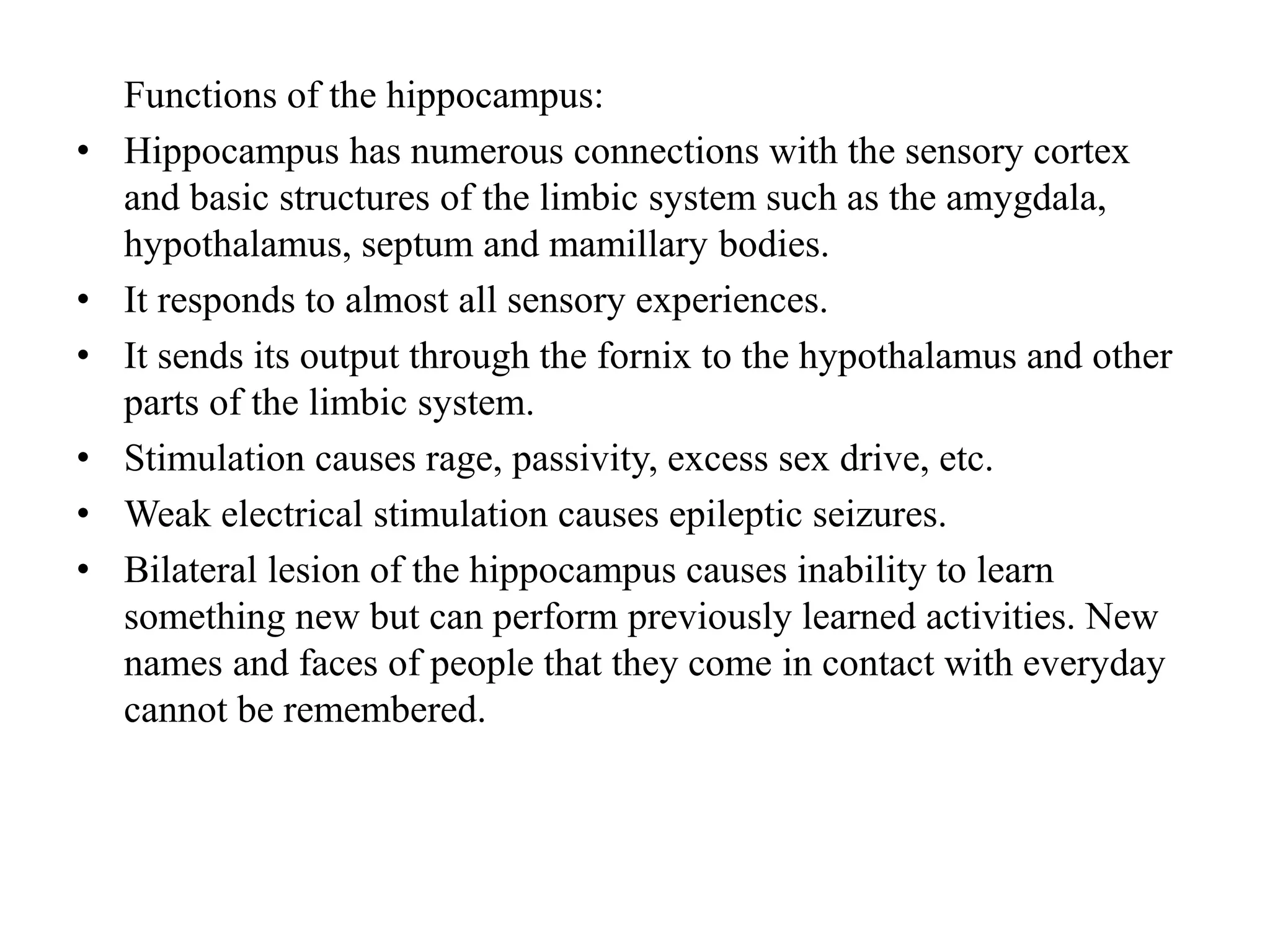
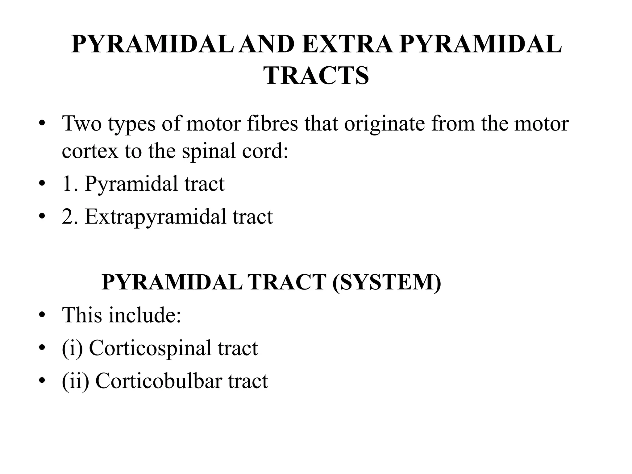
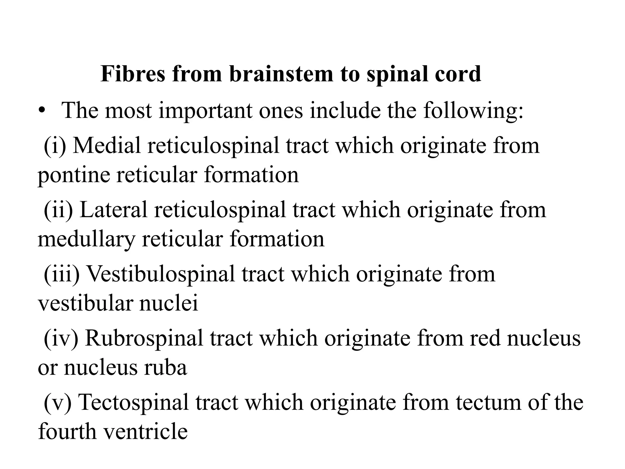
The document discusses the motor system and neurophysiology. It covers topics such as motor centres in the brain and spinal cord, motor neurons, motor units, factors that control muscle tension, neuronal networks, reflexes including monosynaptic and polysynaptic reflexes, the muscle spindle, ventral horn cells, the reticular formation and reticular activating system, electroencephalography, and human sleep patterns including REM and non-REM sleep.
The document discusses reflexes and the nervous system. It defines reflexes as rapid automatic responses to specific stimuli. It describes the five steps in a neural reflex arc: 1) stimulus activates a receptor, 2) activation of a sensory neuron, 3) information processing in the central nervous system, 4) activation of a motor neuron, 5) response of a peripheral effector. Reflexes can be classified based on their development, processing site, response type, and complexity of the neural circuit involved. The stretch tendon reflex is provided as a detailed example of a polysynaptic reflex. The basal ganglia are also summarized as subcortical structures that receive input from and return output to the cerebral cortex to affect motor activity.
The document summarizes key aspects of the peripheral nervous system and reflex activity. It discusses motor endings that release neurotransmitters to activate effectors. It reviews innervation of skeletal and visceral muscle, describing neurotransmitters and responses. It also describes three levels of motor control - segmental, projection, and precommand. Segmental control involves central pattern generators for locomotion. Projection pathways connect to voluntary movement and reflex control. Precommand areas like the cerebellum regulate and coordinate movement. The document also discusses properties of reflex arcs and examples of spinal, stretch, and superficial reflexes. It notes developmental aspects of peripheral nerve distribution and aging changes.
BCM II-Reflex Arc and the reflexes Feb2023.pptxSELDOMWEYUSIA
This document provides an overview of reflex physiology, including:
1. The components and function of the reflex arc, which includes receptors, sensory and motor neurons, and effectors.
2. Types of spinal reflexes like the stretch, myotatic, reciprocal inhibition, and withdrawal reflexes.
3. The role of muscle spindles in the stretch reflex and their operation via gamma motor neurons.
4. Differences between mono- and poly-synaptic reflexes and examples like the knee jerk and withdrawal reflexes.
BCM II-Reflex Arc and the reflexes Feb2023.pptxSELDOMWEYUSIA
This document provides an overview of reflex physiology, including:
1. Defining reflexes as automatic, unconscious responses to internal or external changes that maintain homeostasis.
2. Describing the components of a reflex arc, including receptors, sensory and motor neurons, and effectors.
3. Explaining how muscle spindles function in stretch reflexes via sensory endings that detect muscle stretch and signal motor neurons.
4. Comparing monosynaptic and polysynaptic reflexes, with the stretch reflex being an example of a monosynaptic reflex.
The stretch reflex is an involuntary muscle contraction that occurs when a muscle is stretched. It plays an important role in maintaining posture and control of movement. There are two types of muscle fibers: extrafusal fibers that provide muscle contraction and intrafusal fibers encapsulated in muscle spindles that act as proprioceptors. Muscle spindles contain specialized intrafusal fibers innervated by sensory Ia afferents and motor γ efferents. When a muscle is stretched, the Ia afferents are activated, triggering a monosynaptic reflex that contracts the muscle via α motor neurons, as well as reciprocal inhibition of antagonist muscles. This phasic stretch reflex acts rapidly to correct movement. The tonic
The document summarizes the motor system, including voluntary and involuntary motor functions. It describes various reflexes like the stretch reflex and their components. The motor cortex and corticospinal tract are discussed. The role of upper motor neurons, lower motor neurons, and extrapyramidal tracts in motor control is explained. Various tests to examine the motor system are also mentioned.
1. Spinal shock occurs immediately after complete transection of the spinal cord and is characterized by a complete loss of all reflexes below the level of lesion lasting 2-6 weeks.
2. Early recovery of reflexes begins after spinal shock and includes weak return of the stretch reflex and appearance of new reflexes like the plantar reflex.
3. Full recovery of reflexes may take 6 months as reflexes further strengthen and autonomic functions partially return through reorganization at the spinal level.
Y2 s1 motor system reflexes basal ganglia 2018 comple lecturevajira54
This document summarizes the motor system, including reflexes, the basal ganglia, and motor functions. It describes the stretch reflex and its components like muscle spindles. It discusses the roles of alpha and gamma motor neurons and how they relate to voluntary and involuntary muscle contractions. Clinical tests of reflexes are used to locate lesions in the motor system.
This document discusses motor reflexes and their components. It begins by defining reflexes as involuntary responses to stimuli that do not involve significant brain involvement. It then discusses the stretch reflex in detail, including the roles of muscle spindles, Ia afferent fibers, and alpha and gamma motor neurons. Additional reflexes discussed include withdrawal reflexes, reciprocal innervation, and primitive reflexes seen in infants. The clinical importance of assessing reflexes to localize lesions in the motor system is also mentioned.
The document provides information about the nervous system. It discusses that the nervous system controls all body activities and is divided into the central and peripheral nervous systems. The central nervous system includes the brain and spinal cord and contains gray and white matter. The peripheral nervous system contains nerves that arise from the brain and spinal cord and is divided into the somatic and autonomic systems. Neurons are the basic functional units and transmit signals through electrical impulses known as action potentials.
The document discusses the nervous system, including neurons, the central nervous system, and peripheral nervous system. It describes the structure and function of neurons, including different parts like the cell body, dendrites, and axon. It discusses how neurons transmit electrical signals via action potentials and communicate via synapses. The central nervous system contains the brain and spinal cord, which control and coordinate the body. The peripheral nervous system includes nerves that connect the central nervous system to the rest of the body.
Control and coordination are essential in living organisms. The nervous system provides control and coordination in animals through receptors, nerves, and the brain and spinal cord. In plants, coordination occurs through growth-dependent and independent movements like tropisms and nastic movements in response to stimuli. Hormones play a key role in homeostasis, growth, and development in both animals and plants through feedback mechanisms.
The document provides an overview of the nervous system, including:
1. It describes the organization and main components of the nervous system, including neurons, neuroglia, nerves, and the central and peripheral nervous systems.
2. It explains the functions of the nervous system in sensation, motor control, and higher cognitive processes. It also describes the types of sensory receptors and motor responses.
3. It provides details on the structure and function of neurons, neurotransmission, and the generation and propagation of nerve impulses through neurons.
Reflexes are automatic responses to stimuli that involve a reflex arc consisting of sensory receptors, sensory neurons, processing in the central nervous system, motor neurons, and effectors. Reflexes maintain homeostasis, carry out automatic actions like swallowing and sneezing, and maintain balance and posture through spinal reflexes controlling trunk and limb muscles. There are different types of reflexes including flexor reflexes which cause limb flexion in response to stimuli, and crossed extensor reflexes which cause the opposite limb to extend about 0.2 to 0.5 seconds after a flexor reflex is elicited in one limb. Reciprocal inhibition occurs when a stretch reflex excites one muscle while simultaneously inhibiting the antagonist muscle through reciprocal innervation.
This document provides an overview of applied physiology of the nervous system. It begins with an introduction to the central nervous system, including its organization and main parts like the brain and spinal cord. Key concepts covered include the structure and functions of neurons and neuroglia. The roles of gray and white matter are discussed. Main functions of the central nervous system include temperature regulation, spinal cord reflexes, memory and learning, and voluntary movement. Imaging techniques for the central nervous system are also mentioned. The document goes on to explain topics like resting membrane potential, action potentials, synaptic transmission, and the reflex arc.
PY3.2 Describe the types, functions & properties of nerve fiber.pptxShikha Saxena
1. The document describes the different types, functions, and properties of neurons and nerve fibers. It discusses the three main types of neurons based on their poles - unipolar, bipolar, and multipolar.
2. Neurons are also classified based on their function into motor and sensory neurons. Additionally, nerve fibers are classified based on their diameter and conduction velocity.
3. The key properties of nerve fibers discussed include excitability, conductivity, summation, accommodation, unfatigability, all-or-none response, and refractory periods. The functions of different parts of the neuron are also summarized.
This document discusses different types of spinal reflexes and their components. It describes autonomic reflexes which control organs and glands, and somatic reflexes which control muscles. Specific examples of reflex tests are provided, including the patellar reflex test for somatic reflexes and pupillary light reflex test for autonomic reflexes. Sensory receptors like muscle spindles, Golgi tendon organs, and their roles in reflexes are also explained.
The document discusses the neuromuscular junction and muscle contraction physiology. It defines the neuromuscular junction as the connection between motor neurons and muscle fibers that initiates muscle contraction. The structure and function of the neuromuscular junction is described, including the roles of acetylcholine, receptors, and acetylcholinesterase. The sliding filament model of muscle contraction is introduced. Different muscle fiber types, properties of muscle tissue, and the sarcomere as the contractile unit are defined.
The document discusses the stretch reflex and muscle tone. It defines the stretch reflex as a monosynaptic reflex where stimulation of muscle spindles by stretching a muscle leads to contraction of the same muscle via alpha motor neurons. It describes the roles of gamma motor neurons in maintaining sensitivity of muscle spindles. Disorders of muscle tone like spasticity, rigidity, and hypotonia are explained in relation to lesions in the pyramidal and extrapyramidal systems. Clinical examination techniques for assessing muscle tone are also outlined.
Nerve and muscle.pptx radiology anatomy yes62pwff8nsh
The document provides information about the nervous system and muscles. It discusses the structure and function of neurons, how nerve impulses are transmitted through the nervous system and to muscles, and the different types of muscles in the body. It explains that neurons communicate through electrochemical signals to control and coordinate body functions. When a sensory neuron detects a stimulus, the nervous system integrates the information and may trigger a motor neuron response through synaptic transmission.
The nervous system is organized into two main parts - the central nervous system (CNS) and the peripheral nervous system (PNS). The CNS consists of the brain and spinal cord and acts as the command center that processes sensory input and directs motor output. The PNS connects the CNS to the rest of the body and senses the external environment via sensory receptors. Communication between neurons is mediated by electrical and chemical signals. The nervous system works with the endocrine system to maintain homeostasis via reflexes and other rapid or slower responses.
This document discusses the physiology of reflexes. It defines a reflex as a coordinated involuntary motor response initiated by a stimulus to peripheral receptors. Reflexes are classified based on their clinical presentation, anatomical pathway, number of synapses involved, and functional purpose. Monosynaptic reflexes like the stretch reflex involve only one synapse, while polysynaptic reflexes have multiple synapses. Characteristics of reflexes like summation, irradiation, and fatigue are also described.
A reflex arc includes 5 components: a receptor, afferent nerve, center, efferent nerve, and effector organ. A stimulus activates a receptor, sending a signal along the afferent nerve to the center in the spinal cord or brain. The center then sends a signal back along the efferent nerve to the effector organ, causing it to respond automatically without conscious thought. Reflexes can be classified in various ways, including as inborn or acquired, based on their anatomical location such as spinal or cranial nerves, and as protective or postural reflexes based on their physiological function.
The stretch reflex is a monosynaptic reflex that causes a muscle to contract in response to stretching. It functions to protect muscles from overstretching and helps maintain good posture. The key components of the stretch reflex are: (1) muscle spindles that detect changes in muscle length, (2) the spinal cord which acts as the integrating center, and (3) afferent and efferent nerves which transmit signals between the muscles and central nervous system.
A reflex arc is a neural pathway that controls a reflex response. It consists of a receptor that detects a stimulus, a sensory neuron that conveys this information to the spinal cord, an interneuron that relays the signal to a motor neuron, which then activates an effector such as a muscle or gland. This allows reflex actions to occur quickly through the spinal cord without involving the brain. Common spinal reflexes include the stretch reflex, Golgi tendon reflex, crossed extensor reflex, and withdrawal reflex.
LAND USE LAND COVER AND NDVI OF MIRZAPUR DISTRICT, UPRAHUL
This Dissertation explores the particular circumstances of Mirzapur, a region located in the
core of India. Mirzapur, with its varied terrains and abundant biodiversity, offers an optimal
environment for investigating the changes in vegetation cover dynamics. Our study utilizes
advanced technologies such as GIS (Geographic Information Systems) and Remote sensing to
analyze the transformations that have taken place over the course of a decade.
The complex relationship between human activities and the environment has been the focus
of extensive research and worry. As the global community grapples with swift urbanization,
population expansion, and economic progress, the effects on natural ecosystems are becoming
more evident. A crucial element of this impact is the alteration of vegetation cover, which plays a
significant role in maintaining the ecological equilibrium of our planet.Land serves as the foundation for all human activities and provides the necessary materials for
these activities. As the most crucial natural resource, its utilization by humans results in different
'Land uses,' which are determined by both human activities and the physical characteristics of the
land.
The utilization of land is impacted by human needs and environmental factors. In countries
like India, rapid population growth and the emphasis on extensive resource exploitation can lead
to significant land degradation, adversely affecting the region's land cover.
Therefore, human intervention has significantly influenced land use patterns over many
centuries, evolving its structure over time and space. In the present era, these changes have
accelerated due to factors such as agriculture and urbanization. Information regarding land use and
cover is essential for various planning and management tasks related to the Earth's surface,
providing crucial environmental data for scientific, resource management, policy purposes, and
diverse human activities.
Accurate understanding of land use and cover is imperative for the development planning
of any area. Consequently, a wide range of professionals, including earth system scientists, land
and water managers, and urban planners, are interested in obtaining data on land use and cover
changes, conversion trends, and other related patterns. The spatial dimensions of land use and
cover support policymakers and scientists in making well-informed decisions, as alterations in
these patterns indicate shifts in economic and social conditions. Monitoring such changes with the
help of Advanced technologies like Remote Sensing and Geographic Information Systems is
crucial for coordinated efforts across different administrative levels. Advanced technologies like
Remote Sensing and Geographic Information Systems
9
Changes in vegetation cover refer to variations in the distribution, composition, and overall
structure of plant communities across different temporal and spatial scales. These changes can
occur natural.
Y2 s1 motor system reflexes basal ganglia 2018 comple lecturevajira54
This document summarizes the motor system, including reflexes, the basal ganglia, and motor functions. It describes the stretch reflex and its components like muscle spindles. It discusses the roles of alpha and gamma motor neurons and how they relate to voluntary and involuntary muscle contractions. Clinical tests of reflexes are used to locate lesions in the motor system.
This document discusses motor reflexes and their components. It begins by defining reflexes as involuntary responses to stimuli that do not involve significant brain involvement. It then discusses the stretch reflex in detail, including the roles of muscle spindles, Ia afferent fibers, and alpha and gamma motor neurons. Additional reflexes discussed include withdrawal reflexes, reciprocal innervation, and primitive reflexes seen in infants. The clinical importance of assessing reflexes to localize lesions in the motor system is also mentioned.
The document provides information about the nervous system. It discusses that the nervous system controls all body activities and is divided into the central and peripheral nervous systems. The central nervous system includes the brain and spinal cord and contains gray and white matter. The peripheral nervous system contains nerves that arise from the brain and spinal cord and is divided into the somatic and autonomic systems. Neurons are the basic functional units and transmit signals through electrical impulses known as action potentials.
The document discusses the nervous system, including neurons, the central nervous system, and peripheral nervous system. It describes the structure and function of neurons, including different parts like the cell body, dendrites, and axon. It discusses how neurons transmit electrical signals via action potentials and communicate via synapses. The central nervous system contains the brain and spinal cord, which control and coordinate the body. The peripheral nervous system includes nerves that connect the central nervous system to the rest of the body.
Control and coordination are essential in living organisms. The nervous system provides control and coordination in animals through receptors, nerves, and the brain and spinal cord. In plants, coordination occurs through growth-dependent and independent movements like tropisms and nastic movements in response to stimuli. Hormones play a key role in homeostasis, growth, and development in both animals and plants through feedback mechanisms.
The document provides an overview of the nervous system, including:
1. It describes the organization and main components of the nervous system, including neurons, neuroglia, nerves, and the central and peripheral nervous systems.
2. It explains the functions of the nervous system in sensation, motor control, and higher cognitive processes. It also describes the types of sensory receptors and motor responses.
3. It provides details on the structure and function of neurons, neurotransmission, and the generation and propagation of nerve impulses through neurons.
Reflexes are automatic responses to stimuli that involve a reflex arc consisting of sensory receptors, sensory neurons, processing in the central nervous system, motor neurons, and effectors. Reflexes maintain homeostasis, carry out automatic actions like swallowing and sneezing, and maintain balance and posture through spinal reflexes controlling trunk and limb muscles. There are different types of reflexes including flexor reflexes which cause limb flexion in response to stimuli, and crossed extensor reflexes which cause the opposite limb to extend about 0.2 to 0.5 seconds after a flexor reflex is elicited in one limb. Reciprocal inhibition occurs when a stretch reflex excites one muscle while simultaneously inhibiting the antagonist muscle through reciprocal innervation.
This document provides an overview of applied physiology of the nervous system. It begins with an introduction to the central nervous system, including its organization and main parts like the brain and spinal cord. Key concepts covered include the structure and functions of neurons and neuroglia. The roles of gray and white matter are discussed. Main functions of the central nervous system include temperature regulation, spinal cord reflexes, memory and learning, and voluntary movement. Imaging techniques for the central nervous system are also mentioned. The document goes on to explain topics like resting membrane potential, action potentials, synaptic transmission, and the reflex arc.
PY3.2 Describe the types, functions & properties of nerve fiber.pptxShikha Saxena
1. The document describes the different types, functions, and properties of neurons and nerve fibers. It discusses the three main types of neurons based on their poles - unipolar, bipolar, and multipolar.
2. Neurons are also classified based on their function into motor and sensory neurons. Additionally, nerve fibers are classified based on their diameter and conduction velocity.
3. The key properties of nerve fibers discussed include excitability, conductivity, summation, accommodation, unfatigability, all-or-none response, and refractory periods. The functions of different parts of the neuron are also summarized.
This document discusses different types of spinal reflexes and their components. It describes autonomic reflexes which control organs and glands, and somatic reflexes which control muscles. Specific examples of reflex tests are provided, including the patellar reflex test for somatic reflexes and pupillary light reflex test for autonomic reflexes. Sensory receptors like muscle spindles, Golgi tendon organs, and their roles in reflexes are also explained.
The document discusses the neuromuscular junction and muscle contraction physiology. It defines the neuromuscular junction as the connection between motor neurons and muscle fibers that initiates muscle contraction. The structure and function of the neuromuscular junction is described, including the roles of acetylcholine, receptors, and acetylcholinesterase. The sliding filament model of muscle contraction is introduced. Different muscle fiber types, properties of muscle tissue, and the sarcomere as the contractile unit are defined.
The document discusses the stretch reflex and muscle tone. It defines the stretch reflex as a monosynaptic reflex where stimulation of muscle spindles by stretching a muscle leads to contraction of the same muscle via alpha motor neurons. It describes the roles of gamma motor neurons in maintaining sensitivity of muscle spindles. Disorders of muscle tone like spasticity, rigidity, and hypotonia are explained in relation to lesions in the pyramidal and extrapyramidal systems. Clinical examination techniques for assessing muscle tone are also outlined.
Nerve and muscle.pptx radiology anatomy yes62pwff8nsh
The document provides information about the nervous system and muscles. It discusses the structure and function of neurons, how nerve impulses are transmitted through the nervous system and to muscles, and the different types of muscles in the body. It explains that neurons communicate through electrochemical signals to control and coordinate body functions. When a sensory neuron detects a stimulus, the nervous system integrates the information and may trigger a motor neuron response through synaptic transmission.
The nervous system is organized into two main parts - the central nervous system (CNS) and the peripheral nervous system (PNS). The CNS consists of the brain and spinal cord and acts as the command center that processes sensory input and directs motor output. The PNS connects the CNS to the rest of the body and senses the external environment via sensory receptors. Communication between neurons is mediated by electrical and chemical signals. The nervous system works with the endocrine system to maintain homeostasis via reflexes and other rapid or slower responses.
This document discusses the physiology of reflexes. It defines a reflex as a coordinated involuntary motor response initiated by a stimulus to peripheral receptors. Reflexes are classified based on their clinical presentation, anatomical pathway, number of synapses involved, and functional purpose. Monosynaptic reflexes like the stretch reflex involve only one synapse, while polysynaptic reflexes have multiple synapses. Characteristics of reflexes like summation, irradiation, and fatigue are also described.
A reflex arc includes 5 components: a receptor, afferent nerve, center, efferent nerve, and effector organ. A stimulus activates a receptor, sending a signal along the afferent nerve to the center in the spinal cord or brain. The center then sends a signal back along the efferent nerve to the effector organ, causing it to respond automatically without conscious thought. Reflexes can be classified in various ways, including as inborn or acquired, based on their anatomical location such as spinal or cranial nerves, and as protective or postural reflexes based on their physiological function.
The stretch reflex is a monosynaptic reflex that causes a muscle to contract in response to stretching. It functions to protect muscles from overstretching and helps maintain good posture. The key components of the stretch reflex are: (1) muscle spindles that detect changes in muscle length, (2) the spinal cord which acts as the integrating center, and (3) afferent and efferent nerves which transmit signals between the muscles and central nervous system.
A reflex arc is a neural pathway that controls a reflex response. It consists of a receptor that detects a stimulus, a sensory neuron that conveys this information to the spinal cord, an interneuron that relays the signal to a motor neuron, which then activates an effector such as a muscle or gland. This allows reflex actions to occur quickly through the spinal cord without involving the brain. Common spinal reflexes include the stretch reflex, Golgi tendon reflex, crossed extensor reflex, and withdrawal reflex.
Similar to NEUROPHYSIOLOGY OF THE MOTOR SYSTEM-1.ppt (20)
LAND USE LAND COVER AND NDVI OF MIRZAPUR DISTRICT, UPRAHUL
This Dissertation explores the particular circumstances of Mirzapur, a region located in the
core of India. Mirzapur, with its varied terrains and abundant biodiversity, offers an optimal
environment for investigating the changes in vegetation cover dynamics. Our study utilizes
advanced technologies such as GIS (Geographic Information Systems) and Remote sensing to
analyze the transformations that have taken place over the course of a decade.
The complex relationship between human activities and the environment has been the focus
of extensive research and worry. As the global community grapples with swift urbanization,
population expansion, and economic progress, the effects on natural ecosystems are becoming
more evident. A crucial element of this impact is the alteration of vegetation cover, which plays a
significant role in maintaining the ecological equilibrium of our planet.Land serves as the foundation for all human activities and provides the necessary materials for
these activities. As the most crucial natural resource, its utilization by humans results in different
'Land uses,' which are determined by both human activities and the physical characteristics of the
land.
The utilization of land is impacted by human needs and environmental factors. In countries
like India, rapid population growth and the emphasis on extensive resource exploitation can lead
to significant land degradation, adversely affecting the region's land cover.
Therefore, human intervention has significantly influenced land use patterns over many
centuries, evolving its structure over time and space. In the present era, these changes have
accelerated due to factors such as agriculture and urbanization. Information regarding land use and
cover is essential for various planning and management tasks related to the Earth's surface,
providing crucial environmental data for scientific, resource management, policy purposes, and
diverse human activities.
Accurate understanding of land use and cover is imperative for the development planning
of any area. Consequently, a wide range of professionals, including earth system scientists, land
and water managers, and urban planners, are interested in obtaining data on land use and cover
changes, conversion trends, and other related patterns. The spatial dimensions of land use and
cover support policymakers and scientists in making well-informed decisions, as alterations in
these patterns indicate shifts in economic and social conditions. Monitoring such changes with the
help of Advanced technologies like Remote Sensing and Geographic Information Systems is
crucial for coordinated efforts across different administrative levels. Advanced technologies like
Remote Sensing and Geographic Information Systems
9
Changes in vegetation cover refer to variations in the distribution, composition, and overall
structure of plant communities across different temporal and spatial scales. These changes can
occur natural.
Executive Directors Chat Leveraging AI for Diversity, Equity, and InclusionTechSoup
Let’s explore the intersection of technology and equity in the final session of our DEI series. Discover how AI tools, like ChatGPT, can be used to support and enhance your nonprofit's DEI initiatives. Participants will gain insights into practical AI applications and get tips for leveraging technology to advance their DEI goals.
বাংলাদেশের অর্থনৈতিক সমীক্ষা ২০২৪ [Bangladesh Economic Review 2024 Bangla.pdf] কম্পিউটার , ট্যাব ও স্মার্ট ফোন ভার্সন সহ সম্পূর্ণ বাংলা ই-বুক বা pdf বই " সুচিপত্র ...বুকমার্ক মেনু 🔖 ও হাইপার লিংক মেনু 📝👆 যুক্ত ..
আমাদের সবার জন্য খুব খুব গুরুত্বপূর্ণ একটি বই ..বিসিএস, ব্যাংক, ইউনিভার্সিটি ভর্তি ও যে কোন প্রতিযোগিতা মূলক পরীক্ষার জন্য এর খুব ইম্পরট্যান্ট একটি বিষয় ...তাছাড়া বাংলাদেশের সাম্প্রতিক যে কোন ডাটা বা তথ্য এই বইতে পাবেন ...
তাই একজন নাগরিক হিসাবে এই তথ্য গুলো আপনার জানা প্রয়োজন ...।
বিসিএস ও ব্যাংক এর লিখিত পরীক্ষা ...+এছাড়া মাধ্যমিক ও উচ্চমাধ্যমিকের স্টুডেন্টদের জন্য অনেক কাজে আসবে ...
This slide is special for master students (MIBS & MIFB) in UUM. Also useful for readers who are interested in the topic of contemporary Islamic banking.
How to Build a Module in Odoo 17 Using the Scaffold MethodCeline George
Odoo provides an option for creating a module by using a single line command. By using this command the user can make a whole structure of a module. It is very easy for a beginner to make a module. There is no need to make each file manually. This slide will show how to create a module using the scaffold method.
it describes the bony anatomy including the femoral head , acetabulum, labrum . also discusses the capsule , ligaments . muscle that act on the hip joint and the range of motion are outlined. factors affecting hip joint stability and weight transmission through the joint are summarized.
This presentation was provided by Steph Pollock of The American Psychological Association’s Journals Program, and Damita Snow, of The American Society of Civil Engineers (ASCE), for the initial session of NISO's 2024 Training Series "DEIA in the Scholarly Landscape." Session One: 'Setting Expectations: a DEIA Primer,' was held June 6, 2024.
How to Make a Field Mandatory in Odoo 17Celine George
In Odoo, making a field required can be done through both Python code and XML views. When you set the required attribute to True in Python code, it makes the field required across all views where it's used. Conversely, when you set the required attribute in XML views, it makes the field required only in the context of that particular view.
Strategies for Effective Upskilling is a presentation by Chinwendu Peace in a Your Skill Boost Masterclass organisation by the Excellence Foundation for South Sudan on 08th and 09th June 2024 from 1 PM to 3 PM on each day.
ISO/IEC 27001, ISO/IEC 42001, and GDPR: Best Practices for Implementation and...PECB
Denis is a dynamic and results-driven Chief Information Officer (CIO) with a distinguished career spanning information systems analysis and technical project management. With a proven track record of spearheading the design and delivery of cutting-edge Information Management solutions, he has consistently elevated business operations, streamlined reporting functions, and maximized process efficiency.
Certified as an ISO/IEC 27001: Information Security Management Systems (ISMS) Lead Implementer, Data Protection Officer, and Cyber Risks Analyst, Denis brings a heightened focus on data security, privacy, and cyber resilience to every endeavor.
His expertise extends across a diverse spectrum of reporting, database, and web development applications, underpinned by an exceptional grasp of data storage and virtualization technologies. His proficiency in application testing, database administration, and data cleansing ensures seamless execution of complex projects.
What sets Denis apart is his comprehensive understanding of Business and Systems Analysis technologies, honed through involvement in all phases of the Software Development Lifecycle (SDLC). From meticulous requirements gathering to precise analysis, innovative design, rigorous development, thorough testing, and successful implementation, he has consistently delivered exceptional results.
Throughout his career, he has taken on multifaceted roles, from leading technical project management teams to owning solutions that drive operational excellence. His conscientious and proactive approach is unwavering, whether he is working independently or collaboratively within a team. His ability to connect with colleagues on a personal level underscores his commitment to fostering a harmonious and productive workplace environment.
Date: May 29, 2024
Tags: Information Security, ISO/IEC 27001, ISO/IEC 42001, Artificial Intelligence, GDPR
-------------------------------------------------------------------------------
Find out more about ISO training and certification services
Training: ISO/IEC 27001 Information Security Management System - EN | PECB
ISO/IEC 42001 Artificial Intelligence Management System - EN | PECB
General Data Protection Regulation (GDPR) - Training Courses - EN | PECB
Webinars: https://pecb.com/webinars
Article: https://pecb.com/article
-------------------------------------------------------------------------------
For more information about PECB:
Website: https://pecb.com/
LinkedIn: https://www.linkedin.com/company/pecb/
Facebook: https://www.facebook.com/PECBInternational/
Slideshare: http://www.slideshare.net/PECBCERTIFICATION
A workshop hosted by the South African Journal of Science aimed at postgraduate students and early career researchers with little or no experience in writing and publishing journal articles.
2. MOTOR CENTRES INCLUDE
• Spinal cord
• Brain stem
• Basal Ganglia
• Cerebellum
• Motor Cortex
• Motor system control voluntary muscles.
• Muscle contraction brought about by impulses
from the following sources:
3. • α-motor neurons
• γ-motor neurons
• Interneurons
• Sensory or afferent signals
• Motor system commonly divided into:
• Upper motor neurons
• Lower motor neurons
• Lesion of upper motor neuron causes spastic
paralysis and hyperactive stretch reflexes
• Lesion of lower motor neuron causes flaccid
paralysis, muscular atrophy and absence of reflex
responses.
4. Motor unit
• Definition: This is a motor neuron and all the muscle
fibres it innervates.
• Some motor units contain few muscle fibres while
others contain large number of fibres.
• The function of the muscle determines the number of
fibres per motor unit.
5. Factor involved in the gradation of muscle
tension
• Recruitment of motor units. This is the first factor
required in the gradation of muscle tension. More and
more motor units are brought into play as the required
muscle tension increases.
• Changes in firing frequency of motor units. Different
motor units have different firing frequencies and the
firing frequency of a single motor unit may vary from
time to time as muscle tension varies.
6. • Length of the muscle: The tension developed in a muscle
increases as the length of the muscle increases. Maximum
tension is obtained at the resting length
• Asynchronous discharge of motor units: Muscle fibres
discharge out of phase with one another. Asynchronous
discharge prevents jerky movement, thus allowing for
smooth contraction. Individual muscle fibre response
merge into smooth contraction of the whole muscle
7. NEURONAL NETWORK
• Divergence
• Convergence
• Temporal and spatial facilitation
• Synaptic potentiation
• Occlusion
• Simple inhibitory circuits
• Some neuronal networks boost weak signals while
others suppress over activity.
• Neuronal network recall often in various parts of the
brain.
8. • Divergence
Axons divide into collateral branches that
synapses with few or many other neurons.
Functional importance: (i)Information is made
available simultaneously to all parts of CNS (ii) Boost
incoming signals (iii) Serves as a safety factor that
ensures that information get to their destination.
• Convergence
Some neurons receive input from many other
neurons-this is called convergence
Functional importance is that motor neurons
integrates both excitatory and inhibitory events occuring
at its memebrane.
9. Temporal and spatial facilitation
• Temporal: The effect of several stimuli occuring in quick
succession is greater than the sum of their individual
stimuli.
• It is an increase in excitability brought about by several
EPSPs.
• Spatial: The effect of several stimuli occurring
simultaneously is greater than the sum of their individual
stimuli.
• Similarly, many small IPSPs may add up to produce a
large IPSP.
• EPSPs and IPSPs from different sources may cancel each
other out so that the result is the net sum of both, or even
zero
10. • Synaptic Potentiation:
Repeated use of a synapse leads to
considerable increase in synaptic potential.
Important in learning and memory .
Occurs mainly in the hippocampus.
• Occlusion:
Response of 2 or more stimuli less than sum
of their individuals responses.
12. REFLEXES
• Definition: A stereotyped reaction of the CNS to
sensory stimuli.
• Components: Reflex arc made up of:
-Receptors
-Afferent pathway
-Integration centre
-Efferent pathway
-Effectors
13. Types of Reflexes
• Monosynaptic reflexes
- Have only one central synapse e.g Stretch
reflexes
• Bisynaptic reflexes
- Have 2 central synapses e.g Golgi Tendon Organ
reflex
• Polysynaptic reflexes
- Have more than 2 central synapses e.g.
Withrawal, respiratory, cardiovascular, sexual,
cough, micturition, defeacation reflexes etc,
14. Monosynaptic Reflexes
• Stimulus: Stretch of the muscle
• Receptors: Muscle spindle
• Afferent: Ia and II fibres
• Int. Centre: Spinal cord
• Efferent: Aα motor neurons
• Effectors: Extrafusal fibres of the muscle
• Response: Contraction of the extrafusal muscles
of the affected muscle.
15. Examples of Stretch Reflexes
• Knee jerk reflex
Stimulus: Tapping of the patellar tendon
Response: Extension of the Knee
Efferent: C5- C6 segments of the spinal cord
• Biceps jerk reflex
Stimulus: Tapping the biceps tendon
Response: Extension of the elbow/Twitch of the
biceps
Efferent: C5- C6 segments of the spinal cord
16. • Triceps jerk reflex
Stimulus: Tapping the triceps tendon
Response: Extension of the elbow
Efferent: C6- C8 segments of the spinal cord
• Brachioradialis jerk reflex
Stimulus: Tapping the tendon 4cm above wrist
Response: Flexion of the elbow
Efferent: C5- C6 segments of the spinal cord
• Ankle jerk reflex
Stimulus: Tapping the achilles tendon
Response: Plantarflexion of the foot
Efferent: S1- S2 segments of the spinal cord
19. Structure of the Muscle Spindle
• Muscle spindle made up of 2-10 intrafusal fibres
• Intrafusal fibres lie in parallel with extrafusal
fibres
• 2 Types of intrafusal fibres: (i)Nuclear Bag and
(ii)Nuclear Chain fibres
• Nuclear bag fibres have dilated central portion
that contain many nuclei.
• Nuclear chain fibres are thinner and shorter
• Each intrafusal fibre has contractile ends and
central portion which is not contractile
20. Innervation of the muscle spindle
• Afferent : Groups Ia and II fibres
Ia-Primary/dynamic afferent, have annulospiral
endings on both nuclear bag and nuclear chain fibres
II- Secondary/static afferent, have flower-spray
ending only on nuclear chain fibres
• Efferent: 2 types: β and γ efferents
β-efferent innervate both intrafusal and extrafusal
fibres.
γ-efferent innervate only intrafusal fibres, made up of
dynamic and static gamma efferents.
Dynamicγ innervate mainly nuclear bag fibres while
static γ innervate mainly nuclear chain fibres
21. Central connections of the muscle spindle
• Has only 1 central synapse i.e afferents end
directly on motor neurons supplying extrafusal
fibres
• Time b/w application of stimulus and response is
called reaction time e.g. in humans, reaction time
for knee jerk reflex is 19-24ms
• Central delay: The time taken for the reflex
activity to pass through the spinal cord e.g. central
delay for knee jerk is 0.6-0.9ms
22. Functions of muscle spindle
• Muscle spindle and its reflex connections
constitute a feedback device that operate to
maintain muscle length i.e they detect changes in
muscle length.
• They also detect rate of change in muscle length.
• Ia fibres carry impulses concerning changes and
rate of change in muscle length while group II
fibres carry impulses about changes in muscle
length only.
23. BISYNAPTIC REFLEXES
• Golgi tendon organ reflex is a good example
• GTO reflex also called inverse stretch or jack-knife
reflex
• Stimulus: Muscle tension or load on the muscle
• Receptors: Golgi tendon organ. They lie in series with
extrafusal muscle fibres and also in series with the
load which the muscle bears. Thus, their stretch is
proportional to the tension developed by the muscle.
They have low threshold.
• Afferent: Group Ib fibres
24.
25. • Integrating centre : Spinal cord. Ib fibres end on
inhibitory interneurons in the spinal cord
• Efferents: α motor neurons
• Effectors: Homonymous extrafusal muscle fibres
• Response: Relaxation of the muscle fibres i.e
inhibition of the homonymous extrafusal fibres.
• Function: A tension control system that prevents
overloading of the muscle, thus preventing damage
by forces pulling on them
26. POLYSYNAPTIC REFLEXES
• Withdrawal reflex is a good example
• Stimulus: Painful or noxious stimulus
• Receptors: Free nerve endings
• Afferents: Aδ and C fibres
• Integration centre: Spinal cord
• Efferent: α motor neurons
• Effectors: Extrafusal muscle fibres
• Response:
(i) Contraction of the ipsilateral flexor muscle, thus
producing flexion or withdrawal of part of the body from
the stimulus.
(ii) Inhibition of the ipsilateral extensor muscle through
inhibitory interneurons
27. • Application of a strong stimulus will produce the following
additional responses:
• (iii) contraction of contralateral extensor muscles, hence the
reflex is called cross extensor reflex
• (iv) Relaxation of the contralateral flexor muscle through
inhibitory interneurons
• Responses (iii) and (iv) in turn support the body and
position the subject to run away from the offending stimulus
• Characteristics of Withdrawal reflex
It is protective in function; the limbs are flexed away from
the offending stimulus
Withrawal reflex is pre-potent i.e. they prevent the spinal
pathways from any other reflex activity occuring at that
time.
28. Strong painful stimulus causes after-discharge i.e.
prolonged and repeated firing associated with continous
bombardment of motor neurons by impulses arriving
from complicated and circuitous polysynaptic pathways
Spatial and temporal facilitation may occur
Local sign may occur i.e. exact response depends on the
location of the stimulus
Occlusion may occur
29.
30. VENTRAL HORN CELLS
• These are nerve cells within the spinal cord that are
concerned with motor function
• They are located in the ventral horn of the spinal cord and
are collectively known as the ‘final common pathway’
• They represent the final output from CNS to the skeletal
muscle
• They also serve as terminals for descending pathways from
higher centres and afferents from receptors.
• Distribution of ventral horn cells is as follows:
Medially arranged neurons innervate muscles of the trunk
The most lateral neurons innervate the extremities
The most dorsal neurons innervate the flexors
The most ventral neurons innervate the extensors.
31. RETICULAR FORMATION
• Occupies the mid-ventral portion of medulla and midbrain
• It consists of large number of small neurons arranged in a
complex intertwining network
• Centres located in reticular formation are important for
vegetative functions such as:
Control of blood pressure
Control of respiration
Control of heart rate
Adjustment of endocrine secretions
Regulation of sensory input
Consciousness, etc
32.
33. RETICULAR ACTIVATING SYSTEM
• Acomplex polysynaptic pathway that receives input from the
following sources:
Long ascending sensory tracts
Trigeminal nerve
Auditory system
Visual system
Olfactory system
All these systems funnel and shoot into RAS.
• RAS is non-specific in function, neurons are activated with
equal facility by different sensory stimuli.
• Many neurons converge in RAS i.e. it is a very highly complex
neuronal network
• It is considered to be the brain attention centre and the main
centre for motivation.
34.
35. • RAS is the place where thoughts, internal feelings and the
outside influences converge.
• RAS consists of ascending and descending components.
• The ascending part is connected to the cerebral cortex,
hypothalamus and thalamus.
• The descending component is connected to the
cerebellum and sensory nerves.
• The functions of RAS are under the control of some
cholinergic (acetylcholine) and adrenergic (Adrenaline)
neurotransmitters.
36. • Part of RAS by-pass the thalamus to project profusely to
the cerebral cortex
• Another part of RAS end in intralaminar and related
thalamic nuclei where it projects diffusely and non-
specfically to the cortex.
• RAS is intimately connected with the electrical activity of
the whole cortex, it keeps the brain at an alert state.
37. ELECTROENCEPHALOGRAM (EEG)
• Introduction:
This is background electrical activity in the brain
It is a record of variations in potential and represents the
algebraic summations of the points on the surface at which
measurements are made.
It is a record of potentials resulting from current flow
between the dendrites and the cell bodies in the underlining
brain cells.
It is due to EPSP and IPSP; AP does not contribute to EEG.
The magnitude ot the potential recorded is attenuated
reflecting the fact that electrical changes have been
conducted through a volume conductor.
38. • Recording of EEG
EEG can be recorded extracellularly or intracellularly
In extracellular recording, the recording electrodes are
placed on the skull whereas in intracellular recording, it is
positioned in the brain.
Such recording could be achieved via unipolar or bipolar
recording method.
Bipolar recording shows fluctations in potential between 2
cortical electrodes while unipolar shows the difference
between a cortical electrode and a theoritically indifferent
electrode maintained at zero potential.
39. Records obtained from EEG
1. α waves:
• They are large slow waves with frequency of 8-13Hz
• They have amplitude of about 50mV.
• They are obtained when the individual is at rest, eyes closed
and mind wondering.
• They are most prominent in frontal and occipital lobes.
• They are called ‘synchronized EEG’.
2. β waves:
• They are recorded with the eyes open.
• They have high frequency (30-40Hz) and low amplitude
• They are mostly found in the frontal lobe especially
precentral gyrus
• They are probably harmonics of α waves.
40. 3. Theta waves
• They are seen during sleep
• They have a low frequency of about 4-7Hz and a high
amplitude of about 50mV.
• They occur in normal children.
• They are probably generated in the hippocampus of the limbic
system.
4. δ waves:
• They have high amplitude (50mV) and very low frequency of
about 0.3-2.5Hz.
• They reflect slow changes in membrane potential of cortical
neurons.
41. • The frequency of α waves is reduced by the following:
Low blood glucose level
Low body temperature
High arteria PCO2
Low level of glucocorticoids
Forced hyperventilation
Note: The frequency of α waves is increased when the conditions
above are reversed.
• α- block (Desynchronization of EEG):
This is abolition of α waves .
It occurs when the eyes are open and the α waves are replaced
by fast irregular low voltage waves called ‘α –block’ or
‘desynchronization of EEG’.
Desynchronization is produced by sensory stimulation and
correlated with arousal-alert state.
In terms of neuronal arrangement, desynchronization requires
stimulation up to the level of midbrain
42. Clinical uses of EEG
• Used in the diagnosis of epilepsy
• Useful in detecting tumors in the brain
• Used in detecting accumulation of fluid in the brain e.g.
subdural haematoma.
EEG is also useful in the diagnosis of the following:
• Encephalitis (an inflammation of the brain)
• encephalopathy (a disease that causes brain dysfunction)
• memory problems
• sleep disorders
• Stroke
• dementia
43. Human Sleep
• An awake individual is in active contact with his
environment.
• During sleep, this is greatly interrupted
Sleep Patterns
• There are 2 different sleep patterns:
(i) REM Sleep
(ii) Non-REM Sleep
44. REM Sleep
• This is called paradoxical sleep.
• EEG activity resemble that seen in alert/awake
individuals when their eyes are open, however, sleep is
not interrupted.
• It is marked by intense brain activity.
• Dreams mostly occur during REM sleep.
• REM sleep is thought to play a role in memory
consolidation, the synthesis and organization of cognition,
and mood regulation.
45. Changes that occur during REM sleep:
• Rapid eye movement
• Occurence of large but phasic potential occuring in
groups of 3-5. These potentials originate from the pons,
pass through the geniculate body to the occipital cortex.
Hence they are called ‘Ponto-geniculo-occipital spikes’.
• Hypotonia despite the rapid eye movement. Hypotonia
may be caused by activation of reticular inhibiting area in
the medulla which decreases stretch and polysynaptic
reflexes.
• There is increase in BP, hear rate and respiration.
• REM sleep is frequently associated with penile erection.
46. Non-REM Sleep
• This is divided into 4 stages:
Stage 1: α waves appear in the drowsy state as one goes to sleep.
It is replaced by θ waves of frequency 4-6 Hz with high
amplitude of 50mV.
Stage 2: Marked by fast waves called ‘sleep spindles’ with
frequency of 10-14 Hz and amplitude of 50mV. They appear as
sleep becomes deeper and deeper. HR and body temp. fall.
Stage 3: This is marked by the appearance of high amplitude δ
waves and ‘K-complex’. Also called slow wave sleep.
Stage 4: Maximum slowing of frequency occurs at this stage.
Marked by the presence of δ waves with frequency of 1-2 Hz and
high amplitude of 50mV.
Note: In the course of the night, an individual passes each stage
3-4 times. Thus the characteristics of deep sleep is rythmic slow
waves indicating synchronization
47.
48. CEREBELLUM
Anatomy of Cerebellum:
• Cerebellum sits beside the main sensory and motor systems in
the brain stem.
• It is connected to the brainstem on each side by superior,
middle and inferior peduncles.
• Weighs 10% as much as cerebral cortex; surface area is 75 %
that of cerebral cortex.
• Functionally, cerebellum is divided into 3 parts:
1. Archicerebellum/Vestibulocerebellum/ Flocculonodular lobe:
- This is the oldest part of cerebellum
- It has connection with the vestibular system.
- It is concerned with maintenance of balance and equilibrium
49. 2. Spinocerebellum:
- Receives propioceptive input from the body and motor plan
from the motor cortex.
- It co-ordinates and smoothens ongoing movements; it
achieves this by comparing plans with performance.
3. Neocerebellum:
- This is the newest part of the cerebellum.
- It is highly developed in humans.
- It interacts with motor cortex in the planning and
programming of movements.
50.
51. Histology of the cerebellum
• Cerebellum has a regular and beautiful neuronal structure, hence it is
often described as a ‘neuronal machine’.
• Cerebellar cortex contains 5 types of neurons: Purkinje, Granule,
Basket, Golgi and Stellate cells.
• Purkinje cells are the main cells, they have huge dendrites and about 30
million of them are found in the cerebellar cortex.
• Purkinje cells project to the deep nuclei found in the core of the
cerebellum.
• They are 4 deep nuclei found in the cerebellum: Fastigial, Emboliform,
Globose and Dentate nuclei.
• Deep nuclei have huge receptive areas and Purkinje cells project to
them in a systematic manner:
-Purkinje cells in the medial area project to Fastigial nuclei.
-Purkinje cells on the lateral area project to emboliform nuclei while
those in the intervening area project to globose and dentate nuclei.
• The deep nuclei in turn project to various motor structures such as:
vestibular nuclei, thalamus, cerebral cortex, etc.
52. • Afferents to the cerebellum is via the:
i) Climbing fibres- Specific to cerebellar function, they
originate from the inferior olivary nucleus.
ii) Mossy and Parrellel fibres- Non-specific in function, they
provide propioceptive input from all parts of the body and
motor cortex. They transmit via the pontine nuclei to the
cerebellar cortex
• Granule cell receive input from mossy and parallel fibres and
project to purkinje cells which forms the sole output of the
cerebellar cortex.
• Basket, stellate and golgi cells are examples of inhibitory
interneurons found in the cerebellum. They provide lateral
inhibition and assist in making information sharper.
53.
54. Functions of the Cerebellum
• It is involved in the maintenance of balance and posture. It
receives input from vestibular nuclei and propioceptors and
modulates commands to motor neurons.
• It is primarily concerned with co-ordination, adjustment and
making movements smooth; it has the ability to programme
rapid movements.
• It corrects the course of movements.
• It acts as centre for motor learning. It is involved in adapting
and fine-tuning motor programs to make accurate movements.
• The cerebellum is involved at all levels of muscle control i.e at
the spinal cord, brainstem and cerebral cortex.
• Cerebellum is involved in certain cognitive functions such as
language.
55. Effects of lesion of the cerebellum
1. Difficulty in postural co-ordination such as: standing upright,
Tendency to dizziness, staggering gait while walking.
2. Ataxia: Difficulty in general movements and such movements
are largely unco-ordinated.
3. Hypotonia: Low tone of muscles due to loss of facilitatory
inputs from the cerebellum to the cerebral cortex and brain
stem.
4. Dysmetria/Overshooting/Pastpointing: This is poor prediction
of movements which cause overshooting beyond intended
marks.
5. Adiadochokinesia: This is inability to perform rapid alternating
movements e.g rotation of wrist between pronation and
supination.
6. Intension tremor: This is trembling that appears during
movements, it not seen at rest.
56. 7. Rebound: This slowness to react to changing circumstances.
8. Asynergia: This is inability to achieve balanced activation of
muscle during movements leading to jerky movements.
9. Dysarthria/Scanningor Slurring of speech: This causes jumbled
vocalization leading to speech that is poorly audible and
sometimes unintelligent.
10. Cerebellar Nystagmus: This is tremor of the eyeballs which
occurs when one tries to fixate the eyes on an object by the
side of the eye.
11. Decomposition of movement.
57. BASAL GANGLIA
• Basal ganglia consists of 5 structures on each side of the brain:
Caudate nucleus
Putamen
Globus pallidus
Substantia nigra
Subthalamus
• The caudate nucleus and putamen are collectively called corpus
striatum.
• The putamen and globus pallidus are collectively called lentiform
nucleus
• Globus pallidus is divided into internal and external segments.
• Substantia nigra is divided into pars compacta and pars
reticularis.
• Parts of the thalamus is closely associated with the basal ganglia.
58.
59. The Putamen Circuit
• This is used to describe the various connections of the basal
ganglia.
• The main afferents into the basal ganglia originates from the
cerebral cortex and synapse in the corpus striatum
(corticostriate fibres).
• The corpus striatum also receives afferents from the
centromedial nucleus of the thalamus.
• The various parts of the basal ganglia are connected to one
another.
• The pars compacta of the substantia nigra sends fibres
(nigrostriatal) to the corpus striatum. These fibres release
dopamine i.e dopaminergic fibres.
• The corpus striatum in turn sends fibres that release GABA i.e
GABAergic fibres to the pars reticularis of substantia nigra.
• The corpus striatum also projects fibres to both segments of
60. • The external segment of globus pallidus project to
subthalamus which in turn project to both segments of globus
pallidus.
• The subthalamus also sends fibres to both parts of substantia
nigra.
• The main output (efferent) from the basal ganglia is from the
internal segments of globus pallidus, via the thalamic
fasciculus to the thalamus (centromedial, ventroanterior and
ventrolateral nuclei).
• Substantia nigra also project fibres to the thalamus.
• From these thalamic nuclei, fibres project to the cerebral
cortex, completing the circuit.
Note: Diagram of the putamen circuit
61.
62.
63. Functions of basal ganglia
• Little is known about the precise functions of the basal ganglia,
however, they are associated with the following functions:
1. They participate in the conversion of plans for movement arising in
the association cortex into programmes for movement e.g as in writing
2. They participate in the initiation and perhaps in large scale planning
and execution of movements.
3. They are involved in the process by which abstract thought is
converted into voluntary action.
4. The basal ganglia especially the caudate nucleus plays a role in
cognitive processes. The limbic part of basal ganglia is involved in
motivation and decision making.
5. The basal ganglia is involved in changing the timing and scaling of the
intensity of movements.
6. It is involved in the control of eye movement via impulses from
substantia nigra.
7. The basal ganglia has been suggested to be the gate regulating what
enters and what doesn't enter working memory.
64. Effects of lesion of the Basal ganglia
1. Parkison’s disease (paralysis agitans): The main symptoms
occur if the dopaminergic fibres that connect the substantia
nigra to the corpus striatum particularly the putamen is lesioned
i.e. damage to nigrostriatial fibres. Symptoms of parkison’s
disease include:
a) Akinesia- general poverty of movement as examplified by the
following:
- Absence or impairment of movement
- Expressive movements such as the usual mobility of the face
may be lost giving the face a lifeless and apathetic look.
– There may be loss of associated movements i.e. loss of
movement that occurs with a particular primary activity that is
not strictly necessary e.g swinging of arms when walking.
- The patient blinks less often.
- Shuffling gait often occurs.
- Patient may be slow in walking about (bradykinesia)
65. b) Rigidity or Rigor: This may occur due to increase in muscle
tone and it is present regardless of joint position or movement.
c) Postural difficulty (vague) may be present.
d) Passive tremor (tremor at rest) is usually present.
2. Huntington’s disease (chorea): This is less energetic spontaneous
movement. It is characterized by rapid, involuntary ‘dancing’
movement. It is a hereditary disorder that results in death of
brain cells.
3. Ballismus: This is involuntary movement that affects the proximal
limb muscles. It is characterized by the patient throwing his arm
around in a violent manner when walking. It is caused by lesion
of the subthalamus. When one side of the body is involved, it
leads to hemiballismus.
66. 4. Athetosis: This is characterized by slow, involuntary,
convoluted writhing movement of the fingers, hands,
toes and feet and in some cases arms, legs, neck and
tongue. Lesion of the corpus striatum may be the
possible cause.
5. Fahr Disease- Idiopathic calcification of basal ganglia by
calcium deposits in motor areas of the brain.
67. HYPOTHALAMUS
• Hypothalamus is a structure in the diencephalon which lies the
thalamus.
• It is involved in the control endocrine, autonomic and behavioural
functions of the body.
• It has the highest blood supply in the brain.
• It is divided into 3 parts: Posterior, Anterior and Lateral
hypothalamus. Each of these parts contain several nuclei.
• Posterior hypothalamus: posterior hypothalamus nuclei, dorsomedial
nuclei, perifrontal nucleus, ventromedial nucleus, mamillary body.
• Anterior hypothalamus: Paraventricular nucleus, medial preoptic
nuclei, supraoptic nucleus, posterior preoptic nucleus and anterior
hypothalamic area.
• Lateral hypothalamus: Contain nuclei that control hunger, thirst and
emotions.
68. • Functions of Hypothalamic nuclei:
• Posterior:
Posterior hypothalamic nuclei: Stimulation increases BP, causes
pupillary dilatation, shivering, and corticotrophin release.
Dorsomedial nucleus: GIT stimulation
Perifrontal nucleus: stimulation causes hunger, increase BP and rage
Ventromedial nucleus: Stimulation causes satiety
Mamillary body: stimulation initiates feeding reflexes
• Anterior:
Paraventricular nucleus: Stimulation causes oxytocin release, water
conservation.
Medial preoptic nuclei: Bladder contraction, decrease in HR,
decrease in BP.
Supraoptic nucleus: Stimulation causes ADH release (water
conservation)
Posterior preoptic and anterior hypothalamic area: Controls body
temperature,, panting, sweating and thyrotropin inhibition.
69.
70. • The hypothalamus sends signal in 2 directions:
1. Downwards into the brainstem and reticular formation
2. Upwards into the thalamus, cerebral cortex and limbic cortex
• The hypothalamus is closely related with motor output of the
limbic system
• The hypothalamus controls vegetative functions of the body as
well as many aspects of emotional behaviour.
71. • Summary of the functions of the hypothalamus
The hypothalamus:
• (1) controls the release of 8 major hormones by the
pituitary gland
• It is involved in:
(2) temperature regulation
(3) control of food and water intake
(4) sexual behaviour and reproduction
(5) control of daily cycles in physiological state and
behaviour
(6) mediation of emotional responses.
72. 1. Regulation of feeding:
Feeding is controlled by feeding centre in lateral
hypothalamus while the satiety centre is located in the
ventromedial nucleus.
Stimulation of the feeding centre stimulates hunger
while that of ventromedial nucleus causes satisfaction.
Destruction of the satiety centre causes voracoius
feeding leading to hypothalamic obesity. Whereas
destruction of the feeding centre leads to lethal
starvation called anorexia.
73. 2. Regulation of body water: Hypothalamus regulates body water via 2
ways:
i. Stimulation of thirst receptors which causes the animal to drink
water
ii. Regulation of water excretion in the urine- mainly via the action
of ADH.
3. Regulation of uterine contraction: The PVN secretes oxytocin which
causes uterine contraction and contraction of the breast to empty
milk to the nipple.
4. Control of the Pituitary gland: Regulates anterior pituitary via
releasing and inhibiting hormones. Also regulates posterior
pituitary via hypothalamohypophyseal neural tracts.
5. Regulation of body temperature: Body temperature is regulated in
the anterior hypothalamus especially posterior preoptic area.
6. CVS regulation: Posterior and lateral hypothalamus contain centres
that regulate BP. Regulation is exerted through CV centres and
reticular formation.
74. 7. Regulation of behaviour:
• Hypothalamus is involved in the control of emotional behaviour.
Stimulation of lateral hypothalamus leads to rage and fighting while
stimulation of the preoptic nucleus cuses tranquility.
• Stimulation of certain areas of the hypothalamus stimulates sexual
drive e.g lateral part of medial hypothalamus
• Stimulation of certain areas of the hypothalamus leads to reward and
punishment.
• Reward and punishment can be described as pleasant and
unpleasesant sensation or satisfaction and aversion.
• Stimulation along the course of medial forebrain bundle in lateral
and ventromedial nuclei leads to pleasantness or reward.
• Stimulation of the periventricular areas of the hypothalamus and
thalamus leads to unpleasantness or punishment. Tranquilizers
inhibit both reward and punishment areas, thus decreasing the
affective reactivity of the animal or human.
• Reward and punishment function is important because it drives life
and gives motivation. It helps in learning/ experience.
75. LIMBIC SYSTEM
• This consists of brain structure that lie in the border region between
the hypothalamus and its related structures and the cerebral cortex.
• It consists of the entire basal system of the brain that controls one’s
emotional behaviour and drive e.g. rage, fear, pleasure, sexual
behaviour, control of appetite, etc.
• The limbic system constitute a route whereby emotional influences
can produce autonomic, endocrine or reflex changes.
• The limbic system is made of the following parts:
1. Limbic cortex
2. Subcortical structures
3. Hypothalamus
• Limbic cortex consists of the following: temporal cortex,
orbitofrontal area, cingulate gyrus, parahippocampal gyrus,
pyriform area and uncus.
76. • Subcortical structures include: Preoptic area, anterior thalamic
nuclei, parts of the basal ganglia, hippocampus and amygdala.
• Hypothalamus: Already described.
Afferent and Efferent Connections of the Limbic System (Papez
Circuit):
• There is a closed system of information flow between the limbic
system and the hypothalamus and thalamus, this is called the ‘Papez
circuit’.
• In this circuit, the fornix connects the hippocampus to the mamillary
bodies which in turn project to the anterior nuclei of the thalamus
which in turn sends fibres to the cingulate gyrus which the
completes the circuit by sending fibers to hippocampus. Through
these connections, the limbic system and hypothalamus control
emotional behaviour.
• The functions of various parts of the limbic system have been
obtained via electrical stimulation, ablation or surgical removal or
destruction of such specific areas.
77. Functions of specific parts of the limbic system
1. Limbic cortex: Function as cerebral association area for control of
behaviour.
• Ablation of the temporal cortex causes the animal to develop
consumatory behaviour, have intense sex drive and loses all fear.
• Ablation of the orbitofrontal cortex: Bilateral ablation of this area causes
insomnia and motor restlessness
• Ablation of the subcallosal gyri: Bilateral ablation of this area releases the
rage centre in the septum and hypothalamus and produces fits of rage in
the animal.
2. Functions of the Amygdala:
• The amygdala receives impulses from all portions of the limbic cortex.
• Because of its multiple connections, the amygdala has been referred to as
the window through which the limbic system sees the place of a person in
the world.
• The amygdala in turn transmits signals to: same cortical areas;
hippocampus; septum; thalamus and mostly to the hypothalamus.
• Amygdala patterns the behavioural response of an individual so that is
appropriate for each occasion.
78. • Stimulation of the amygdala elicit the same response as stimulating
the hypothalamus plus additional effects such as: Increase in BP,
Increase in HR, increase or decrease in GIT secretion and motility,
defeacation, micturition, pupillary dilatation and secretion of
anterior pituitary hormones.
• Stimulation of the amygdala also elicits sexual activities such as;
erection, copulatory movements, ejaculation, ovulation, uterine
activity and premature labour.
Bilateral ablation of the amygdala causes Kluver Bucy Syndrome:
• This syndrome was described in 1939 by Kluver and Bucy from
studies done in monkeys.
• It is associated with changes in behaviour which include the
following:
Excessive tendency to examine objects orally
Loss of fear
Decreased aggresiveness
Change in dietary habits e.g. hebivorous animal can become
canivorous
79. Sometimes psychic blindness i.e inability to distinguish between
edible and non-edible objects.
Often excessive sex drive e.g. attempt to copulate with immature
animals, animals of wrong sex or even of different species
80. Functions of the hippocampus:
• Hippocampus has numerous connections with the sensory cortex
and basic structures of the limbic system such as the amygdala,
hypothalamus, septum and mamillary bodies.
• It responds to almost all sensory experiences.
• It sends its output through the fornix to the hypothalamus and other
parts of the limbic system.
• Stimulation causes rage, passivity, excess sex drive, etc.
• Weak electrical stimulation causes epileptic seizures.
• Bilateral lesion of the hippocampus causes inability to learn
something new but can perform previously learned activities. New
names and faces of people that they come in contact with everyday
cannot be remembered.
81. PYRAMIDALAND EXTRA PYRAMIDAL
TRACTS
• Two types of motor fibres that originate from the motor
cortex to the spinal cord:
• 1. Pyramidal tract
• 2. Extrapyramidal tract
PYRAMIDAL TRACT (SYSTEM)
• This include:
• (i) Corticospinal tract
• (ii) Corticobulbar tract
82. CORTICOSPINAL TRACT
• Extends from the motor cortex to the spinal cord as a
single fibre i.e without synapsing
• At the level of the medulla, 80% of the fibres decussate
and run in large pyramids or paired bundles near the
ventral sulcus.
• These structures are referred to as pyramids, hence the
name pyramidal tract.
• The fibres that decussate form the lateral corticospinal
tract.
• The remaining 20% (ipsilateral) fibres form the anterior
or ventral corticospinal tract. They end on spinal on spinal
motor neurones on the same side of the body.
83. • Anterior corticospinal tract control the axial and proximal
limb muscles.
• Anterior corticospinal tract end in either the cervical or
thoracic segments of the spinal cord.
• Lateral corticospinal tract runs through the entire length
of the spinal cord.
• Most of the pyramidal tract fibres end on interneurones
with only a few ending directly on motor neurones.
• The pyramidal tract is the primary pathway for
movement, forced commands are sent to the spinal cord
through it.
84. CORTICOBULBAR TRACT
• They extends from the motor cortex to the brain stem
• They synapse on interneurones which in turn synapse on
motor nuclei of cranial nerves
• Note: The ventral corticospinal tract do not run through
the pyramids in the medulla. Hence, there is no rigid
division between pyramidal and extrapyramidal tracts.
85. Functions of pyramidal tract
• The pyramidal tract is concerned with fine-skilled
voluntary movement, particlarly those associated with
manipulation e.g cloth weaving
• Forced commands are sent to the spinal cord through
the pyramidal tract.
86. EXTRAPYRAMIDAL TRACT
• This is used clinically to denote those part of the motor
system that are not part of the direct corticospinal tract.
• These include pathways through the basal ganglia,
reticular formation, vestibular and red nuclei.
• Extrapyramidal tract contain 2 types of fibres:
(1) Fibres from motor cortex to brain stem
(2) Fibres from brain stem to spinal cord
87. Fibres from motor cortex to brain stem
The 4 most important ones include:
(i) Fibres from motor cortex directly to brain stem
through internal capsule.
(ii) Fibres that synapse in corpus striatum before going
to the brain stem
(iii) Those that synapse in globus pallidus before going
to the brain stem
(iv) Those that first synapse in corpus striatum, then in
globus pallidus before going to the brain stem.
These fibres synapse on the same side of the brain stem,
decussation only occurs after they have synapsed.
88. Fibres from brainstem to spinal cord
• The most important ones include the following:
(i) Medial reticulospinal tract which originate from
pontine reticular formation
(ii) Lateral reticulospinal tract which originate from
medullary reticular formation
(iii) Vestibulospinal tract which originate from
vestibular nuclei
(iv) Rubrospinal tract which originate from red nucleus
or nucleus ruba
(v) Tectospinal tract which originate from tectum of the
fourth ventricle
89. Their terminations
• Medial reticulospinal tract end in the medial part of spinal
cord
• Lateral reticulospinal tract end in dorsal /posterior part of
the spinal cord
• Vestibulospinal tract end on the ventral part of the spinal
cord
• Tectospinal tract on the ventral part of the spinal cord
• Rubrospinal tract end on the lateral part of the spinal
cord.
• Most of these fibres end on interneurones with only a few
ending directly on motor neurones
90. Functions of the Extrapyramidal tract
• Both medial and lateral reticulospinal tract function in the
control of basic instinctual reactions e.g. startled reaction
to gun shot. They are also involved in the control of
posture.
• Vestibulospinal tract is involved in the control of posture.
They also support the body against gravity. The tract is
associated with the cerebellum which may control the
spinal cord through it.
• Tectospinal tract act as integration centre for coordination
of vision and learning especially in orienting responses.
• The function of rubrospinal tract is not clear, however,
stimulating it electrically produces flexion i.e it may be
involved in the control of flexor muscles