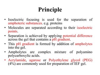
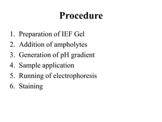
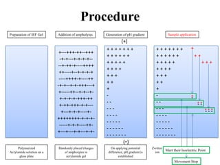
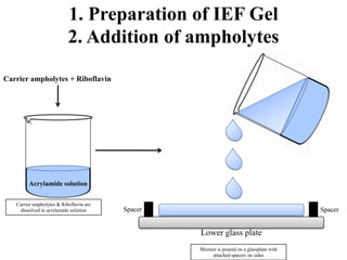
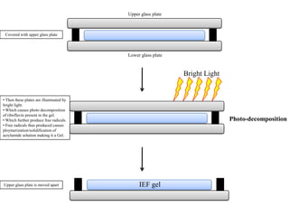
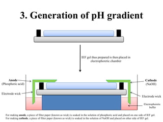
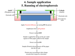
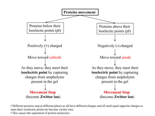
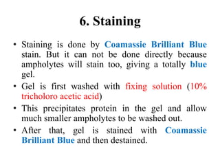
Isoelectric Focusing (IEF)
Amandeep Singh
Assistant Professor,
Department of Biotechnology,
GSSDGS Khalsa College,
Patiala
Principle
• Isoelectric focusing is used for the separation of
amphoteric substances. e.g. proteins
• Molecules are separated according to their isoelectric
points.
• Separation is achieved by applying potential difference
across the gel that contains a pH gradient.
• This pH gradient is formed by addition of ampholytes
into the gel.
• Ampholytes are complex mixture of polyamino
polycarboxylic acids.
• Acrylamide, agarose or Polyethylene glycol (PEG)
(4%) are commonly used for preparation of IEF gel.
Procedure
1. Preparation of IEF Gel
2. Addition of ampholytes
3. Generation of pH gradient
4. Sample application
5. Running of electrophoresis
6. Staining
+---+++-++--+++
-+-+---+-+-+-+--
--+-++-+----++++
++----+--+-+-+-+
----++++-+----++
-+-+--+-+-+++---+
+-----++--+-+--+-
+-+-+-+-+++-++
+-++-+++-+-+---
+---+-+-+----+-
++++++++-+-+-+-
-----+-+-+----++-
+---+---+-+---+-+-
+ + + + + + +
+ + + + + +
+ + + + +
+ + + +
+ + +
+ +
+
-
- -
- - -
- - - -
- - - - -
- - - - - -
- - - - - - -
+ + + + + + +
+ + + + + + + +
+ + + + + + + +
+ + + +
+ + +
+ +
+
-
- -
- - -
- - - -
- - - - -
- - - - - -
- - - - - - -
+-
+ +- -
+ + +- - -
+
Meet their Isoelectric Point
Movement Stop
Preparation of IEF Gel Addition of ampholytes Generation of pH gradient Sample application
Zwittor
ion
(+)
(-)
Procedure
Polymerized
Acrylamide solution on a
glass plate
Randomly placed charges
of ampholytes in
acrylamide gel
On applying potential
difference, pH gradient is
established
1. Preparation of IEF Gel
2. Addition of ampholytes
Carrier ampholytes + Riboflavin
Acrylamide solution
Lower glass plate
SpacerSpacer
Carrier ampholytes & Riboflavin are
dissolved in acrylamide solution
Mixture is poured on a glassplate with
attached spacers on sides
Lower glass plate
Upper glass plate
Bright Light
Photo-decomposition
Covered with upper glass plate
• Then these plates are illuminated by
bright light.
• Which causes photo decomposition
of riboflavin present in the gel.
• Which further produce free radicals.
• Free radicals thus produced causes
ploymerization/solidification of
acrylamide solution making it a Gel.
IEF gelUpper glass plate is moved apart
3. Generation of pH gradient
Electrode wick
Electrode wick
Anode
(Phosphoric acid)
Cathode
(NaOH)
IEF gel thus prepared is then placed in
electrophoretic chamber
For making anode, a piece of filter paper (known as wick) is soaked in the solution of phosphoric acid and placed on one side of IEF gel.
For making cathode, a piece of filter paper (known as wick) is soaked in the solution of NaOH and placed on other side of IEF gel.
Electrophoretic
buffer
4. Sample application
5. Running of electrophoresis
Apply Potential difference by turning ON the power
Ampholytes form a pH gradient
Power is turned OFF
Sample is applied by laying on the gel, small
squares of filter paper soaked in the sample.
Power is turned ON
Sample (Protein) electrophoresis off the paper
into the IEF gel
Protein move according to their charge
towards their respective electrodes
Electrode wick
Electrode wick
Anode
(Phosphoric acid)
Cathode
(NaOH)
Electrophoretic
buffer
Proteins movement
Proteins below their
Isoelectic points (pI)
Proteins above their
Isoelectic points (pI)
Positively (+) charged Negatively (-) charged
Move toward cathode Move toward anode
As they move, they meet their
isoelectric point by capturing
charges from ampholytes
present in the gel
As they move, they meet their
isoelectric point by capturing
charges from ampholytes
present in the gel
Movement Stop
(become Zwittor ion)
Movement Stop
(become Zwittor ion)
• Different proteins stop at different places as all have different charges and all need equal opposite charges to
meet their isoelectric points (to become zwitter ion).
• This causes the separation of protein molecules.
6. Staining
• Staining is done by Coamassie Brilliant Blue
stain. But it can not be done directly because
ampholytes will stain too, giving a totally blue
gel.
• Gel is first washed with fixing solution (10%
tricholoro acetic acid)
• This precipitates protein in the gel and allow
much smaller ampholytes to be washed out.
• After that, gel is stained with Coamassie
Brilliant Blue and then destained.

Isoelectric focusing

  • 1.
    Isoelectric Focusing (IEF) AmandeepSingh Assistant Professor, Department of Biotechnology, GSSDGS Khalsa College, Patiala
  • 2.
    Principle • Isoelectric focusingis used for the separation of amphoteric substances. e.g. proteins • Molecules are separated according to their isoelectric points. • Separation is achieved by applying potential difference across the gel that contains a pH gradient. • This pH gradient is formed by addition of ampholytes into the gel. • Ampholytes are complex mixture of polyamino polycarboxylic acids. • Acrylamide, agarose or Polyethylene glycol (PEG) (4%) are commonly used for preparation of IEF gel.
  • 3.
    Procedure 1. Preparation ofIEF Gel 2. Addition of ampholytes 3. Generation of pH gradient 4. Sample application 5. Running of electrophoresis 6. Staining
  • 4.
    +---+++-++--+++ -+-+---+-+-+-+-- --+-++-+----++++ ++----+--+-+-+-+ ----++++-+----++ -+-+--+-+-+++---+ +-----++--+-+--+- +-+-+-+-+++-++ +-++-+++-+-+--- +---+-+-+----+- ++++++++-+-+-+- -----+-+-+----++- +---+---+-+---+-+- + + ++ + + + + + + + + + + + + + + + + + + + + + + + + - - - - - - - - - - - - - - - - - - - - - - - - - - - - + + + + + + + + + + + + + + + + + + + + + + + + + + + + + + + + + - - - - - - - - - - - - - - - - - - - - - - - - - - - - +- + +- - + + +- - - + Meet their Isoelectric Point Movement Stop Preparation of IEF Gel Addition of ampholytes Generation of pH gradient Sample application Zwittor ion (+) (-) Procedure Polymerized Acrylamide solution on a glass plate Randomly placed charges of ampholytes in acrylamide gel On applying potential difference, pH gradient is established
  • 5.
    1. Preparation ofIEF Gel 2. Addition of ampholytes Carrier ampholytes + Riboflavin Acrylamide solution Lower glass plate SpacerSpacer Carrier ampholytes & Riboflavin are dissolved in acrylamide solution Mixture is poured on a glassplate with attached spacers on sides
  • 6.
    Lower glass plate Upperglass plate Bright Light Photo-decomposition Covered with upper glass plate • Then these plates are illuminated by bright light. • Which causes photo decomposition of riboflavin present in the gel. • Which further produce free radicals. • Free radicals thus produced causes ploymerization/solidification of acrylamide solution making it a Gel. IEF gelUpper glass plate is moved apart
  • 7.
    3. Generation ofpH gradient Electrode wick Electrode wick Anode (Phosphoric acid) Cathode (NaOH) IEF gel thus prepared is then placed in electrophoretic chamber For making anode, a piece of filter paper (known as wick) is soaked in the solution of phosphoric acid and placed on one side of IEF gel. For making cathode, a piece of filter paper (known as wick) is soaked in the solution of NaOH and placed on other side of IEF gel. Electrophoretic buffer
  • 8.
    4. Sample application 5.Running of electrophoresis Apply Potential difference by turning ON the power Ampholytes form a pH gradient Power is turned OFF Sample is applied by laying on the gel, small squares of filter paper soaked in the sample. Power is turned ON Sample (Protein) electrophoresis off the paper into the IEF gel Protein move according to their charge towards their respective electrodes Electrode wick Electrode wick Anode (Phosphoric acid) Cathode (NaOH) Electrophoretic buffer
  • 9.
    Proteins movement Proteins belowtheir Isoelectic points (pI) Proteins above their Isoelectic points (pI) Positively (+) charged Negatively (-) charged Move toward cathode Move toward anode As they move, they meet their isoelectric point by capturing charges from ampholytes present in the gel As they move, they meet their isoelectric point by capturing charges from ampholytes present in the gel Movement Stop (become Zwittor ion) Movement Stop (become Zwittor ion) • Different proteins stop at different places as all have different charges and all need equal opposite charges to meet their isoelectric points (to become zwitter ion). • This causes the separation of protein molecules.
  • 10.
    6. Staining • Stainingis done by Coamassie Brilliant Blue stain. But it can not be done directly because ampholytes will stain too, giving a totally blue gel. • Gel is first washed with fixing solution (10% tricholoro acetic acid) • This precipitates protein in the gel and allow much smaller ampholytes to be washed out. • After that, gel is stained with Coamassie Brilliant Blue and then destained.