Importance
• Can bea sign of serious diseases
• Can be seen in other specialties
• Hard to diagnose because it integrates
several organs and systems together and the
underlying cause is not clear.
• Very common, but hard to deal with.
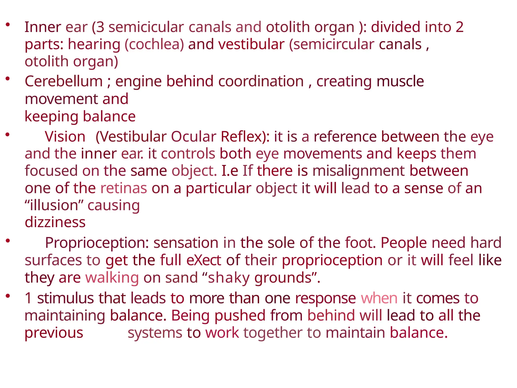
• Inner ear(3 semicicular canals and otolith organ ): divided into 2
parts: hearing (cochlea) and vestibular (semicircular canals ,
otolith organ)
• Cerebellum ; engine behind coordination , creating muscle
movement and
keeping balance
• Vision (Vestibular Ocular Reflex): it is a reference between the eye
and the inner ear. it controls both eye movements and keeps them
focused on the same object. I.e If there is misalignment between
one of the retinas on a particular object it will lead to a sense of an
“illusion” causing
dizziness
• Proprioception: sensation in the sole of the foot. People need hard
surfaces to get the full eXect of their proprioception or it will feel like
they are walking on sand “shaky grounds”.
• 1 stimulus that leads to more than one response when it comes to
maintaining balance. Being pushed from behind will lead to all the
previous systems to work together to maintain balance.
Physiology
Function of vestibular
system:
•“Input” resulting from a stimulus that needs to be corrected through the
vestibular system such as falling down. An “output” results from
responses of the vestibular system to the input such as the eyes,
cerebellum .. Etc.
• The physical stimulus (input) will be transformed into a biological
stimulas in the brain stem which will in turn be sent afterwards to the
corresponding areas in the vestibular system.
• Transform of the forces associate with head acceleration and gravity into a
biological signals that the brain can use to develop subjective awareness of
head position in space (orientation)
• produce motor reflexes that will maintain posture and ocular stability to
prevent the
feeling of dizziness.
• If there is a defect in the input and output processes the patient will
present with vertigo, defects in the gait or ocular distortions.
8.
It is notsurprisingly that vestibular lesion
cause:
• Imbalance
• posture and gait
imbalance
• visual distortion (oscillopsia
9.
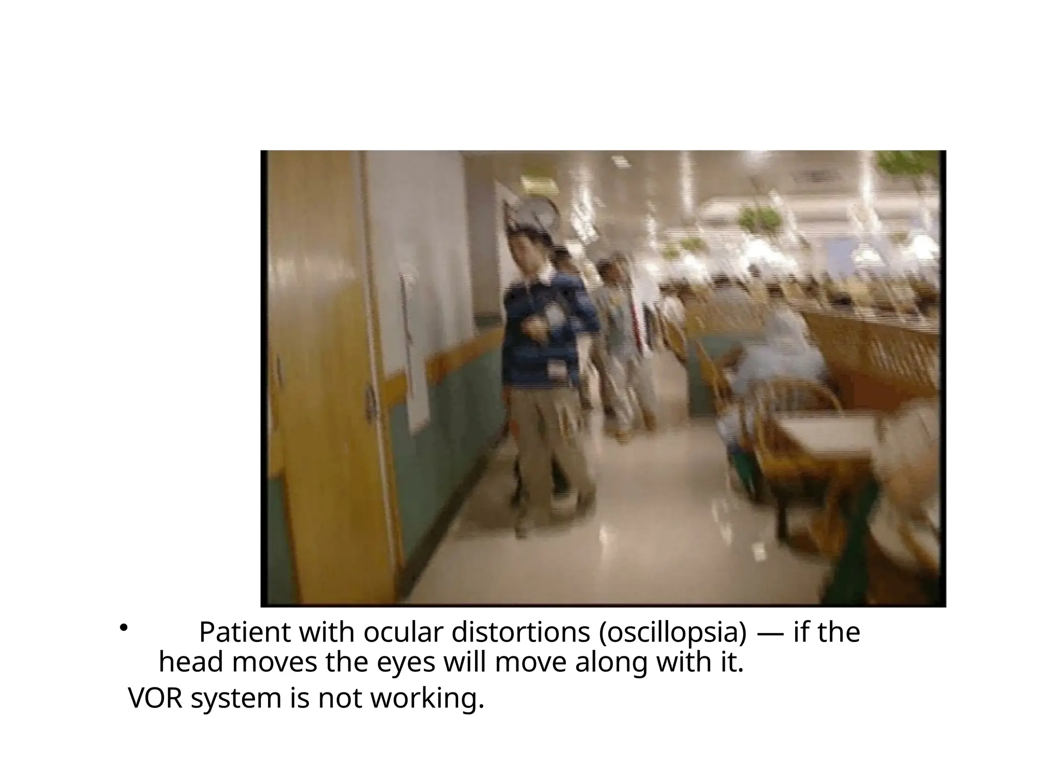
• Patient withocular distortions (oscillopsia) — if the
head moves the eyes will move along with it.
VOR system is not working.
VERTI
• The word"vertigo" comes from the
Latin "vertere", to turn + the su0ix "-
igo", a condition = a condition of
turning about).
• It is an allusion of being moving or
the world is moving too.
12.
What are thequestions to ask in history ?
• Onset (acute/chronic)
• Frequency — how often
• Duration
• Associated auditory symptoms
• Aggravating and relieving factors
• Ear disease or ear surgery — tinnitus?
• Trauma
• Migraine
• Ototoxic drug intake — (chemotherapy,
aminoglycosides, methotrexate)
• Family history
• Motion sickness
13.
Differential diagnosis
A) peripheralvestibular loss — up to the
vestibular
nerve.
B) central vestibular loss —above the level of
the vestibular nerve and towards the brain.
14.
What are thecauses of
peripheral vestibular
loss ?
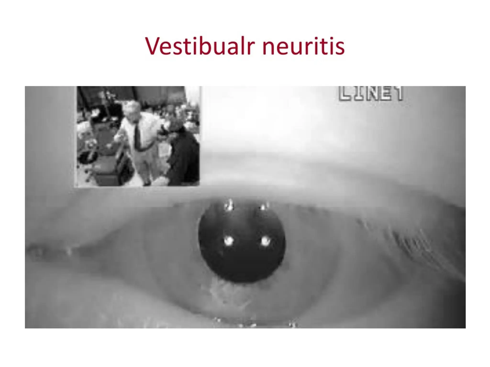
Vestibuiar Neuritis
• Viralinfection of vestibular organ
• Affect all ages but rare in children — mostly adults
• Añected patient presents acutely with spontaneous
nystagmus ,vertigo and nausea &vomiting stays for hours and
sometimes days.
• Patient requires only symptomatic treatment
• It takes 3 weeks to recover from vestibular neuritis
• Diagnosis — no other tool other than history.
• Recent study studies show that giving steroids decreases the 3
week recovery period.
18.
BPPV( benign
paroxysmal
positional vertigo
•Its provoked by certain positions.
• Pathophysiology:
• Calcium carbonate particles shear oñ and enter the canal
leading to brief episodes of vertigo.
C*n
M
Canalithiasi
s Cupulolithiasis
Vestibulithiasls
19.
BPPV
• The mostcommon cause of vertigo in
patient >
40 years
• Repeated attacks of vertigo usually of
short duration less than a minute .
• Provoked by certain positions (rolling in
beds, looking up ,and head rotations)
• Not associated with any hearing
impairment
20.
BPPV
Diagnosis
• History
• Dix-HaIpikemaneuver : putting the patient in a
certain position to stimulate the arack, and to look at
the eye (causes nystagmus) to see which canal is mostly
aFected by trying to push the particles inside the canal
and inducing the sense of dizziness.
• Treatment: repositioning of the head to get particles
out of the canal (Epley or particle repositioning
maneuver) . No medical or surgical treatment needed.
• Epley's maneuver could even be done at home.
21.
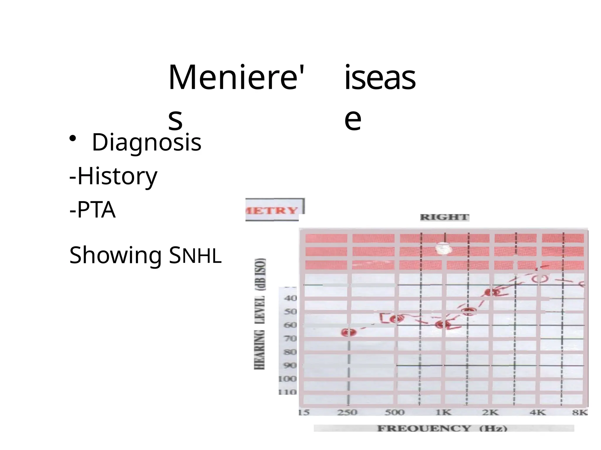
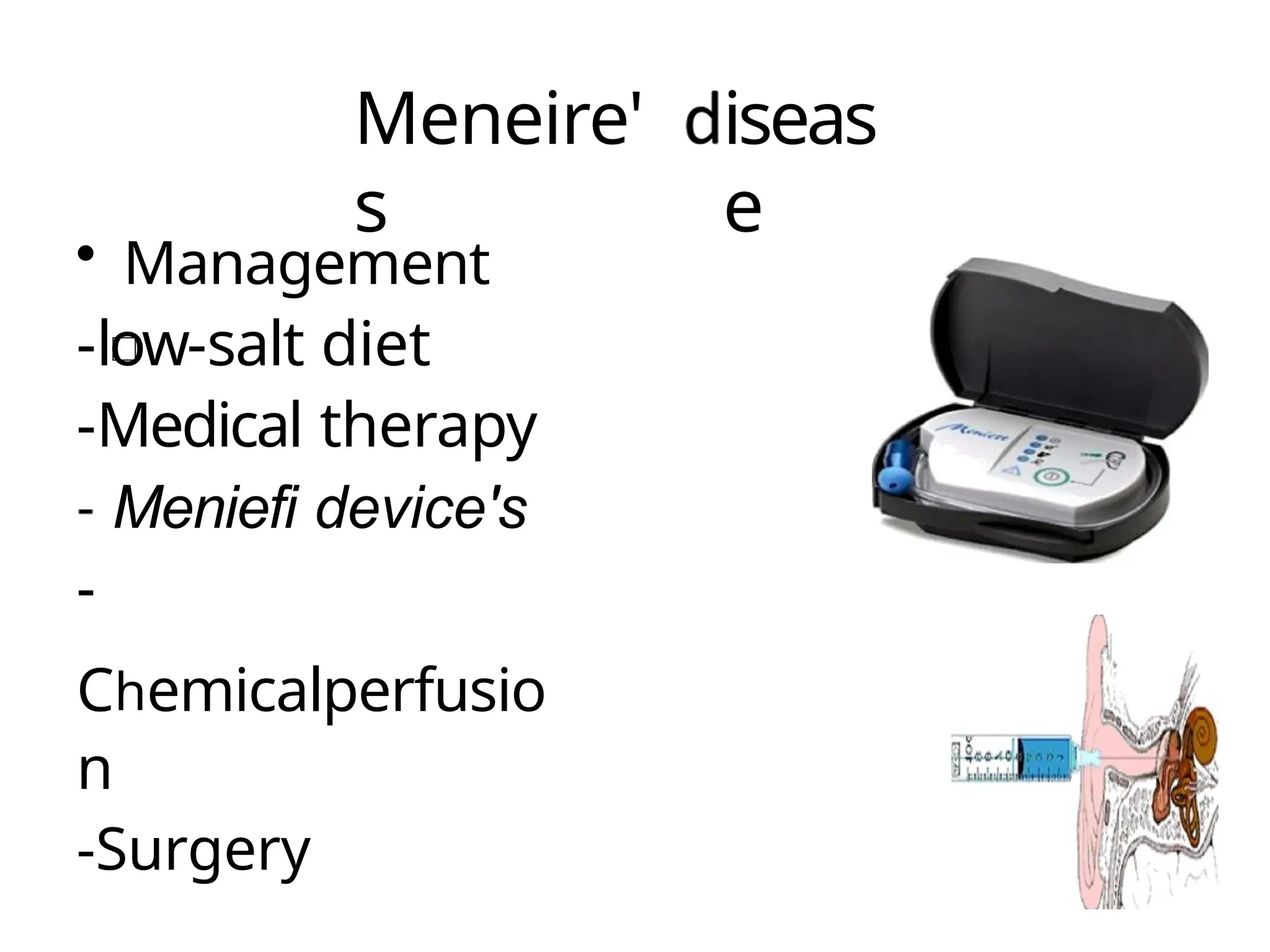
Endolymphatic
hydrop (Meneire's
disease)
• vertigo(minutes to hours )
• Low frequency fluctuating SNHL
• Tinnitus and fullness in the ear.
• In 10 - 20% of cases the disease
later
involves the opposite ear
22.
Endolymphatic
hydrop (Meneire's
disease)
• vertigo(minutes to hours )
• Low frequency fluctuating SNHL
• Tinnitus and fullness in the ear.
• In 10 - 20% of cases the disease
later
involves the opposite ear
I
I
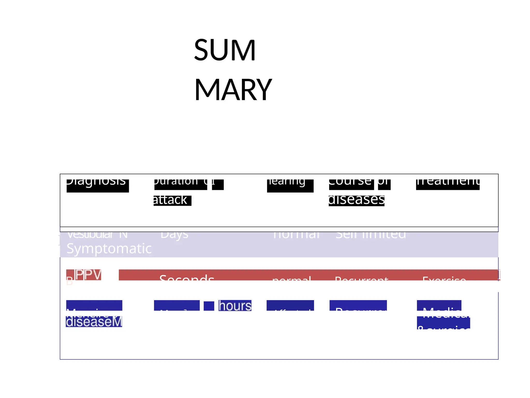
Diagnosis Duration OÎhearing Course of Treatment
attack diseases
Vestibular N Days normal Self limited
Symptomatic
B Seconds normal Recurrent Exercise
Meneire s M;‹les IO Affected Recurrent Medicaï
&surgical
SU
MARY
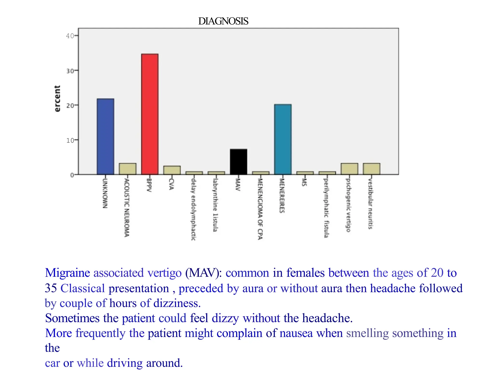
40
10
DIAGNOSIS
Migraine associated vertigo(MAV): common in females between the ages of 20 to
35 Classical presentation , preceded by aura or without aura then headache followed
by couple of hours of dizziness.
Sometimes the patient could feel dizzy without the headache.
More frequently the patient might complain of nausea when smelling something in
the
car or while driving around.
Central
• CVA (Cerebrovascular accident)-
most
common
• Brain tumor ( acoustic neuroma )
• Multiple sclerosis
30.
CV
A
• Elderly patientwith chronic
disease like (DM ,HTN) with
sudden attack of vertigo
+neuroiogicaI symptoms
31.
Acoustic
tumor
• Benign tumor
•Arise from vestibular division of
VIII Clinical presentation:
• Unilateral tinnitus
• Hearing loss
• Dizziness
• The only way to differentiate between
Meniere's
disease and the Acoustic tumor is by MRI.
Scenario #
1
The patientwho is having a first
ever attack of acute spontaneous
vertigo.
• Acute vestibular neuritis
• cerebellar infarction.
How to differentiate ?
- Clinically ( General appearance of patient
/nystagmus/head impulse test)
- Radiology
37.
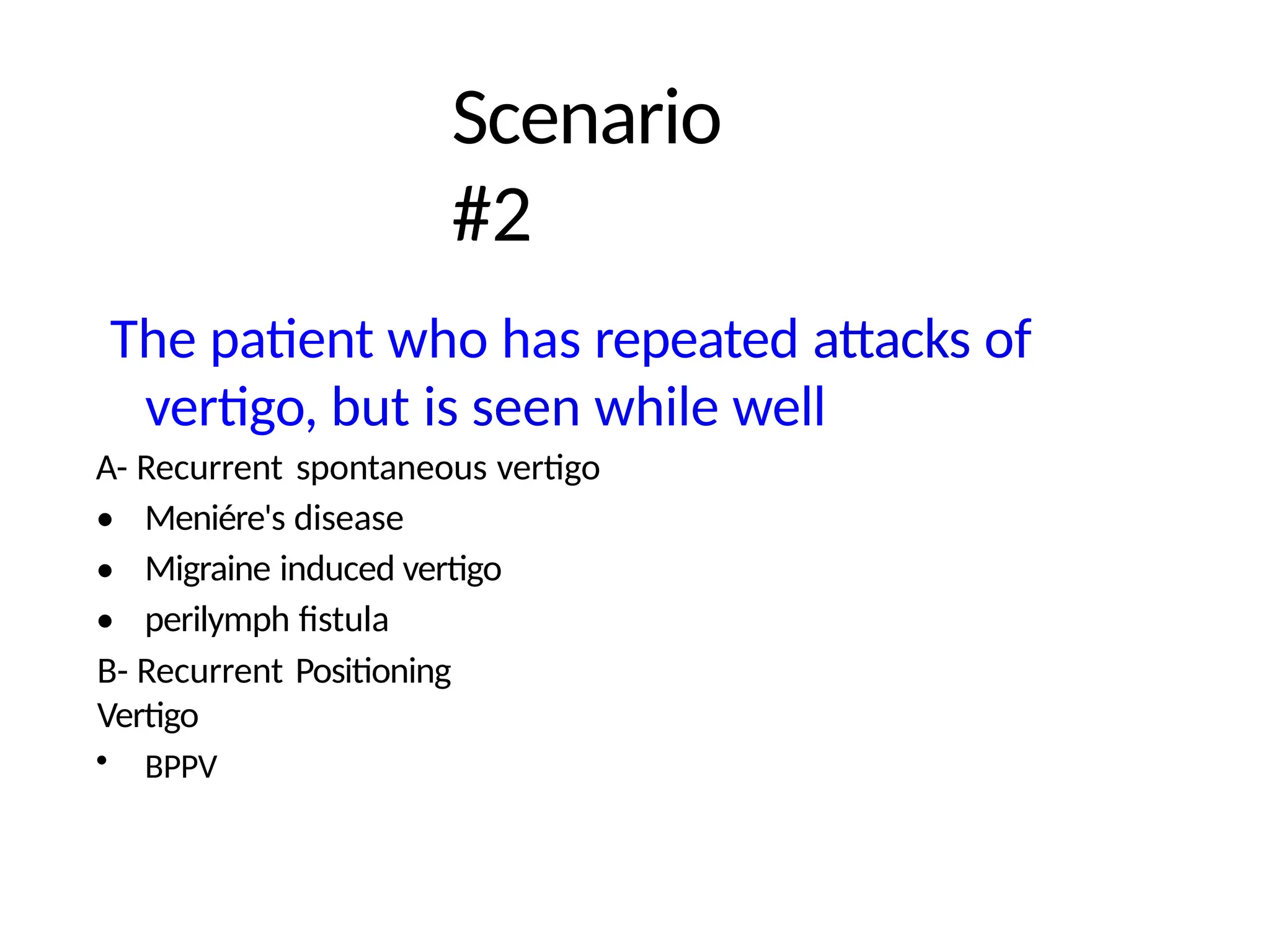
Scenario
#2
The patient whohas repeated attacks of
vertigo, but is seen while well
A- Recurrent spontaneous vertigo
•
•
•
Meniére's disease
Migraine induced vertigo
perilymph fistula
B- Recurrent Positioning
Vertigo
• BPPV
38.
Scenario
#3
The patient whois off balance
• Bilateral vestibulopathy — could be
due to streptomycin
• posterior fossa tumour