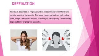
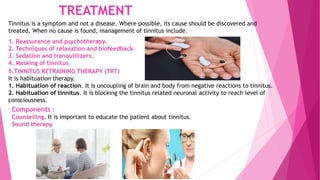
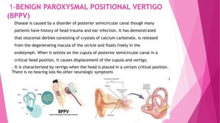
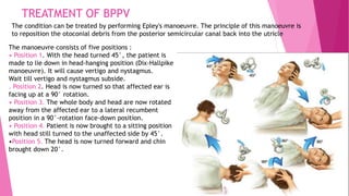
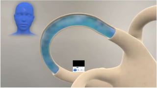
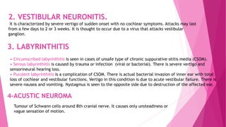
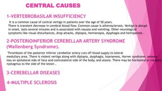
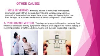
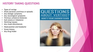
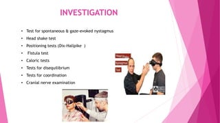
TINNITIS AN VERTIGO
PRESENTED BY
DR AWAIS IRSHAD
LEARNING OBJECTIVES
TINNITUS
• Definition
• Classification
• Causes of tinnitus
• Treatment of tinnitus
VERTIGO
• Definition of vertigo
• It’s Causes
• Specific Question for History
• Differential diagnosis
• Investigation
• Management Plan
DEFFINATION
Tinnitus is described as ringing sound or noises in ears when there is no
outside source of the sounds. The sound ranges varies from high to low
pitch, single tone to multi-tonal, or having no tonal quality. Tinnitus may
begin suddenly or progress gradually.
SYMPTOMS
Tinnitus symptoms include the below types of noises in
ears:
. Ringing
. Buzzing
. Roaring
. Clicking
. Hissing
TYPES OF TINNITUS
There are two broad types of tinnitus:
1-Subjective tinnitus is tinnitus which only hear by patient. This is the most
common type of tinnitus. It can be caused by ear problems in your outer, middle or
inner ear.
2-Objective tinnitus is tinnitus in which doctor can hear when he or she does
an examination. This is rare type and it may be caused due to blood vessel problem,
an inner ear bone condition or muscle contractions.
CAUSES OF SUBJECTIVE TINNITUS
1 Otologic
• Impacted wax
• Fluid in middle ear
• Acute otitis media
• Chronic otitis media
• Ménière’s disease
• Presbycusis
• Noise-induced hearing loss
• Idiopathic sudden SNHL
• Acoustic neuroma
2 Metabolic
• Hypothyroidism
• Hyperthyroidism
• Obesity
• Hyperlipidemia
• Vitamin deficiency
(e.g. B12)
3 Neurologic
• Head injury (labyrinthine
• Temporal bone fractures
• Whiplash injury
• Multiple sclerosis
• Brain hemorrhage
• Brain infarct
4 Cardiovascular
• Hypertension
• Hypotension
• Anemia
• Cardiac arrhythmias
• Arteriosclerosis
5 Pharmacologic
• All ototoxic drugs
6 Psychogenic
• Anxiety/depression
CAUSES OF OBJECTIVE TINNITUS
• Vascular
• AV shunts
– Congenital AV malformations
– Glomus tumor of middle ear
• Arterial bruit
– Carotid aneurysm
– Carotid stenosis
– Vascular loop pressing on VIIIth nerve in
internal auditory canal
– High-riding carotid artery
– Persistent stapedial artery
• Venous hum
– Dehiscent jugular bulb
• Patulous Eustachian tube
• Palatal myoclonus
• Idiopathic stapedius or
tensor tympani myoclonus
• Dental
• Clicking of TM joint
TREATMENT
Tinnitus is a symptom and not a disease. Where possible, its cause should be discovered and
treated. When no cause is found, management of tinnitus include.
1. Reassurance and psychotherapy.
2. Techniques of relaxation and biofeedback.
3. Sedation and tranquillizers.
4. Masking of tinnitus.
5.TINNITUS RETRAINING THERAPY (TRT)
It is habituation therapy.
1. Habituation of reaction. It is uncoupling of brain and body from negative reactions to tinnitus.
2. Habituation of tinnitus. It is blocking the tinnitus related neuronal activity to reach level of
consciousness.
Components :
Counselling. It is important to educate the patient about tinnitus.
Sound therapy
VERTIGO
A feeling in which the external world seems to revolve around the
individual or in which the individual itself seems to revolve in space.
Not a disease, But a symptom.
CAUSES OF VERTIGO
Disorder of vestibular system causes vertigo and are divided into
1-Peripheral vestibular disorders
2-Central vestibular disorders
1. Peripheral, which involve vestibular end organs and their 1st order neurons
(i.e. the vestibular nerve). The cause lies in the internal ear or the VIIIth nerve.
They are responsible for 85% of all cases of vertigo.
2. Central, which involve central nervous system after the entrance of vestibular
nerve in the brainstem and involve vestibulo-ocular, vestibulospinal and other
central nervous system pathways.
VESTIBULAR DISORDER
PERIPHERAL
• Meniere's disease
• Benign paroxysmal positional vertigo
• Vestibular neuronitis
• Labyrinthitis
• Vestibulotoxic drugs
• Head trauma
• Perilymph fistula
• Syphilis
• Acoustic neuroma
CENTRAL
• Vertebrobasilar insufficiency
• Posterior inferior cerebellar artery
syndrome
• Basilar migraine
• Cerebellar disease
• Multiple sclerosis
• Tumors of brainstem and fourth
ventricle
• Epilepsy
• Cervical vertigo
1-BENIGN PAROXYSMAL POSITIONAL VERTIGO
(BPPV)
Disease is caused by a disorder of posterior semicircular canal though many
patients have history of head trauma and ear infection. It has demonstrated
that otoconial derbies consisting of crystals of calcium carbonate, is released
from the degenerating macula of the utricle and floats freely in the
endolymph. When it settles on the cupula of posterior semicircular canal in a
critical head position, it causes displacement of the cupula and vertigo.
. It is characterized by vertigo when the head is placed in a certain critical position.
There is no hearing loss No other neurologic symptoms
TREATMENT OF BPPV
The condition can be treated by performing Epley's manoeuvre. The principle of this manoeuvre is
to reposition the otoconial debris from the posterior semicircular canal back into the utricle
The manoeuvre consists of five positions :
• Position 1. With the head turned 45°, the patient is
made to lie down in head-hanging position (Dix-Hallpike
manoeuvre). It will cause vertigo and nystagmus.
Wait till vertigo and nystagmus subside.
. Position 2. Head is now turned so that affected ear is
facing up at a 90° rotation.
• Position 3. The whole body and head are now rotated
away from the affected ear to a lateral recumbent
position in a 90°-rotation face-down position.
• Position 4. Patient is now brought to a sitting position
with head still turned to the unaffected side by 45°.
•Position 5. The head is now turned forward and chin
brought down 20°.
2. VESTIBULAR NEURONITIS.
It is characterized by severe vertigo of sudden onset with no cochlear symptoms. Attacks may last
from a few days to 2 or 3 weeks. It is thought to occur due to a virus that attacks vestibular
ganglion.
3. LABYRINTHITIS
• Circumscribed labyrinthitis is seen in cases of unsafe type of chronic suppurative otitis media (CSOM).
• Serous labyrinthitis is caused by trauma or infection (viral or bacterial). There is severe vertigo and
sensorineural hearing loss.
• Purulent labyrinthitis is a complication of CSOM. There is actual bacterial invasion of inner ear with total
loss of cochlear and vestibular functions. Vertigo in this condition is due to acute vestibular failure. There is
severe nausea and vomiting. Nystagmus is seen to the opposite side due to destruction of the affected ear.
Tumour of Schwann cells around 8th cranial nerve. It causes only unsteadiness or
vague sensation of motion.
4-ACUSTIC NEUROMA
2-POSTERIOINFERIOR CEREBELLAR ARTERY SYNDROME
(Wallenberg Syndrome).
Thrombosis of the posterior inferior cerebellar artery cuts off blood supply to lateral
medullary area. There is violent vertigo along with diplopia, dysphagia, hoarseness, Horner syndrome, sensory
loss on ipsilateral side of face and contralateral side of the body, and ataxia. There may be horizontal or rotatory
nystagmus to the side of the lesion .
It is a common cause of central vertigo in patients over the age of 50 years.
There is transient decrease in cerebral blood flow. Common cause is atherosclerosis. Vertigo is abrupt
in onset, lasts several minutes and is associated with nausea and vomiting. Other neurological
symptoms like visual disturbances, drop attacks, diplopia, hemianopia, dysphagia and hemiparesis .
1-VERTEBROBASILAR INSUFFICIENCY
3-CEREBELLAR DISEASES
4-MULTIPLE SCLEROSIS
CENTRAL CAUSES
OTHER CAUSES
1. OCULAR VERTIGO. Normally, balance is maintained by integrated
information received from the eyes, labyrinths and somatosensory system. A
mismatch of information from any of these organs causes vertigo and in this case
from the eyes. i.e acute extraocular muscle paresis or high errors of refraction
2. PSYCHOGENIC VERTIGO. This diagnosis is suspected in patients suffering from
emotional tension and anxiety. Symptom of vertigo is often vague in the form of floating or
swimming sensation or light headedness. Caloric test shows an exaggerated response.
HISTORY TAKING QUESTIONS
• Types of vertigo
• Onset,duration,continous or episodic
• Any otologic symptoms
• Any neurological symptoms
• Tinnitus( unilateral bilateral)
• Gait ataxia or imbalance
• Vomiting/nausea
• Any visual disturbances
• Head position and headache
• Stress history
• Any drug intake
DIFFERENTIAL DIAGNOSIS
PERIPHERAL:
• BPPV
• Perilymphatic fistula
• Ménière's disease
• Vestibular neuronitis
• Basilar artery migraine
• Acoustic neuroma
• Hypothyroidism
• Hypoglycemia
CENTRAL:
• CNS infection (BS,cerebellum)
• Tumor (Benign or Neoplastic)
• Labyrinthitis
• Cerebellar hemorrhage/infarct
• Vertebrobasilar insufficiency
• Multiple sclerosis
• Traumatic( post traumatic syn)
INVESTIGATION
• Test for spontaneous & gaze-evoked nystagmus
• Head shake test
• Positioning tests (Dix-Hallpike )
• Fistula test
• Caloric tests
• Tests for disequilibrium
• Tests for coordination
• Cranial nerve examination
CHANGE IN DIET AND LIFESTYLE
• salt restriction
• avoiding alcohol
• Bed rest to avoid vertigo
• Reducing your high sugar intake
• Exercises prescribed by physiotherapists involve moving the eye balls up and
down and sideways in a supine or sitting position. Balance exercises help to
regain normal activities faster
TREATMENT OF VERTIGO
1. Reassurance/Psychological Support
2. Pharmacotherapy
3. Vestibular suppressive drugs
4. Adaptation exercises
5. Intratympanic antibiotic injections
6. Surgery
• Conservative
• Destructive
Treatment is according to the specific causes of vertigo
And the symptoms occurring
CBL-9
A 60 years old diabetic patient has presented with a complaint of
sudden brief period of imbalance on head movement specially getting
out of the bed. There is no history of any hearing loss.
A similar episode had occurred about 4 years back.
What specific questions will you ask to reach the
diagnosis?
Give differential diagnosis?
Give management plan of your diagnosis?
What complications can develop?
Write a prescription for your patient?
THANKS

TINNITUS AND VERTIGO

  • 1.
  • 2.
    LEARNING OBJECTIVES TINNITUS • Definition •Classification • Causes of tinnitus • Treatment of tinnitus VERTIGO • Definition of vertigo • It’s Causes • Specific Question for History • Differential diagnosis • Investigation • Management Plan
  • 3.
    DEFFINATION Tinnitus is describedas ringing sound or noises in ears when there is no outside source of the sounds. The sound ranges varies from high to low pitch, single tone to multi-tonal, or having no tonal quality. Tinnitus may begin suddenly or progress gradually.
  • 4.
    SYMPTOMS Tinnitus symptoms includethe below types of noises in ears: . Ringing . Buzzing . Roaring . Clicking . Hissing
  • 5.
    TYPES OF TINNITUS Thereare two broad types of tinnitus: 1-Subjective tinnitus is tinnitus which only hear by patient. This is the most common type of tinnitus. It can be caused by ear problems in your outer, middle or inner ear. 2-Objective tinnitus is tinnitus in which doctor can hear when he or she does an examination. This is rare type and it may be caused due to blood vessel problem, an inner ear bone condition or muscle contractions.
  • 6.
    CAUSES OF SUBJECTIVETINNITUS 1 Otologic • Impacted wax • Fluid in middle ear • Acute otitis media • Chronic otitis media • Ménière’s disease • Presbycusis • Noise-induced hearing loss • Idiopathic sudden SNHL • Acoustic neuroma 2 Metabolic • Hypothyroidism • Hyperthyroidism • Obesity • Hyperlipidemia • Vitamin deficiency (e.g. B12) 3 Neurologic • Head injury (labyrinthine • Temporal bone fractures • Whiplash injury • Multiple sclerosis • Brain hemorrhage • Brain infarct 4 Cardiovascular • Hypertension • Hypotension • Anemia • Cardiac arrhythmias • Arteriosclerosis 5 Pharmacologic • All ototoxic drugs 6 Psychogenic • Anxiety/depression
  • 7.
    CAUSES OF OBJECTIVETINNITUS • Vascular • AV shunts – Congenital AV malformations – Glomus tumor of middle ear • Arterial bruit – Carotid aneurysm – Carotid stenosis – Vascular loop pressing on VIIIth nerve in internal auditory canal – High-riding carotid artery – Persistent stapedial artery • Venous hum – Dehiscent jugular bulb • Patulous Eustachian tube • Palatal myoclonus • Idiopathic stapedius or tensor tympani myoclonus • Dental • Clicking of TM joint
  • 8.
    TREATMENT Tinnitus is asymptom and not a disease. Where possible, its cause should be discovered and treated. When no cause is found, management of tinnitus include. 1. Reassurance and psychotherapy. 2. Techniques of relaxation and biofeedback. 3. Sedation and tranquillizers. 4. Masking of tinnitus. 5.TINNITUS RETRAINING THERAPY (TRT) It is habituation therapy. 1. Habituation of reaction. It is uncoupling of brain and body from negative reactions to tinnitus. 2. Habituation of tinnitus. It is blocking the tinnitus related neuronal activity to reach level of consciousness. Components : Counselling. It is important to educate the patient about tinnitus. Sound therapy
  • 9.
    VERTIGO A feeling inwhich the external world seems to revolve around the individual or in which the individual itself seems to revolve in space. Not a disease, But a symptom.
  • 10.
    CAUSES OF VERTIGO Disorderof vestibular system causes vertigo and are divided into 1-Peripheral vestibular disorders 2-Central vestibular disorders 1. Peripheral, which involve vestibular end organs and their 1st order neurons (i.e. the vestibular nerve). The cause lies in the internal ear or the VIIIth nerve. They are responsible for 85% of all cases of vertigo. 2. Central, which involve central nervous system after the entrance of vestibular nerve in the brainstem and involve vestibulo-ocular, vestibulospinal and other central nervous system pathways.
  • 11.
    VESTIBULAR DISORDER PERIPHERAL • Meniere'sdisease • Benign paroxysmal positional vertigo • Vestibular neuronitis • Labyrinthitis • Vestibulotoxic drugs • Head trauma • Perilymph fistula • Syphilis • Acoustic neuroma CENTRAL • Vertebrobasilar insufficiency • Posterior inferior cerebellar artery syndrome • Basilar migraine • Cerebellar disease • Multiple sclerosis • Tumors of brainstem and fourth ventricle • Epilepsy • Cervical vertigo
  • 12.
    1-BENIGN PAROXYSMAL POSITIONALVERTIGO (BPPV) Disease is caused by a disorder of posterior semicircular canal though many patients have history of head trauma and ear infection. It has demonstrated that otoconial derbies consisting of crystals of calcium carbonate, is released from the degenerating macula of the utricle and floats freely in the endolymph. When it settles on the cupula of posterior semicircular canal in a critical head position, it causes displacement of the cupula and vertigo. . It is characterized by vertigo when the head is placed in a certain critical position. There is no hearing loss No other neurologic symptoms
  • 13.
    TREATMENT OF BPPV Thecondition can be treated by performing Epley's manoeuvre. The principle of this manoeuvre is to reposition the otoconial debris from the posterior semicircular canal back into the utricle The manoeuvre consists of five positions : • Position 1. With the head turned 45°, the patient is made to lie down in head-hanging position (Dix-Hallpike manoeuvre). It will cause vertigo and nystagmus. Wait till vertigo and nystagmus subside. . Position 2. Head is now turned so that affected ear is facing up at a 90° rotation. • Position 3. The whole body and head are now rotated away from the affected ear to a lateral recumbent position in a 90°-rotation face-down position. • Position 4. Patient is now brought to a sitting position with head still turned to the unaffected side by 45°. •Position 5. The head is now turned forward and chin brought down 20°.
  • 15.
    2. VESTIBULAR NEURONITIS. Itis characterized by severe vertigo of sudden onset with no cochlear symptoms. Attacks may last from a few days to 2 or 3 weeks. It is thought to occur due to a virus that attacks vestibular ganglion. 3. LABYRINTHITIS • Circumscribed labyrinthitis is seen in cases of unsafe type of chronic suppurative otitis media (CSOM). • Serous labyrinthitis is caused by trauma or infection (viral or bacterial). There is severe vertigo and sensorineural hearing loss. • Purulent labyrinthitis is a complication of CSOM. There is actual bacterial invasion of inner ear with total loss of cochlear and vestibular functions. Vertigo in this condition is due to acute vestibular failure. There is severe nausea and vomiting. Nystagmus is seen to the opposite side due to destruction of the affected ear. Tumour of Schwann cells around 8th cranial nerve. It causes only unsteadiness or vague sensation of motion. 4-ACUSTIC NEUROMA
  • 16.
    2-POSTERIOINFERIOR CEREBELLAR ARTERYSYNDROME (Wallenberg Syndrome). Thrombosis of the posterior inferior cerebellar artery cuts off blood supply to lateral medullary area. There is violent vertigo along with diplopia, dysphagia, hoarseness, Horner syndrome, sensory loss on ipsilateral side of face and contralateral side of the body, and ataxia. There may be horizontal or rotatory nystagmus to the side of the lesion . It is a common cause of central vertigo in patients over the age of 50 years. There is transient decrease in cerebral blood flow. Common cause is atherosclerosis. Vertigo is abrupt in onset, lasts several minutes and is associated with nausea and vomiting. Other neurological symptoms like visual disturbances, drop attacks, diplopia, hemianopia, dysphagia and hemiparesis . 1-VERTEBROBASILAR INSUFFICIENCY 3-CEREBELLAR DISEASES 4-MULTIPLE SCLEROSIS CENTRAL CAUSES
  • 17.
    OTHER CAUSES 1. OCULARVERTIGO. Normally, balance is maintained by integrated information received from the eyes, labyrinths and somatosensory system. A mismatch of information from any of these organs causes vertigo and in this case from the eyes. i.e acute extraocular muscle paresis or high errors of refraction 2. PSYCHOGENIC VERTIGO. This diagnosis is suspected in patients suffering from emotional tension and anxiety. Symptom of vertigo is often vague in the form of floating or swimming sensation or light headedness. Caloric test shows an exaggerated response.
  • 18.
    HISTORY TAKING QUESTIONS •Types of vertigo • Onset,duration,continous or episodic • Any otologic symptoms • Any neurological symptoms • Tinnitus( unilateral bilateral) • Gait ataxia or imbalance • Vomiting/nausea • Any visual disturbances • Head position and headache • Stress history • Any drug intake
  • 19.
    DIFFERENTIAL DIAGNOSIS PERIPHERAL: • BPPV •Perilymphatic fistula • Ménière's disease • Vestibular neuronitis • Basilar artery migraine • Acoustic neuroma • Hypothyroidism • Hypoglycemia CENTRAL: • CNS infection (BS,cerebellum) • Tumor (Benign or Neoplastic) • Labyrinthitis • Cerebellar hemorrhage/infarct • Vertebrobasilar insufficiency • Multiple sclerosis • Traumatic( post traumatic syn)
  • 20.
    INVESTIGATION • Test forspontaneous & gaze-evoked nystagmus • Head shake test • Positioning tests (Dix-Hallpike ) • Fistula test • Caloric tests • Tests for disequilibrium • Tests for coordination • Cranial nerve examination
  • 21.
    CHANGE IN DIETAND LIFESTYLE • salt restriction • avoiding alcohol • Bed rest to avoid vertigo • Reducing your high sugar intake • Exercises prescribed by physiotherapists involve moving the eye balls up and down and sideways in a supine or sitting position. Balance exercises help to regain normal activities faster
  • 22.
    TREATMENT OF VERTIGO 1.Reassurance/Psychological Support 2. Pharmacotherapy 3. Vestibular suppressive drugs 4. Adaptation exercises 5. Intratympanic antibiotic injections 6. Surgery • Conservative • Destructive Treatment is according to the specific causes of vertigo And the symptoms occurring
  • 23.
    CBL-9 A 60 yearsold diabetic patient has presented with a complaint of sudden brief period of imbalance on head movement specially getting out of the bed. There is no history of any hearing loss. A similar episode had occurred about 4 years back. What specific questions will you ask to reach the diagnosis? Give differential diagnosis? Give management plan of your diagnosis? What complications can develop? Write a prescription for your patient?
  • 24.