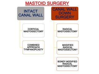
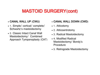
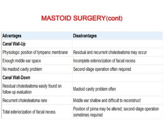
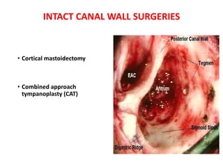
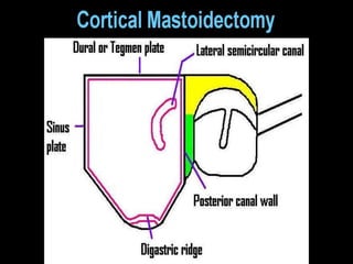
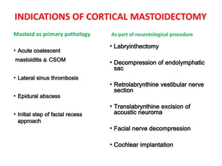
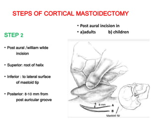
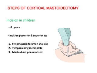
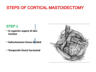
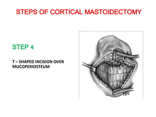
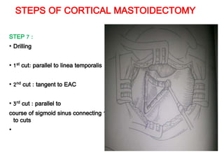
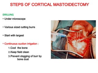
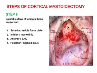
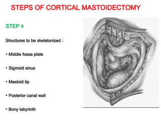
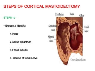
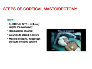
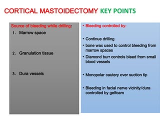
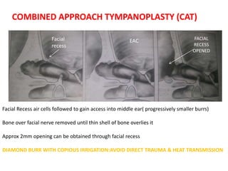
CSOM SURGERIES
Dr Harjitpal Singh
Assistant Professor(ENT),
Dr RKGMC, Hamirpur
CHRONIC SUPPURATIVE OTITIS MEDIA
CSOM is a long standing infection of a part or
whole of the middle ear cleft, characterized by
ear discharge and a permanent perforation
Types of CSOM:
1. Tubotympanic
2. Atticoantral
OBJECTIVES OF SURGERY FOR CSOM
• Eradication of disease
• Prevention of recurrence
• Prevention of complications that can occur due to CSOM
• Restoration of hearing
TREATMENT CSOM- UNSAFE
•Surgical treatment
•Reconstructive surgery
MASTOID SURGERY
INTACT
CANAL WALL
CORTICAL
MASTOIDECTOMY
COMBINED
APPROACH
TYMPANOPLASTY
CANAL WALL
DOWN
SURGERY
RADICAL
MASTOIDECTOMY
MODIFIED
RADICAL
MASTOIDECTOMY
BONDY MODIFIED
RADICAL
MASTOIDECTOMY
MASTOID SURGERY(cont)
• CANAL WALL UP (CWU)
• 1. Simple/ cortical/ complete/
Schwartz's mastoidectomy
• 2. Classic Intact Canal Wall
Mastoidectomy/ Combined
Approach Tympanoplasty (CAT)
• CANAL WALL DOWN (CWD)
• 1. Atticotomy
• 2. Atticoantrotomy
• 3. Radical Mastoidectomy
• 4. Modified Radical
Mastoidectomy/ Bondy’s
Procedure
• 5. Retrograde Mastoidectomy
MASTOID SURGERY(cont)
CHOICE OF SURGERY
• Extent of disease
• Health
• Status of contralateral ear
• Surgeons experience
• Patient preference
MASTOID SURGERY(cont)
INTACT CANAL WALL SURGERIES
• Cortical mastoidectomy
• Combined approach
tympanoplasty (CAT)
PRE-OPERATIVE EVALUATION
Diagnosis of cholesteatoma:
1. Well taken history
2. Examination under microscope
OTHERS:
• Hearing assessment (poorer ear operated 1st)
• Radiograph & imaging studies(suspected complications)
• Patient counselling
CORTICAL MASTOIDECTOMY
Other terminologies:
• Simple mastoidectomy
• Schwartz mastoidectomy
• Conservative mastoidectomy
• Complete mastoidectomy
STEPS OF CAT
1. Cortical mastoidectomy
2. Anterior Tympanotomy : via tympanomeatal flap
3. Posterior Tympanotomy: via facial recess approach
4. Tympanoplasty
HISTORY OF CORTICAL MASTOIDECTOMY
•Hippocrates proposed the idea
•16th century: Ambrose Pare advised mastoid exploration
for young king of France
•1649:Rialon : described mastoid surgery
•1736: Jean Petit of paris performed it successfully
HISTORY OF CORTICAL MASTOIDECTOMY
•1853 : William Wilde recommended incision
•1873 : schwartze described indications & technique of
simple mastoidectomy
•1950: Janssen described Intact canal wall
mastoidectomy.
INDICATIONS OF CORTICAL MASTOIDECTOMY
Mastoid as primary pathology
• Acute coalescent
mastoiditis & CSOM
• Lateral sinus thrombosis
• Epidural abscess
• Initial step of facial recess
approach
As part of neurotological procedure
• Labryinthectomy
• Decompression of endolymphatic
sac
• Retrolabrynthine vestibular nerve
section
• Translabrynthine excision of
acoustic neuroma
• Facial nerve decompression
• Cochlear implantation
ANAESTHESIA
General Anaesthesia( preferred) /Local
anaesthesia
Muscle Relaxants avoided( if facial nerve monitoring)
Nitrous oxide stopped 30 minutes before; if graft
has to be kept
POSITIONING OF THE PATIENT
•Patient is placed closed to the edge of the table,
•Patient’s body strapped on table with both arms
padded and tucked closed to body.
•Head turned approx 120 degrees away from surgeon
and is supported with a folded towel placed b/t table and
contralateral cheek.
•Operating table which can rotate along its long axis.
•Hydraulic chair.
PATIENT PREPARATION & POSITIONING
• Hair shaved 2cm superior & 1cm
posterior to auricle
• Surgical site prepared with alcohol
& betadine solution
• Square off site with clear
adhesive drapes.
• Perioperative / prophylactic
antibiotic use debated
STEPS OF CORTICAL MASTOIDECTOMY
STEP 1
2% lidocaine with
1:100,000 epinephrine
infiltrated
STEPS OF CORTICAL MASTOIDECTOMY
STEP 2
• Post aural /william wilde
incision
• Superior: root of helix
• Inferior : to lateral surface
of mastoid tip
• Posterior: 8-10 mm from
post auricular groove
• Post aural incision in
• a)adults b) children
STEPS OF CORTICAL MASTOIDECTOMY
• <2 years
• Incision posterior & superior as:
1. Stylomastoid foramen shallow
2. Tympanic ring incomplete
3. Mastoid not pneumatized
Incision in children
STEPS OF CORTICAL MASTOIDECTOMY
STEP 3
• In superior aspect of skin
incision
• Subcutaneous tissue divided
• Temporalis fascia harvested
STEPS OF CORTICAL MASTOIDECTOMY
STEP 4
T – SHAPED INCISION OVER
MUCOPERIOSTEUM
STEPS OF CORTICAL MASTOIDECTOMY
STEP 5
Mucoperiosteum
overlying mastoid elevated with
periosteum elevator
SPINE OF HENLE
STEPS OF CORTICAL MASTOIDECTOMY
STEP 6
• Surgical landmarks on lateral
cortical wall identified
• MacEwen’s triangle identified
Macewen’s/Suprameatal triangle/ fossa mastoidea
OUTLINED BY : line parallel to Linea temporalis
line perpendicular to linea temoralis posterior to EAC
line tangential to EAC coursing through spine of henle
STEPS OF CORTICAL MASTOIDECTOMY
STEP 7 :
• Drilling
• 1st cut: parallel to linea temporalis
• 2nd cut : tangent to EAC
• 3rd cut : parallel to
course of sigmoid sinus connecting 1st
to cuts
•
STEPS OF CORTICAL MASTOIDECTOMY
DRILLING
• Under microscope
• Various sized cutting burrs
• Start with largest
• Continuous suction irrigation :
1.Cool the bone
2.Keep field clean
3.Prevent clogging of burr by
bone dust
STEPS OF CORTICAL MASTOIDECTOMY
STEP 8
Lateral surface of temporal bone
saucerized:
1. Superior: middle fossa plate
2. Inferior : mastoid tip
3. Anterior : EAC
4. Posterior : sigmoid sinus
BOUNDARIES OF MASTOID CAVITY
• Superior: Dural or Tegmen plate
• Anterior: Posterior wall of external auditory canal
• Inferior: Digastric ridge
• Posterior: Sigmoid sinus plate
• Medially: Lateral semicircular canal
STEPS OF CORTICAL MASTOIDECTOMY
STEP 9
Structures to be skeletonized :
• Middle fossa plate
• Sigmoid sinus
• Mastoid tip
• Posterior canal wall
• Bony labyrinth
STEPS OF CORTICAL MASTOIDECTOMY
STEPS 10
• Expose & identify:
1.incus
2.Aditus ad antrum
3.Fossa incudis
4. Course of facial nerve
STEPS OF CORTICAL MASTOIDECTOMY
STEP 11
• SURGICAL SITE : profusely
irrigate mastoid cavity
• Haemostasis ensured
• Wound site closed in layers
• Mastoid dressing/ Glasscock
pressure dressing applied
CORTICAL MASTOIDECTOMY KEY POINTS
Source of bleeding while drilling:
1. Marrow space
2. Granulation tissue
3. Dura vessels
• Bleeding controlled by:
• Continue drilling
• bone wax used to control bleeding from
marrow spaces
• Diamond burr controls bleed from small
blood vessels
• Monopolar cautery over suction tip
• Bleeding in facial nerve vicinity/dura
controlled by gelfoam
COMBINED APPROACH TYMPANOPLASTY (CAT)
OTHER NAMES:
• Intact canal wall tympanoplasty with mastoidectomy
• Closed cavity tympanoplasty with mastoidectomy
• Facial recess approach
• Posterior tympanotomy
COMBINED APPROACH TYMPANOPLASTY (CAT)
IDEAL CANDIDATE
WELL AERATED MIDDLE EAR CLEFT
• FUNCTIONAL EUSTACHIAN TUBE
• LARGE PNEUMATIZED MASTOID
• FOLLOW UP POSSIBLE
• LIMITED DISEASE
STEPS OF FACIAL RECESS APPROACH
• Cortical mastoidectomy
• Attention turned to facial recess
Medial: Facial nerve (mastoid
segment)
Lateral: Chorda tympani
Superior :Fossa incudis
COMBINED APPROACH TYMPANOPLASTY (CAT)
Facial
recess
EAC FACIAL
RECESS
OPENED
Facial Recess air cells followed to gain access into middle ear( progressively smaller burrs)
Bone over facial nerve removed until thin shell of bone overlies it
Approx 2mm opening can be obtained through facial recess
DIAMOND BURR WITH COPIOUS IRRIGATION:AVOID DIRECT TRAUMA & HEAT TRANSMISSION
COMBINED APPROACH TYMPANOPLASTY (CAT)
Facial nerve or cholesteatoma?
Probe with needle
• Facial nerve bounces back
• Cholesteatoma/ mucosa doesn’t bounce
STRUCTURE VISIBLE THROUGH FACIAL RECESS
• INCUS
• INCUDOSTAPEDIAL
JOINT
• STAPES SUPRASTRUCTURE
• PROMONTORY
• ROUND WINDOW NICHE
STRUCTURE NOT VISUALIZED THROUGH FACIAL RECESS
• Oval window
• Stapes footplate
• Anterior epitympanum
• Sinus tympani
ACCESSED
THROUGH
EAR CANAL
FACIAL RECESS & SINUS TYMPANI
LATERAL
MEDIAL
c
t
SINUS TYMPANI
COMBINED APPROACH TYMPANOPLASTY (CAT)
• IF INCUS ERODED BY CHOLESTEATOMA-> INCUS REMOVED
• BONY BRIDGE ON INFERIOR ASPECT OF FOSSA INCUDIS
REMOVED
• HANDLE OF MALLEUS REMOVED
• VISUALIZE & REMOVE DISEASE FROM ANTERIOR
EPITYMPANUM
COMBINED APPROACH TYMPANOPLASTY (CAT)
•Plastic sheeting & gel foam placed in middle ear
•Tympanic membrane reconstructed
•Wound site closed in layers
CANAL WALL DOWN SURGERY
• Radical mastoidectomy
• Modified radical mastoidectomy
• Bondy’s mastoidectomy
HISTORY OF CANAL WALL DOWN MASTOID SURGERY
• 1873: Von Troltsch
• 1873: Jansen: MRM
• 1889: Radical term for posterior canal wall removal(Von
Bergmann)
• 1893: Zaufal & Stacke; radical mastoidectomy
• 1910: Bondy’s modification
• 1940: Boettcher used drill
RADICAL MASTOIDECTOMY
• Radical mastoidectomy is a canal wall down mastoidectomy
• Performed to eradicate disease from middle ear cleft.
• Mastoid cavity, tympanum and EAC are converted into a
common cavity exteriorised through the EAC.
• Wherein the structures of tympanic cavity (remnants of the
incus and malleus, and the drum remnant) are removed.
RADICAL MASTOIDECTOMY
INDICATIONS:
•Unresectable cholesteatoma extending down the
Eustachian tube or into the petrous apex
•Promontory cochlear fistula caused by cholesteatoma
•Chronic perilabyrinthine osteitis or cholesteatoma that
cannot be removed and must be cleaned or inspected
periodically
•Resection of temporal bone neoplasms with periodic
monitoring
RADICAL MASTOIDECTOMY
• Perform cortical mastoidectomy.
• Lower facial ridge & break facial bridge.
• Remove cholesteatoma & granulations
• Remove normal middle ear mucosa, T.M. remnant & ossicles (except
stapes footplate).
• Close Eustachian tube opening
• Mastoid, E.A.C. & middle ear become single cavity.
MODIFIED RADICAL MASTOIDECTOMY
•It is done where the disease is localized to attic and
mastoid antrum.
•MRM is a surgical procedure where the disease
process is eradicated from the middle ear cleft.
•Followed by converting the mastoid cavity, middle
ear and EAC into a single, smooth, self-cleansing
cavity exteriorized through EAC.
•In this operation healthy and normal middle ear
structures such as mucosa, ossicles and remnant of
tympanic membrane are preserved with no
compromise on the removal of disease.
MODIFIED RADICAL MASTOIDECTOMY
ABSOLUTE INDICATIONS:
1. Unresectable disease
2. Unreconstructable Posterior canal wall
3. Failure of first stage CWU procedure because of
poor E T function.
4. Inadequate Patient Follow up.
MODIFIED RADICAL MASTOIDECTOMY
RELATIVE INDICATIONS:
1. Disease in only hearing ear or in a dead ear.
2. Medical illness
3. Severe otologic or CNS complications
4. Neoplasms
5. Poor E T function.
MODIFIED RADICAL MASTOIDECTOMY
CONTRAINDICATIONS:
1. Chronic otitis media without cholesteatoma
2. Acute otitis media with coalescent mastoiditis,
3. persistent secretory otitis media, or
4. Chronic allergic otitis media.
5. Tuberculous otitis media.
MODIFIED RADICAL MASTOIDECTOMY
• Intact canal wall mastoidectomy is performed.
• Then lowering of canal wall is performed.
• Area of canal wall to be lowered consists of:
 Anterior buttress : tympanosquamous suture
 Posterior buttress: facial ridge & intervening bridge
 Scutum : lateral epitympanic wall
MODIFIED RADICAL MASTOIDECTOMY
anterior buttress lowered to anterior canal wall
posterior buttress removed to level of facial nerve
MODIFIED RADICAL MASTOIDECTOMY
Drilling over posterior canal wall:
• Large cutting burr
• Bone removal begun inside of
EAC on anterosuperior surface
• Direction :parallel to nerve
superior to inferior
MODIFIED RADICAL MASTOIDECTOMY
• As bone overlying malleus, incus & facial
nerve is thinned
AVOID:
1. Inadverent drilling
2. Inadverent pressure medially
• The last bridge of the posterior canal wall
is removed while using a curette to
protect the ossicles and facial nerve.
MODIFIED RADICAL MASTOIDECTOMY
• Bone overlying vertical segment
of facial nerve: facial ridge
lowered
• Remove cholesteatoma and
granulation tissue from
mastoid and middle ear
cavity.
• Preserve healthy mucosa,
TM remnant and ossicles.
• Perform tympanoplasty.
• Perform concho-
meatoplasty.
MODIFIED RADICAL MASTOIDECTOMY KEY POINTS
In addition to adequate disease removal 4 basic steps for trouble free
mastoid:
1. Adequate saucerization(no overhang)
2. Adequately lowered facial ridge
3. Mastoid tip management: if pneumatized(drill or amputate)
4. Adequate meatoplasty
MEATOPLASTY
•One percent lidocaine with 1:100,000 epinephrine is
infiltrated into the conchal bowl.
•With a finger in the conchal bowl, a semilunar incision
is made into the cartilage posteriorly until the knife tip
is felt through the anterior skin.
MEATOPLASTY
• This crescent-shaped cartilage
measures about 1.5 x 2 cm
BONDY’S MODIFIED RADICAL MASTOIDECTOMY
• Superior and posterior EAC
removed to exteriorize
cholesteatoma in mastoid &
attic to EAC
• HALLMARK :
Tympanic membrane & middle
ear not disturbed.
Completed Bondy-modified
radical mastoidectomy.
BONDY’S MODIFIED RADICAL MASTOIDECTOMY
DISEASE TO BE REMOVED FROM:
• FACIAL RECESS
• SINUS TYMPANI
• SINODURAL ANGLE
• ANTERIOR ATTIC
• ALONG FACIAL NERVE
• ALONG STAPES
• MASTOID TIP
ATTICOTOMY
INDICATION:
• Limited attic cholesteatoma confined to central epitympanic area.
• Edges of scutal defect drilled with diamond burr & gradually widened.
• Superior wall thinned
• Margins of cholesteatoma sac identified & exteriorized.
ATTICOANTROSTOMY
• It is an extension of the
atticotomy in a posterior
direction through the
transmeatal route, in which
lateral attic and aditus walls are
removed, and the antrum is
entered.
• It can be performed through the
transcortical route, but is usually
performed through a transmeatal
route.
POST OPERATIVE CARE
•Foul smelling discharge: remove pack. Send for C/S
•Remove stitches on 7th POD & pack on 10th POD
•Keep ear dry
•Regular follow up after discharge.
•PODry
•Regular follow up after discharge
FOLLOW UP
• 6-10wks time period for cavity to heal.
• Every 2-3 wks cavity debrided & granulation tissue removed
• Then follow up every 6mths – 1 yr depending on condition of mastoid
.
DISCHARGING CAVITY-CAUSES
1. Inadequate concho-meatoplasty
2. Recurrence of cholesteatoma
3. Persistent infection: petrositis, T.B., sinusitis
4. Persistent allergy
5. Retained foreign body: cotton ball
6. Persistent extra-dural abscess
7. Residual cholesteatoma: facial ridge, facial bridge, anterior &
posterior buttress, mastoid tip, sinus tympani, anterior
epitympanum.
COMPLICATIONS OF SURGERY
• Injury to facial nerve
• Injury to HSCC
• Injury to sigmoid sinus
• Injury to dura
• SNHL
• RECURRENCE/RESIDUAL DISEASE discharging cavity
• Perichonderitis
INJURY TO FACIAL NERVE
• Intra op FN trauma: decompress proximal & distal
• Post op <immediate>: make sure FN was identified + integrity tested intra
op
yes
• Observe few hrs  effect of LA vanishes
• Tight mastoid dressing may press on exposed FN remove pack
• Incomplete palsy  put on steroids
• Progression to complete palsy re-exploration
NOTE: assistance with experienced colleague must!!
INJURY TO DURA
• MINOR: concealed by surrounding arachnoid tissue
• Remove surrounding 5mm bone to inspect dura & brain
• Fascia graft between normal dura & surrounding bone
• Macerated muscle
• Hydroxypatite bone cement with fascia over it.
INJURY TO DURA
INJURY TO SIGMOID SINUS
• Press with finger immediately
• Remove suction aspirator
• Small laceration : bipolar cautery / thrombin soaked gelfoam
• Large laceration: large gelfoam over surface
opening; if surrounding bony cover present ;bone wax used .
INJURY TO SIGMOID SINUS
INJURY TO HSCC
IMMEDIATELY SEAL WITH :
• Facial plug
• Bone wax
• Muscle plug
IATROGENIC LABRYNTHINE FISTULA:
• LSCC
• Stapes footplate
SNHL
Etiology:
• Opening labyrinth
• Manipulation of ossicles
• Drilling on ossicles
• Unidentified causes
RECONSTRUCTIVE SURGERIES
MYRINGOPLASTY
Procedure used to repair a perforated tympanic
membrane using a graft material, without need to
examine the middle ear.
Advantages:-
1. Restoring the hearing loss
2. Checking repeated infections from EAC and ET
3. Checking aeroallergens reaching the exposed middle
ear mucosa leading to persistent ear discharge.
SURGICAL APPROACHES FOR MYRINGOPLASTY
1. Post aural approach
2. Endomeatal or transcanal approach
3. Endaural approach
ENDOMEATAL OR TRANSCANAL APPROACH
ROSEN’S INCISION
ENDAURAL APPROACH
POST AURAL / WILDE’S INCISION
TYMPANOTOMIES
• Opening the tympanic cavity by elevating a tympanomeatal flap
together with the fibrous annulus.
• Tympanotomies can be divided into:
1. Posterior tympanotomy- 12–o’clock to 6-o’clock posteriorly
(rosen incision).
2. Inferior tympanotomy – 9-o’clock to 3-o’clock incision inferiorly.
3. Anterior tympanotomy – 12-o’clock to the 6-o’clock incision
about 5mm lateral to annulus anteriorly.
4. Superior tympanotomy - 9-o’clock to 3-o’clock incision about
5mm lateral to shrapnell’s membrane.
GRAFT MATERIALS
AUTOGRAFT (AUTOGENOUS GRAFT) – graft from same person.
These include:
1. Temporalis muscle fascia
2. Tragal perichondrium
3. Conchal perichondrium
4. Tragal or conchal cartilage
5. Periosteum (mastoid process and temporal squama)
6. Vein ( great saphenous vein, cubital vein)
7. Fatty tissue (ear lobule)
8. Subcutaneous tissue
9. Fascia lata
10.Ear canal skin
GRAFTING TECHNIQUES
• OVERLAY TECHNIQUE
• UNDERLAY TECHNIQUE
OVERLAY TECHNIQUE
1.This technique is used when there is no remnant of the tympanic
membrane.
2.The graft rests over the anterior and the posterior tympanic sulcus
and underneath the malleus handle.
3.The edges of the graft are covered by meatal skin.
COMPLICATIONS OF OVERLAY TECHNIQUE
1.Blunting of anterior sulcus.
2.Epithelial pearls – they are epidermal cyst, when squamous
epithelium is buried under the graft.
3.Lateralisation of graft- graft loses contact from the malleus
handle resulting in conductive loss.
UNDERLAY TECHNIQUE
• The presence of an anterior remnant of the tympanic membrane is required for this
type of fascial graft.
• The graft is placed under the anterior remnant of the tympanic membrane and over
the posterior tympanic sulcus.
• The graft lies under the malleus handle.
COMPLICATIONS OF UNDERLAY TECHNIQUE
1. Middle ear becomes narrow.
2. Graft may get adherent to promontory.
3. Anteriorly graft may loose contact from remnant of
tympanic membrane leading to anterior perforation.
FAT GRAFT MYRINGOPLASTY
OTHER TECHNIQUES
OSSICULOPLASTY
•Aim is to surgically optimize the hearing
mechanism
•Performed in otherwise healthy ears or in
conjunction with tympanoplasty and mastoidectomy
for chronic ear disease
TYMPANOPLASTY
TYMPANOPLASTY
TYMPANOPLASTY includes :
• Canaloplasty (widening of bony part of the external auditory canal)
• Myringoplasty (closure of the eardrum perforation in cases with a normal ossicular chain and
without any other surgical procedures in the tympanic cavity or middle ear)
• Ossiculoplasty (reconstruction of ossicular chain)
AIMS OF TYMPANOPLASTY
1. Eradication of disease
2. Restoration of tympanic membrane
3. Reconstruction of a sound transformer mechanism
OBJECTIVES OF TYMPANOPLASTY
IN DECREASING ORDER OF PRIORITY
•elimination of disease to produce a safe and dry ear;
•alteration of anatomy to prevent recurrent disease, and
to optimize cleaning and otologic monitoring;
• reconstruction of the middle ear to achieve
serviceable and stable postoperative hearing
INDICATIONS FOR TYMPANOPLASTY
•Tympanic membrane perforations and associated
hearing loss with or without middle ear pathology
such as tympanosclerosis , small retraction pockets
, and cholesteatomas.
CONTRAINDICATIONS
ABSOLUTE:
•Poor general health
•Malignant tumours of outer / middle ear
•Uncontrolled cholesteatoma
•Unusual infections like malignant otitis externa
•Complications of chronic ear disease such as
meningitis , brain abscess ,or lateral sinus
thrombosis
•If it is the only or significantly better hearing ear.
CONTRAINDICATIONS
RELATIVE:
•Nonfunctioning eustachian tube
•Nasal allergy
•Chronic Otitis externa
•Acute exacerbation of chronic otitis media , chronic
mucoid discharge associated with allergic
rhinosinusitis
PREOPERATIVE EVALUATION
•Complete history and head and neck
examination
•Otoscopic examination , best accomplished by
operating microscope
•Audiogram ,including PTA and air bone
conduction thresholds as well as speech
discrimination scores.
ANAESTHESIA
1. GENERAL ANESTHESIA :-
•Extensive removal of tympanic cavity mucosa or
tympanic cavity cholesteatoma
•Any surgery in the anterior tympanum or tympanic
orifice of the Eustachian tube
•Cases requiring mastoidectomy or reconstruction
of the ear canal
•Children
ANAESTHESIA
•Uncooperative adults, apprehensive adults
•Patients who spontaneously prefer or request GA
•Any surgery lasting more than 1 ½ - 2 hours.
•Revision tympanoplasties where major pieces of
temporal muscle fascia have already been
harvested previously
2. LOCAL ANESTHESIA :–
• Limited to cooperative adults with dry,
noninfected ears and no evidence of mastoid
disease.
TYPES OF TYMPANOPLASTY
ACCORDING TO WULLSTEIN (1968)
TYPE I - TYMPANOPLASTY
•TYPE I – perforation in tympanic membrane repaired
with a graft. Intact ossicular chain . Myringoplasty
TYPE II - TYMPANOPLASTY
• TYPE II – defective or absent malleus handle, but intact incudostapedial
joint. The fascia is placed on the lenticular process of the incus.
Myringoincudopexy
• TYPE III – malleus and incus are absent. Graft is placed directly on the
stapes head. Myringostapediopexy producing a shallow middle ear and a
collumella effect.
TYPE III - TYMPANOPLASTY
TYPE IV - TYMPANOPLASTY
• TYPE IV – only the foot plate of stapes is present . It is exposed to the
external ear and graft is placed between the oval and round windows. A
narrow middle ear (cavum minor) is thus created, to have an air pocket
around the round window
TYPE V - TYMPANOPLASTY
• TYPE V – stapes footplate is fixed but round window is functioning. Another
window is created on horizontal SCC and covered with a graft. Fenestration
Operation
TECHNIQUES OF TYMPANOPLASTY
(A) Type I. Repair of tympanic membrane (TM) with temporalis fascia.
(B) Type III: minor columella. Ossicular strut or partial ossicular
replacement prosthesis (PORP) is placed between stapes head and
manubrium/TM.
(C) Type III: major columella. Total ossicular
replacement prosthesis (TORP) is placed from stapes
footplate to the manubrium/TM.
(D) Type III: Stapes columella. Performed with canal wall-
down (CWD) mastoidectomy and obliteration of mastoid.
Thin cartilage disk and temporalis fascia are placed on
stapes head.
(E) Type IV. Round window is acoustically shielded by thick
cartilage and temporalis fascia while footplate is covered
with thin skin graft. Also performed with CWD
mastoidectomy
(F) Type V. Similar to type IV, except for total
stapedectomy and footplate replacement by an adipose
graft.
POSTOPERATIVE CARE
• PRECAUTIONS
Do not drive home after discharged the next morning
No air travel until 4 weeks after surgery
Do not blow nose until ear is healed
When sneezing, keep mouth open
Avoid water entering ear canal
• Oral antibiotics
• First postoperative visit after one week
• The gelfoam over graft is gently suctioned away, if still present , at
the second visit 3 to 4 weeks later.
• Improvement in hearing can be noticed 6 to 8 weeks after
surgery, but maximum may take 4 to 6 months
COMPLICATIONS OF TYMPANOPLASTY
•Intraoperative Bleeding
•Facial Nerve Injury
•Wound infection / Perichondritis
•Wound Hematoma
•Chorda Tympani Nerve Injury
•Tympanoplasty Failure
•Recurrent / Residual Middle Ear Cholesteatoma
•Sensorineural Hearing Loss / Dizziness.
THANK YOU

CSOM SURGERIES

  • 1.
    CSOM SURGERIES Dr HarjitpalSingh Assistant Professor(ENT), Dr RKGMC, Hamirpur
  • 2.
    CHRONIC SUPPURATIVE OTITISMEDIA CSOM is a long standing infection of a part or whole of the middle ear cleft, characterized by ear discharge and a permanent perforation Types of CSOM: 1. Tubotympanic 2. Atticoantral
  • 3.
    OBJECTIVES OF SURGERYFOR CSOM • Eradication of disease • Prevention of recurrence • Prevention of complications that can occur due to CSOM • Restoration of hearing
  • 4.
    TREATMENT CSOM- UNSAFE •Surgicaltreatment •Reconstructive surgery
  • 5.
    MASTOID SURGERY INTACT CANAL WALL CORTICAL MASTOIDECTOMY COMBINED APPROACH TYMPANOPLASTY CANALWALL DOWN SURGERY RADICAL MASTOIDECTOMY MODIFIED RADICAL MASTOIDECTOMY BONDY MODIFIED RADICAL MASTOIDECTOMY
  • 6.
    MASTOID SURGERY(cont) • CANALWALL UP (CWU) • 1. Simple/ cortical/ complete/ Schwartz's mastoidectomy • 2. Classic Intact Canal Wall Mastoidectomy/ Combined Approach Tympanoplasty (CAT) • CANAL WALL DOWN (CWD) • 1. Atticotomy • 2. Atticoantrotomy • 3. Radical Mastoidectomy • 4. Modified Radical Mastoidectomy/ Bondy’s Procedure • 5. Retrograde Mastoidectomy
  • 7.
    MASTOID SURGERY(cont) CHOICE OFSURGERY • Extent of disease • Health • Status of contralateral ear • Surgeons experience • Patient preference
  • 8.
  • 9.
    INTACT CANAL WALLSURGERIES • Cortical mastoidectomy • Combined approach tympanoplasty (CAT)
  • 10.
    PRE-OPERATIVE EVALUATION Diagnosis ofcholesteatoma: 1. Well taken history 2. Examination under microscope OTHERS: • Hearing assessment (poorer ear operated 1st) • Radiograph & imaging studies(suspected complications) • Patient counselling
  • 11.
    CORTICAL MASTOIDECTOMY Other terminologies: •Simple mastoidectomy • Schwartz mastoidectomy • Conservative mastoidectomy • Complete mastoidectomy
  • 12.
    STEPS OF CAT 1.Cortical mastoidectomy 2. Anterior Tympanotomy : via tympanomeatal flap 3. Posterior Tympanotomy: via facial recess approach 4. Tympanoplasty
  • 13.
    HISTORY OF CORTICALMASTOIDECTOMY •Hippocrates proposed the idea •16th century: Ambrose Pare advised mastoid exploration for young king of France •1649:Rialon : described mastoid surgery •1736: Jean Petit of paris performed it successfully
  • 14.
    HISTORY OF CORTICALMASTOIDECTOMY •1853 : William Wilde recommended incision •1873 : schwartze described indications & technique of simple mastoidectomy •1950: Janssen described Intact canal wall mastoidectomy.
  • 15.
    INDICATIONS OF CORTICALMASTOIDECTOMY Mastoid as primary pathology • Acute coalescent mastoiditis & CSOM • Lateral sinus thrombosis • Epidural abscess • Initial step of facial recess approach As part of neurotological procedure • Labryinthectomy • Decompression of endolymphatic sac • Retrolabrynthine vestibular nerve section • Translabrynthine excision of acoustic neuroma • Facial nerve decompression • Cochlear implantation
  • 16.
    ANAESTHESIA General Anaesthesia( preferred)/Local anaesthesia Muscle Relaxants avoided( if facial nerve monitoring) Nitrous oxide stopped 30 minutes before; if graft has to be kept
  • 17.
    POSITIONING OF THEPATIENT •Patient is placed closed to the edge of the table, •Patient’s body strapped on table with both arms padded and tucked closed to body. •Head turned approx 120 degrees away from surgeon and is supported with a folded towel placed b/t table and contralateral cheek. •Operating table which can rotate along its long axis. •Hydraulic chair.
  • 18.
    PATIENT PREPARATION &POSITIONING • Hair shaved 2cm superior & 1cm posterior to auricle • Surgical site prepared with alcohol & betadine solution • Square off site with clear adhesive drapes. • Perioperative / prophylactic antibiotic use debated
  • 19.
    STEPS OF CORTICALMASTOIDECTOMY STEP 1 2% lidocaine with 1:100,000 epinephrine infiltrated
  • 20.
    STEPS OF CORTICALMASTOIDECTOMY STEP 2 • Post aural /william wilde incision • Superior: root of helix • Inferior : to lateral surface of mastoid tip • Posterior: 8-10 mm from post auricular groove • Post aural incision in • a)adults b) children
  • 21.
    STEPS OF CORTICALMASTOIDECTOMY • <2 years • Incision posterior & superior as: 1. Stylomastoid foramen shallow 2. Tympanic ring incomplete 3. Mastoid not pneumatized Incision in children
  • 22.
    STEPS OF CORTICALMASTOIDECTOMY STEP 3 • In superior aspect of skin incision • Subcutaneous tissue divided • Temporalis fascia harvested
  • 23.
    STEPS OF CORTICALMASTOIDECTOMY STEP 4 T – SHAPED INCISION OVER MUCOPERIOSTEUM
  • 24.
    STEPS OF CORTICALMASTOIDECTOMY STEP 5 Mucoperiosteum overlying mastoid elevated with periosteum elevator
  • 25.
  • 26.
    STEPS OF CORTICALMASTOIDECTOMY STEP 6 • Surgical landmarks on lateral cortical wall identified • MacEwen’s triangle identified
  • 27.
    Macewen’s/Suprameatal triangle/ fossamastoidea OUTLINED BY : line parallel to Linea temporalis line perpendicular to linea temoralis posterior to EAC line tangential to EAC coursing through spine of henle
  • 28.
    STEPS OF CORTICALMASTOIDECTOMY STEP 7 : • Drilling • 1st cut: parallel to linea temporalis • 2nd cut : tangent to EAC • 3rd cut : parallel to course of sigmoid sinus connecting 1st to cuts •
  • 29.
    STEPS OF CORTICALMASTOIDECTOMY DRILLING • Under microscope • Various sized cutting burrs • Start with largest • Continuous suction irrigation : 1.Cool the bone 2.Keep field clean 3.Prevent clogging of burr by bone dust
  • 30.
    STEPS OF CORTICALMASTOIDECTOMY STEP 8 Lateral surface of temporal bone saucerized: 1. Superior: middle fossa plate 2. Inferior : mastoid tip 3. Anterior : EAC 4. Posterior : sigmoid sinus
  • 31.
    BOUNDARIES OF MASTOIDCAVITY • Superior: Dural or Tegmen plate • Anterior: Posterior wall of external auditory canal • Inferior: Digastric ridge • Posterior: Sigmoid sinus plate • Medially: Lateral semicircular canal
  • 32.
    STEPS OF CORTICALMASTOIDECTOMY STEP 9 Structures to be skeletonized : • Middle fossa plate • Sigmoid sinus • Mastoid tip • Posterior canal wall • Bony labyrinth
  • 33.
    STEPS OF CORTICALMASTOIDECTOMY STEPS 10 • Expose & identify: 1.incus 2.Aditus ad antrum 3.Fossa incudis 4. Course of facial nerve
  • 34.
    STEPS OF CORTICALMASTOIDECTOMY STEP 11 • SURGICAL SITE : profusely irrigate mastoid cavity • Haemostasis ensured • Wound site closed in layers • Mastoid dressing/ Glasscock pressure dressing applied
  • 35.
    CORTICAL MASTOIDECTOMY KEYPOINTS Source of bleeding while drilling: 1. Marrow space 2. Granulation tissue 3. Dura vessels • Bleeding controlled by: • Continue drilling • bone wax used to control bleeding from marrow spaces • Diamond burr controls bleed from small blood vessels • Monopolar cautery over suction tip • Bleeding in facial nerve vicinity/dura controlled by gelfoam
  • 36.
    COMBINED APPROACH TYMPANOPLASTY(CAT) OTHER NAMES: • Intact canal wall tympanoplasty with mastoidectomy • Closed cavity tympanoplasty with mastoidectomy • Facial recess approach • Posterior tympanotomy
  • 37.
    COMBINED APPROACH TYMPANOPLASTY(CAT) IDEAL CANDIDATE WELL AERATED MIDDLE EAR CLEFT • FUNCTIONAL EUSTACHIAN TUBE • LARGE PNEUMATIZED MASTOID • FOLLOW UP POSSIBLE • LIMITED DISEASE
  • 39.
    STEPS OF FACIALRECESS APPROACH • Cortical mastoidectomy • Attention turned to facial recess Medial: Facial nerve (mastoid segment) Lateral: Chorda tympani Superior :Fossa incudis
  • 40.
    COMBINED APPROACH TYMPANOPLASTY(CAT) Facial recess EAC FACIAL RECESS OPENED Facial Recess air cells followed to gain access into middle ear( progressively smaller burrs) Bone over facial nerve removed until thin shell of bone overlies it Approx 2mm opening can be obtained through facial recess DIAMOND BURR WITH COPIOUS IRRIGATION:AVOID DIRECT TRAUMA & HEAT TRANSMISSION
  • 41.
    COMBINED APPROACH TYMPANOPLASTY(CAT) Facial nerve or cholesteatoma? Probe with needle • Facial nerve bounces back • Cholesteatoma/ mucosa doesn’t bounce
  • 42.
    STRUCTURE VISIBLE THROUGHFACIAL RECESS • INCUS • INCUDOSTAPEDIAL JOINT • STAPES SUPRASTRUCTURE • PROMONTORY • ROUND WINDOW NICHE
  • 43.
    STRUCTURE NOT VISUALIZEDTHROUGH FACIAL RECESS • Oval window • Stapes footplate • Anterior epitympanum • Sinus tympani ACCESSED THROUGH EAR CANAL
  • 44.
    FACIAL RECESS &SINUS TYMPANI LATERAL MEDIAL c t SINUS TYMPANI
  • 45.
    COMBINED APPROACH TYMPANOPLASTY(CAT) • IF INCUS ERODED BY CHOLESTEATOMA-> INCUS REMOVED • BONY BRIDGE ON INFERIOR ASPECT OF FOSSA INCUDIS REMOVED • HANDLE OF MALLEUS REMOVED • VISUALIZE & REMOVE DISEASE FROM ANTERIOR EPITYMPANUM
  • 46.
    COMBINED APPROACH TYMPANOPLASTY(CAT) •Plastic sheeting & gel foam placed in middle ear •Tympanic membrane reconstructed •Wound site closed in layers
  • 47.
    CANAL WALL DOWNSURGERY • Radical mastoidectomy • Modified radical mastoidectomy • Bondy’s mastoidectomy
  • 48.
    HISTORY OF CANALWALL DOWN MASTOID SURGERY • 1873: Von Troltsch • 1873: Jansen: MRM • 1889: Radical term for posterior canal wall removal(Von Bergmann) • 1893: Zaufal & Stacke; radical mastoidectomy • 1910: Bondy’s modification • 1940: Boettcher used drill
  • 49.
    RADICAL MASTOIDECTOMY • Radicalmastoidectomy is a canal wall down mastoidectomy • Performed to eradicate disease from middle ear cleft. • Mastoid cavity, tympanum and EAC are converted into a common cavity exteriorised through the EAC. • Wherein the structures of tympanic cavity (remnants of the incus and malleus, and the drum remnant) are removed.
  • 50.
    RADICAL MASTOIDECTOMY INDICATIONS: •Unresectable cholesteatomaextending down the Eustachian tube or into the petrous apex •Promontory cochlear fistula caused by cholesteatoma •Chronic perilabyrinthine osteitis or cholesteatoma that cannot be removed and must be cleaned or inspected periodically •Resection of temporal bone neoplasms with periodic monitoring
  • 51.
  • 52.
    RADICAL MASTOIDECTOMY • Performcortical mastoidectomy. • Lower facial ridge & break facial bridge. • Remove cholesteatoma & granulations • Remove normal middle ear mucosa, T.M. remnant & ossicles (except stapes footplate). • Close Eustachian tube opening • Mastoid, E.A.C. & middle ear become single cavity.
  • 53.
    MODIFIED RADICAL MASTOIDECTOMY •Itis done where the disease is localized to attic and mastoid antrum. •MRM is a surgical procedure where the disease process is eradicated from the middle ear cleft. •Followed by converting the mastoid cavity, middle ear and EAC into a single, smooth, self-cleansing cavity exteriorized through EAC. •In this operation healthy and normal middle ear structures such as mucosa, ossicles and remnant of tympanic membrane are preserved with no compromise on the removal of disease.
  • 54.
    MODIFIED RADICAL MASTOIDECTOMY ABSOLUTEINDICATIONS: 1. Unresectable disease 2. Unreconstructable Posterior canal wall 3. Failure of first stage CWU procedure because of poor E T function. 4. Inadequate Patient Follow up.
  • 55.
    MODIFIED RADICAL MASTOIDECTOMY RELATIVEINDICATIONS: 1. Disease in only hearing ear or in a dead ear. 2. Medical illness 3. Severe otologic or CNS complications 4. Neoplasms 5. Poor E T function.
  • 56.
    MODIFIED RADICAL MASTOIDECTOMY CONTRAINDICATIONS: 1.Chronic otitis media without cholesteatoma 2. Acute otitis media with coalescent mastoiditis, 3. persistent secretory otitis media, or 4. Chronic allergic otitis media. 5. Tuberculous otitis media.
  • 57.
    MODIFIED RADICAL MASTOIDECTOMY •Intact canal wall mastoidectomy is performed. • Then lowering of canal wall is performed. • Area of canal wall to be lowered consists of:  Anterior buttress : tympanosquamous suture  Posterior buttress: facial ridge & intervening bridge  Scutum : lateral epitympanic wall
  • 58.
    MODIFIED RADICAL MASTOIDECTOMY anteriorbuttress lowered to anterior canal wall posterior buttress removed to level of facial nerve
  • 59.
    MODIFIED RADICAL MASTOIDECTOMY Drillingover posterior canal wall: • Large cutting burr • Bone removal begun inside of EAC on anterosuperior surface • Direction :parallel to nerve superior to inferior
  • 60.
    MODIFIED RADICAL MASTOIDECTOMY •As bone overlying malleus, incus & facial nerve is thinned AVOID: 1. Inadverent drilling 2. Inadverent pressure medially • The last bridge of the posterior canal wall is removed while using a curette to protect the ossicles and facial nerve.
  • 61.
    MODIFIED RADICAL MASTOIDECTOMY •Bone overlying vertical segment of facial nerve: facial ridge lowered • Remove cholesteatoma and granulation tissue from mastoid and middle ear cavity. • Preserve healthy mucosa, TM remnant and ossicles. • Perform tympanoplasty. • Perform concho- meatoplasty.
  • 62.
    MODIFIED RADICAL MASTOIDECTOMYKEY POINTS In addition to adequate disease removal 4 basic steps for trouble free mastoid: 1. Adequate saucerization(no overhang) 2. Adequately lowered facial ridge 3. Mastoid tip management: if pneumatized(drill or amputate) 4. Adequate meatoplasty
  • 63.
    MEATOPLASTY •One percent lidocainewith 1:100,000 epinephrine is infiltrated into the conchal bowl. •With a finger in the conchal bowl, a semilunar incision is made into the cartilage posteriorly until the knife tip is felt through the anterior skin.
  • 64.
    MEATOPLASTY • This crescent-shapedcartilage measures about 1.5 x 2 cm
  • 68.
    BONDY’S MODIFIED RADICALMASTOIDECTOMY • Superior and posterior EAC removed to exteriorize cholesteatoma in mastoid & attic to EAC • HALLMARK : Tympanic membrane & middle ear not disturbed. Completed Bondy-modified radical mastoidectomy.
  • 69.
    BONDY’S MODIFIED RADICALMASTOIDECTOMY DISEASE TO BE REMOVED FROM: • FACIAL RECESS • SINUS TYMPANI • SINODURAL ANGLE • ANTERIOR ATTIC • ALONG FACIAL NERVE • ALONG STAPES • MASTOID TIP
  • 70.
    ATTICOTOMY INDICATION: • Limited atticcholesteatoma confined to central epitympanic area. • Edges of scutal defect drilled with diamond burr & gradually widened. • Superior wall thinned • Margins of cholesteatoma sac identified & exteriorized.
  • 71.
    ATTICOANTROSTOMY • It isan extension of the atticotomy in a posterior direction through the transmeatal route, in which lateral attic and aditus walls are removed, and the antrum is entered. • It can be performed through the transcortical route, but is usually performed through a transmeatal route.
  • 72.
    POST OPERATIVE CARE •Foulsmelling discharge: remove pack. Send for C/S •Remove stitches on 7th POD & pack on 10th POD •Keep ear dry •Regular follow up after discharge. •PODry •Regular follow up after discharge
  • 73.
    FOLLOW UP • 6-10wkstime period for cavity to heal. • Every 2-3 wks cavity debrided & granulation tissue removed • Then follow up every 6mths – 1 yr depending on condition of mastoid .
  • 74.
    DISCHARGING CAVITY-CAUSES 1. Inadequateconcho-meatoplasty 2. Recurrence of cholesteatoma 3. Persistent infection: petrositis, T.B., sinusitis 4. Persistent allergy 5. Retained foreign body: cotton ball 6. Persistent extra-dural abscess 7. Residual cholesteatoma: facial ridge, facial bridge, anterior & posterior buttress, mastoid tip, sinus tympani, anterior epitympanum.
  • 75.
    COMPLICATIONS OF SURGERY •Injury to facial nerve • Injury to HSCC • Injury to sigmoid sinus • Injury to dura • SNHL • RECURRENCE/RESIDUAL DISEASE discharging cavity • Perichonderitis
  • 76.
    INJURY TO FACIALNERVE • Intra op FN trauma: decompress proximal & distal • Post op <immediate>: make sure FN was identified + integrity tested intra op yes • Observe few hrs  effect of LA vanishes • Tight mastoid dressing may press on exposed FN remove pack • Incomplete palsy  put on steroids • Progression to complete palsy re-exploration NOTE: assistance with experienced colleague must!!
  • 77.
    INJURY TO DURA •MINOR: concealed by surrounding arachnoid tissue • Remove surrounding 5mm bone to inspect dura & brain • Fascia graft between normal dura & surrounding bone • Macerated muscle • Hydroxypatite bone cement with fascia over it.
  • 78.
  • 79.
    INJURY TO SIGMOIDSINUS • Press with finger immediately • Remove suction aspirator • Small laceration : bipolar cautery / thrombin soaked gelfoam • Large laceration: large gelfoam over surface opening; if surrounding bony cover present ;bone wax used .
  • 80.
  • 81.
    INJURY TO HSCC IMMEDIATELYSEAL WITH : • Facial plug • Bone wax • Muscle plug IATROGENIC LABRYNTHINE FISTULA: • LSCC • Stapes footplate
  • 82.
    SNHL Etiology: • Opening labyrinth •Manipulation of ossicles • Drilling on ossicles • Unidentified causes
  • 83.
  • 84.
    MYRINGOPLASTY Procedure used torepair a perforated tympanic membrane using a graft material, without need to examine the middle ear. Advantages:- 1. Restoring the hearing loss 2. Checking repeated infections from EAC and ET 3. Checking aeroallergens reaching the exposed middle ear mucosa leading to persistent ear discharge.
  • 85.
    SURGICAL APPROACHES FORMYRINGOPLASTY 1. Post aural approach 2. Endomeatal or transcanal approach 3. Endaural approach
  • 86.
    ENDOMEATAL OR TRANSCANALAPPROACH ROSEN’S INCISION
  • 87.
  • 88.
    POST AURAL /WILDE’S INCISION
  • 89.
    TYMPANOTOMIES • Opening thetympanic cavity by elevating a tympanomeatal flap together with the fibrous annulus. • Tympanotomies can be divided into: 1. Posterior tympanotomy- 12–o’clock to 6-o’clock posteriorly (rosen incision). 2. Inferior tympanotomy – 9-o’clock to 3-o’clock incision inferiorly. 3. Anterior tympanotomy – 12-o’clock to the 6-o’clock incision about 5mm lateral to annulus anteriorly. 4. Superior tympanotomy - 9-o’clock to 3-o’clock incision about 5mm lateral to shrapnell’s membrane.
  • 90.
    GRAFT MATERIALS AUTOGRAFT (AUTOGENOUSGRAFT) – graft from same person. These include: 1. Temporalis muscle fascia 2. Tragal perichondrium 3. Conchal perichondrium 4. Tragal or conchal cartilage 5. Periosteum (mastoid process and temporal squama) 6. Vein ( great saphenous vein, cubital vein) 7. Fatty tissue (ear lobule) 8. Subcutaneous tissue 9. Fascia lata 10.Ear canal skin
  • 91.
    GRAFTING TECHNIQUES • OVERLAYTECHNIQUE • UNDERLAY TECHNIQUE
  • 92.
    OVERLAY TECHNIQUE 1.This techniqueis used when there is no remnant of the tympanic membrane. 2.The graft rests over the anterior and the posterior tympanic sulcus and underneath the malleus handle. 3.The edges of the graft are covered by meatal skin.
  • 93.
    COMPLICATIONS OF OVERLAYTECHNIQUE 1.Blunting of anterior sulcus. 2.Epithelial pearls – they are epidermal cyst, when squamous epithelium is buried under the graft. 3.Lateralisation of graft- graft loses contact from the malleus handle resulting in conductive loss.
  • 95.
    UNDERLAY TECHNIQUE • Thepresence of an anterior remnant of the tympanic membrane is required for this type of fascial graft. • The graft is placed under the anterior remnant of the tympanic membrane and over the posterior tympanic sulcus. • The graft lies under the malleus handle.
  • 96.
    COMPLICATIONS OF UNDERLAYTECHNIQUE 1. Middle ear becomes narrow. 2. Graft may get adherent to promontory. 3. Anteriorly graft may loose contact from remnant of tympanic membrane leading to anterior perforation.
  • 98.
  • 99.
  • 100.
    OSSICULOPLASTY •Aim is tosurgically optimize the hearing mechanism •Performed in otherwise healthy ears or in conjunction with tympanoplasty and mastoidectomy for chronic ear disease
  • 101.
  • 102.
    TYMPANOPLASTY TYMPANOPLASTY includes : •Canaloplasty (widening of bony part of the external auditory canal) • Myringoplasty (closure of the eardrum perforation in cases with a normal ossicular chain and without any other surgical procedures in the tympanic cavity or middle ear) • Ossiculoplasty (reconstruction of ossicular chain)
  • 103.
    AIMS OF TYMPANOPLASTY 1.Eradication of disease 2. Restoration of tympanic membrane 3. Reconstruction of a sound transformer mechanism
  • 104.
    OBJECTIVES OF TYMPANOPLASTY INDECREASING ORDER OF PRIORITY •elimination of disease to produce a safe and dry ear; •alteration of anatomy to prevent recurrent disease, and to optimize cleaning and otologic monitoring; • reconstruction of the middle ear to achieve serviceable and stable postoperative hearing
  • 105.
    INDICATIONS FOR TYMPANOPLASTY •Tympanicmembrane perforations and associated hearing loss with or without middle ear pathology such as tympanosclerosis , small retraction pockets , and cholesteatomas.
  • 106.
    CONTRAINDICATIONS ABSOLUTE: •Poor general health •Malignanttumours of outer / middle ear •Uncontrolled cholesteatoma •Unusual infections like malignant otitis externa •Complications of chronic ear disease such as meningitis , brain abscess ,or lateral sinus thrombosis •If it is the only or significantly better hearing ear.
  • 107.
    CONTRAINDICATIONS RELATIVE: •Nonfunctioning eustachian tube •Nasalallergy •Chronic Otitis externa •Acute exacerbation of chronic otitis media , chronic mucoid discharge associated with allergic rhinosinusitis
  • 108.
    PREOPERATIVE EVALUATION •Complete historyand head and neck examination •Otoscopic examination , best accomplished by operating microscope •Audiogram ,including PTA and air bone conduction thresholds as well as speech discrimination scores.
  • 109.
    ANAESTHESIA 1. GENERAL ANESTHESIA:- •Extensive removal of tympanic cavity mucosa or tympanic cavity cholesteatoma •Any surgery in the anterior tympanum or tympanic orifice of the Eustachian tube •Cases requiring mastoidectomy or reconstruction of the ear canal •Children
  • 110.
    ANAESTHESIA •Uncooperative adults, apprehensiveadults •Patients who spontaneously prefer or request GA •Any surgery lasting more than 1 ½ - 2 hours. •Revision tympanoplasties where major pieces of temporal muscle fascia have already been harvested previously 2. LOCAL ANESTHESIA :– • Limited to cooperative adults with dry, noninfected ears and no evidence of mastoid disease.
  • 111.
  • 112.
    TYPE I -TYMPANOPLASTY •TYPE I – perforation in tympanic membrane repaired with a graft. Intact ossicular chain . Myringoplasty
  • 113.
    TYPE II -TYMPANOPLASTY • TYPE II – defective or absent malleus handle, but intact incudostapedial joint. The fascia is placed on the lenticular process of the incus. Myringoincudopexy
  • 114.
    • TYPE III– malleus and incus are absent. Graft is placed directly on the stapes head. Myringostapediopexy producing a shallow middle ear and a collumella effect. TYPE III - TYMPANOPLASTY
  • 115.
    TYPE IV -TYMPANOPLASTY • TYPE IV – only the foot plate of stapes is present . It is exposed to the external ear and graft is placed between the oval and round windows. A narrow middle ear (cavum minor) is thus created, to have an air pocket around the round window
  • 116.
    TYPE V -TYMPANOPLASTY • TYPE V – stapes footplate is fixed but round window is functioning. Another window is created on horizontal SCC and covered with a graft. Fenestration Operation
  • 117.
    TECHNIQUES OF TYMPANOPLASTY (A)Type I. Repair of tympanic membrane (TM) with temporalis fascia.
  • 118.
    (B) Type III:minor columella. Ossicular strut or partial ossicular replacement prosthesis (PORP) is placed between stapes head and manubrium/TM.
  • 119.
    (C) Type III:major columella. Total ossicular replacement prosthesis (TORP) is placed from stapes footplate to the manubrium/TM.
  • 120.
    (D) Type III:Stapes columella. Performed with canal wall- down (CWD) mastoidectomy and obliteration of mastoid. Thin cartilage disk and temporalis fascia are placed on stapes head.
  • 121.
    (E) Type IV.Round window is acoustically shielded by thick cartilage and temporalis fascia while footplate is covered with thin skin graft. Also performed with CWD mastoidectomy
  • 122.
    (F) Type V.Similar to type IV, except for total stapedectomy and footplate replacement by an adipose graft.
  • 123.
    POSTOPERATIVE CARE • PRECAUTIONS Donot drive home after discharged the next morning No air travel until 4 weeks after surgery Do not blow nose until ear is healed When sneezing, keep mouth open Avoid water entering ear canal • Oral antibiotics • First postoperative visit after one week • The gelfoam over graft is gently suctioned away, if still present , at the second visit 3 to 4 weeks later. • Improvement in hearing can be noticed 6 to 8 weeks after surgery, but maximum may take 4 to 6 months
  • 124.
    COMPLICATIONS OF TYMPANOPLASTY •IntraoperativeBleeding •Facial Nerve Injury •Wound infection / Perichondritis •Wound Hematoma •Chorda Tympani Nerve Injury •Tympanoplasty Failure •Recurrent / Residual Middle Ear Cholesteatoma •Sensorineural Hearing Loss / Dizziness.
  • 125.

Editor's Notes

  • #11 Hearing tests : role in counselling Imaging CT : to look 4 mastoid air cell system;bony erosion,soft tissue shadow Complication like labrynthine fistula or abnormal facial nerve
  • #17 Nitros oxide relatively insoluble so its uptake & washout from blood is rpid, diffuses into middle ear & can displace the graft
  • #20 Infilterated into postaural region& medial and lateral part of EAC Infilterated into postaural region& medial and lateral part of EAC
  • #22 Avoid injury to 7 CN
  • #23 If continuan as CAT
  • #24 C shape in revision cases
  • #27 M
  • #28 Overlies mastoid antrum 10 -15 mm deep to it lies antrum Cribrosa area due to small blood vessels
  • #29 Dril under microscope with various sized round cutting burrs M
  • #33 Dura : pinkish hue & change in sound of drill Sinu: bluish hue & change in sound of burr Labryinth : compact smooth amber color
  • #36 Diamond burr clogs blood vessels with bone dust Used to control bleeding by gentle pushing stroke with minimal or no irrigation.
  • #38 As intact canal wall doesnot address the problem of mastoid aeration
  • #40 SEAT FOR CHOLESTEATOMA ESPECIALLY IF PERFORTION BELOW POSTERIOR MALLEAL FOLD
  • #41 HORIZONTAL CROSS SECTION OF TEMPORAL BONE LOOKING UPPER SECTION FROM BELOW
  • #47 Plastic sheet to prevent adhesions between raw surface of tm & denuded area of facial recess
  • #53 Nasopharyngeal reflux can moist cavity
  • #63 Mastoid tip:mastoid air cells lateral to digastric drilled away Mayos sissors
  • #75 Recurrrent ds + occurs after complete removal;due to formation of retraction pockets in posterosup quadrant of tm Residual ds = persistant cholesteatoma in mid ear or mastoid following incomplete removal
  • #78 Plus lumbur puncture to reduce ICP & head end elaevated