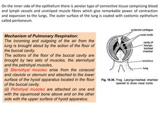
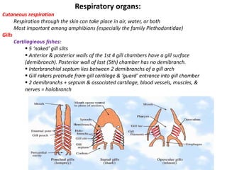
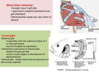
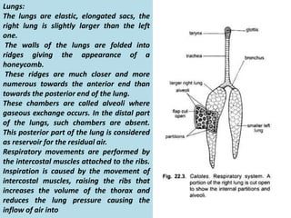
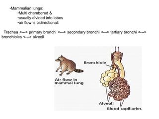
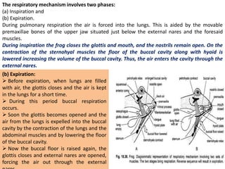
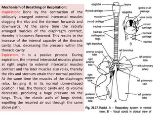
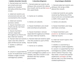
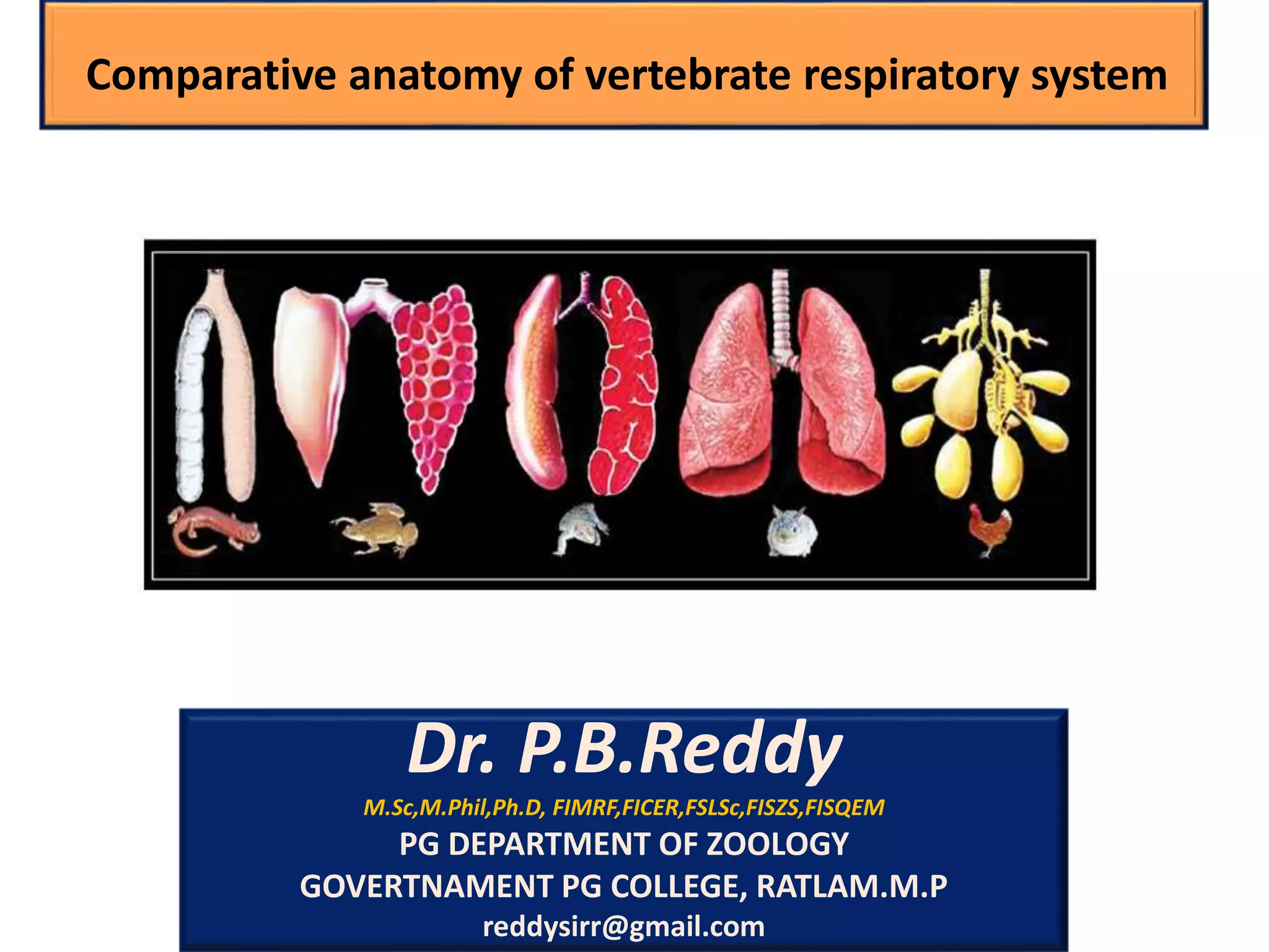
Dr. P.B.Reddy provides an overview of the comparative anatomy of the vertebrate respiratory system. The document discusses why animals need to breathe and defines internal and external respiration. It then describes the key characteristics and functions of respiratory organs across different vertebrate groups, including fish, amphibians, reptiles and others. Specific structures are examined such as gills, lungs and the mechanisms of gas exchange that occur.
Evolutionary change in heart of vertebrates
Heart is situated ventral to the oseophagus in the pericardial section of the coelom.
Heart is a highly muscular pumping organ that pumps blood into arteries and sucks it back through the veins.
In vertebrates it has undergone transformation by twisting from a straight tube to a complex multi-chambered organ.
. There has been an increase in the number of chambers in heart during evolution of vertebrates.
The heart is covered by a transparent protective covering, called pericardium. It is a single layer in fish.
Within pericardium there is a pericardial fluid, protects the heart from the external injury.
The evolution of the heart is based on the separation of oxygenated blood from deoxygenated blood for efficient oxygen transport.
Physiology of Respiration in InvertebratesPRANJAL SHARMA
In physiology, respiration is the movement of oxygen from the outside environment to the cells within tissues, and the removal of carbon dioxide in the opposite direction. In these slides you will get to know about Physiology of Respiration in Invertibrates.
The primitive blueprint for the heart and circulatory system emerged with the arrival of the third mesodermal germ layer in bilaterians. Since then, hearts in animals have evolved from a single layered tube to a multiple chambered heart in due course of time.
INTRODUCTION
The term urogenital refers to something that has both urinary and genital origins. The word urogenital is used because the urinary and reproductive systems in males merge.
These are grouped together because of their proximity to each other, their common embryological origin and the use of common pathways (ex. urethra).
Kidneys and urinary ducts form the urinary system.
The Urinary system performs two important homeostatic processes like excretion and osmoregulation. This system is intimately associated both anatomically, and in terms of embryonic origin with the genital system.
The genital system includes the gonads which generate gametes and the genital ducts that serve as passages for the gametes.
Though functionally different the two organ systems the urinary and the genital system are treated together as the urino- genital system, since both develop from the same segmental blocks of trunk mesoderm or adjacent tissues and share many of the ducts.
Thus although the two systems have nothing common functionally they are closely associated in their use of common ducts and are studied under the broad heading of urinogenital system.
The function of the excretory system is crucial in considering the possible environment of the ‘vertebrate life ’. Several main functions can be attributed to all vertebrate excretory systems:
Excretion of nitrogenous waste products.
Maintaining homeostasis with regard to ions (i.e. salt balance).
Regaining valuable substances (glucose, salts, amino acids, etc.)
Maintaining a physiological osmotic value (i.e. water balance).
The excretory system is formed by a series of paired, segmental nephrons that begin with a nephrostome opening into the coelomic cavity.
A pair of glomeruli per segment, supplied by branches from the aorta, projects into the coelomic cavity close to these nephrostomes.
At a later stage of development, the glomerulus/nephrostome area becomes separated from the rest of the coelomic cavity by an epithelial fold.
The nephrons connect to a duct that is formed by caudal growth of the most anterior nephric tubules. These paired urinary ducts open near the anal region.
INTRODUCTION
The jaw (Upper and lower) is any opposable articulated structure at the entrance of the mouth.
It is typically used for grasping and manipulating food.
Jaw suspension means the fusion of upper jaw and lower jaw or skull for efficient biting.
There are different ways in which these attachments are attained depending upon the modifications in visceral arches in vertebrates.
In most vertebrates, the jaws are bony or cartilaginous and oppose vertically.
The vertebrate jaw is derived from the most anterior two pharyngeal arches supporting the gills, and usually bears numerous teeth.
The vertebrate jaw probably originally evolved in the Silurian period and appeared in the Placoderm fish which further diversified in the Devonian.
It is believed that the hyoid system suspends the jaw from the brain case of the skull, permitting great mobility of the jaws.
The original selective advantage offered by the jaw may not be related to feeding, but rather to increased respiration efficiency.
The jaws were used in the buccal pump (observable in modern fish and amphibians) that pumps water across the gills of fish or air into the lungs in the case of amphibians.
Over evolutionary time the more familiar use of jaws (to humans), in feeding, was selected for and became a very important function in vertebrates. Many teleost fish have substantially modified jaws for suction feeding and jaw protrusion, resulting in highly complex jaws with dozens of bones involved.
Jaw Suspension or Suspensoria:
The method by which the upper and lower jaws are suspended or attached from the chondrocranium is known as jaw suspension or suspensorium.
Amongst the visceral arches, the first (mandibular) arch consists of
= a dorsal palato pterygoquadrate bar forming the upper jaw,
= and ventral Meckel’s cartilage forms the lower jaw.
The second (hyoid) arch consists of = a dorsal hyomandibular supporting and suspending the jaws with the cranium, and a ventral hyoid.
The remaining visceral arches support the gills and are, hence, called branchial arches. Thus, splanchnocranium forms the jaws and suspends them with the chondrocranium.
ORIGIN OF CHORDATES
Animal kingdom is basically divided into two sub kingdoms:
Non-chordata- including animals without notochord.
Chordata- This comprising animals having notochord or chorda dorsalis.
Chordates were evolved sometime 500 million years ago during Cambrian period (invertebrates were also began to evolve in this period) .
Chamberlain (1900) pointed out that all modern chordates possess glomerular kidneys that are designed to remove excess water from body.
It is believed that Chordates have originated from invertebrates.
It is difficult to determine from which invertebrate group the chordates were developed.
Chordate ancestors were soft bodied animals. Hence they were not preserved as Fossils.
However, early fossils of chordates have all been recovered from marine sediments and even modern protochordates are all marine forms.
Also glomerular kidneys are also found in some marine forms such as myxinoids and sharks. That makes the marine origin of chordates more believable.
Chordates evolved from some deuterostome ancestor (echinoderms, hemichordates, pogonophorans etc.) as they have similarities in embryonic development, type of coelom and larval stages.
Many theories infers origin of chordates, hemichordates and echinoderms from a common ancestor.
The vertebrate brain
The vertebrate brain is the main part of the central nervous system. The brain and the spinal cord make up the central nervous system,
In most of the vertebrates the brain is at the front, in the head. It is protected by the skull and close to the main sense organs.
Brains are extremely complex and the part of human and animal body. The brain controls the other organs of the body, either by activating muscles or by causing secretion of chemicals such as hormones and neurotransmitters.
Muscular action allows rapid and coordinated responses to changes in the environment.
The brain of an adult human weights about 1300–1400 grams .
In vertebrates, the spinal cord by itself can cause reflex responses as well as simple movement such as swimming or walking. However, sophisticated control of behaviour requires a centralized brain.
The structure of all vertebrate brains is basically the same.
At the same time, during the course of evolution, the vertebrate brain has undergone changes, and become more effective.
In so-called 'lower' animals, most or all of the brain structure is inherited, and therefore their behaviour is mostly instinctive.
In mammals, and especially in man, the brain is developed further during life by learning. This has the benefit of helping them fit better into their environment. The capacity to learn is seen best in the cerebral cortex.
Three principles
The brain and nervous system is essentially a system which makes connections. It has input from sense organs and output to muscles. It is connected in several ways with the endocrine system, which makes hormones, and the digestive system and sex system. Hormones work slowly, so those changes are gradual.
The brain is a kind of department store. It has, all inter-connected, departments which do different things. They all help each other gather senses.
Much of what the body does is not conscious. Basically, much of the body runs on automatic (breathing, heart beat, hungry, hair growth) adjusted by the autonomic nervous system. The brain, too, does much of its work without a person noticing it. The unconscious mind refers to the brain activities which are hardly ever noticed.
The integumentary system comprises the skin and its appendages. Skin + derivatives= Integument.
It aims to protect the body from various kinds of damage, such as loss of water or damages from outside.
The integumentary system in chordates includes hair, scales, feathers, hooves, and nails.
It may serve to water proof, and protect the deeper tissues.
Excrete wastes, and regulate body temperature.
It is the attachment site for sensory receptors to detect pain, sensation, pressure, and temperature.
The main function of gills is respiration...In gills, there are many hair like projections called gill filaments..in gill filaments, there are number of lamella, from transfer of gases and water occur..
Origin of the Lateral Line System
Lateral line is a canal along the side of a fish containing pores that open into tubes supplied with sense organs sensitive to low vibrations.
Robert H. Denison explained the origin of the lateral line system. He explained that early vertebrates had a pore-canal system in the dermis which functioned as a primitive sensory system in detecting water movement.
Through the evidences from fossils, embryology and comparative anatomy, Denison (1966) established that the inner ear is closely related to the lateral line system. He found a distinct relationship between the pore canal system and the lateral line in Osteotraci.
The inner ear and the lateral line are developed from ectodermal thickenings, called dorso-lateral placodes. These have a number of similarities, including receptors with sensory hairs, and are both innervated by fibers in the acoustico-lateral area of the brain.
The pore canal system is present and developed in Osteostraci (ostracoderm).
It is also present in Heterostraci which is another group of ostracoderms and includes early vertebrates such as lungfishes and crossopterygians.
As its presence is extensive, it is reasonable to suggest that the pore canal system was a primitive character in early vertebrates .
In transverse sections also , it is very difficult to differentiate the pore canal system from a lateral line canal.
Structure of the Lateral Line System
Epidermal structures called neuromasts form the peripheral area of the lateral line.
Neuromasts consist of two types of cells, hair cells and supporting cells.
Hair cells have an epidermal origin and each hair cell has one high kynocyle (5-10 μm) and 30 to 150 short stereocilia (2-3 μm).
The number of hair cells in each neuromast depends on its size, and they can range from dozens to thousands.
Hair cells can be oriented in two opposite directions with each hair cell surrounded by supporting cells.
At the basal part of each hair cell, there are synaptic contacts with afferent and efferent nerve fibers. Afferent fibers, transmit signals to the neural centres of the lateral line and expand at the neuromast base. The regulation of hair cells is achieved by the action of efferent fibers.
Stereocilia and kinocilium of hair cells are immersed into a cupula and are located above the surface of the sensory epithelium.
The cupula is created by a gel-like media, which is secreted by non-receptor cells of the neuromast.
Presentation on Organ & Mechanism of Respiration in Pisces And Amphibiansvskgondia
This is Powerpoint presentation helpful for students and teachers. It includes Defination of Respiration & Function of respiratory system. Also contains mechanism of respiration and various repiratory organs of pisces and amphibians, their structures and fuctions.
Water pollution is a threat to aquatic organisms, including fish, and is a global concern for aquatic biodiversity and ecosystem integrity. This study evaluates the effect of waterborne pollutants on Gangetic Mystus (Mystus cavasius) collected from Chambal River at Nagda, Ujjain. Mystus appeared to be a useful biomarker to assess the impact of toxicity of water pollution.
Zooplankton are the animal component of the plankton community. They are heterotrophic, meaning they can't make their own food and must eat other organisms. In particular, they eat phytoplankton, which are generally smaller than zooplankton.11 species of zooplankton were found in the Shivna River. The most abundant species were copepods Oithona similis, Paracalanus sp., and Calanus sinicus.The species composition of zooplankton varies by season. The highest number of species were found in winter, followed by autumn, summer, and spring. The highest abundance of zooplankton was found in summer, and the lowest in post-monsoon.
This presentation explores how climate change alters the pursuit of economic development: the transformation of poor economies and their people into prosperous ones.
This is hardly the first attempt to reconcile the climate agenda with that of economic development. The United Nations’ Sustainable Development Goals are significant for defining a dual agenda where development targets for people and planet sit alongside each other in a unifying framework.1 Much commentary focuses on the compatibility of the two agendas. A radical and specious view pits progress on climate change and economic development as strict substitutes and calls for no less than the unravelling of economic development to save the planet.2 Cooler heads point instead to their complementarity: the critical role of economic development in supporting adaptation and the recognition that investments in the green transition will propel economies rather than sacrifice living standards.3
In contrast, this essay takes as its starting point that the goals and salience of economic development are immutable. The question posed here is how the quest for economic development changes in a world gripped by a changing climate. The essay argues that climate change will force three major changes: a reappraisal of the causes of and prospects for development, the rebirth of the economics of transition, and a reformulation of the problem development is trying to solve. In a final section, it asks what these changes could mean for international security and for the community of national and global actors who set policy and strategy in this field.
Climate change refers to long-term shifts in temperatures and weather patterns. Such shifts can be natural, due to changes in the sun’s activity or large volcanic eruptions. But since the 1800s, human activities have been the main driver of climate change, primarily due to the burning of fossil fuels like coal, oil and gas.
Burning fossil fuels generates greenhouse gas emissions that act like a blanket wrapped around the Earth, trapping the sun’s heat and raising temperatures.
The main greenhouse gases that are causing climate change include carbon dioxide and methane. These come from using gasoline for driving a car or coal for heating a building, for example. Clearing land and cutting down forests can also release carbon dioxide. Agriculture, oil and gas operations are major sources of methane emissions. Energy, industry, transport, buildings, agriculture and land use are among the main sectors causing greenhouse gases.
In India, bacteria that cause common infections, such as urinary tract and bloodstream infections, are becoming resistant to nearly all antibiotics. This resistance is due to a combination of factors: uncontrolled access to antibiotics, gaps in infection prevention and control (IPC) practices, and high rates of communicable diseases. Antibiotic resistance, or AR, is a serious problem throughout the country, and threatens to reduce the usefulness of antibiotics both in India and around the world.
Because of this emerging threat, India is committed to slowing the spread of AR. Two institutions within India’s Ministry of Health – the Indian Council of Medical Research and National Centre for Disease Control – each developed national networks of public and private hospitals to measure AR trends, prevent healthcare-associated infections (HAIs), and enhance appropriate use of antibiotics. The All India Institute of Medical Sciences is coordinating HAI measurement and prevention efforts in both networks. In addition, efforts in the state of Tamil Nadu focus on building district-level IPC capacity to prevent HAIs, focusing on maternal and neonatal patients.
The Indian Governamnet is is working closely with partners at the national and state level to:
Detect AR pathogens, including novel strains, by developing lab networks and lab expertise.
Use standardized surveillance to monitor and track AR infections in healthcare to learn how often these infections occur and to help develop strategies to prevent them.
Implement focused IPC activities and training.
Optimize use and reduce misuse of critical antibiotics through antibiotic stewardship programs.
Physiological and histopathological effects of Bisphenol A .Bisphenol A is less soluble in water. For that reason, dimethyl sulfoxide (DMSO) was used as a medium to obtain proper distribution in the test solution (Chen, J., et al, 2015). Working solution of commercial grade Bisphenol A (97% pure) was prepared by dilution of stock solution double distilled water immediately prior to experimental use. Serial dilutions of the stock solution were prepared using previously aerated, copper free and stored tap water. The water was continuously aerated. This was prepared by dissolving BPA (50mg) in 100ml of DMSO and the desired concentrations of BPA in tap water were prepared by adding appropriate volumes of this stock solution into test aquarium. A static non-renewable bioassay was conducted in triplicate for each concentration with four animals in each tub. No water exchange was done and the fishes were not fed during the period of the experiment. Percentage mortality was recorded at 12, 24, 48, 72 and 96 h interval. Control group was subjected to acetone at the maximum acetone volume used in the dilution of the dose concentrations. The range of LC50 for H.fossilis (mean wt. 36.78 g) under given conditions was determined to lie between 5 and 10 mg/L for BPA. Hence, for the definitive test, concentrations such as 2, 4, 6, 8, 10, 12, and 14 mg/L of BPA concentration were selected. The test was conducted in triplicate for each concentration with 10 fishes in each tank. At the end of 96 h, the fishes that had survived were anesthetized with clove oil at 100 mg/L, sampled for blood, and processed for hematological analysis. The data obtained from the experiment was processed by probit analysis using a Microsoft Excel computer program.
When pollutants are discharged from a specific location such as a drain pipe carrying industrial effluents discharged directly into a water body it represents point source pollution.
In contrast, non-point sources include discharge of pollutants from diffused sources or from a larger area such as runoff from agricultural fields, grazing lands, construction sites, abandoned mines and pits, etc.
Targets of Sustainable Development Goal 3
WHO Framework Convention on Tobacco Control; support research, development and universal access to affordable vaccines and medicines; increase health financing and support health workforce in developing countries
INTRODUCTION
Toxicology is the science of the poisons. It also studies the nature, effects, detection, assessment and treatment of their effects on biological material.
Toxicology is a multidisciplinary science. The ultimate objective of the combined research is to determine how an organism is affected by exposure to an agent.
This includes an understanding of:
How the agent moves and interact with living cells and tissues of the organism;
What parts of the organism are affected by its presence and health outcomes of this exposure.
Evaluation of the toxicity of substances whose biological effects may not have been well characterized.
The influence of chemical toxicity is mainly
determined by the dosage, duration of exposure,
route of exposure, species, age, sex, and environment.
The goal of toxicology is to contribute to the
general knowledge and harmful actions of
chemical substances.
2. to study their mechanisms of action,
3. and to estimate their possible risks to humans
HISTORY
Dioscorides, a Greek physician in the court of the Roman emperor Nero, made the first attempt to classify plants according to their toxic and therapeutic effect. Poisonous plants and animals were recognized and their extracts used for hunting or in warfare.
In 1500 BC people used hemlock, opium, arrow poisons, and certain metals to poison enemies or for state executions.
Theophrastus Phillipus Auroleus Bombastus von Hohenheim (1493–1541) (also referred to as Paracelsus, a Roman physician from the first century) is considered "the father" of toxicology.
He stated that "All things are poisonous and nothing is without poison; only the dose makes a thing not poisonous.“
Mathieu Orfila (1813) is considered the modern father of toxicology.
In 1850, Jean Stas became the first person to successfully isolate plant poisons from human tissue.
Hippolyte Visart de Bocarmé used nicotine to kill his brother-in-law. He extracted nicotine from tobacco leaves.
The 20th and 21st Centuries have marked by great advancements in the level of understanding of toxicology. DNA and various biochemicals that maintain body functions have been discovered. Our level of knowledge of toxic effects on organs and cells has expanded to the molecular level.
Introduction
Gnathostomata are the jawed vertebrates. (gnathos= "jaw" + (stoma)="mouth".
It comprises roughly 60,000 species. (99% of all living vertebrates).
Living gnathostomes have teeth, and paired appendages.
A horizontal semicircular canal is present in the inner ear.
Myelin sheaths is present on the neurons.
Adaptive immune system uses V(D) J recombination ( it is the mechanism of somatic recombination that occurs only in developing lymphocytes during the early stages of T and B cell maturation. VDJ recombination is the process by which T cells and B cells randomly assemble different gene segments – known as variable (V), diversity (D) and joining (J) genes – in order to generate unique receptors (known as antigen receptors) that can collectively recognize many different types of molecule. While Agnatha (petromyzon and hagfish) use genetic recombination in the variable lymphocyte receptor gene.
It is now assumed that Gnathostomata evolved from ancestors that already possessed a pair of both pectoral and pelvic fins.
In addition to this, some placoderms were shown to have a third pair of paired appendages, that had been modified to claspers in males and basal plates in females—a pattern not seen in any other vertebrate group.
It is believed that the jaws evolved from anterior gill support arches that had acquired a new role, being modified to pump water over the gills by opening and closing the mouth more effectively – the buccal pump mechanism.
Presence of Calcified, bony skull and vertebra are the characteristic features of Gnathostomata (fishes, amphibians, reptiles, birds and mammals).
Pelvic fins are situated just in front of the anus.
Interventrals and basiventrals present in the backbone. These are the elements of the backbone which lie under the notochord, and match the basidorsals and interdorsals respectively.
Gill arches which lie internally to the gills and branchial blood vessels, contrary to the gill arches of all jawless craniates, which are external to the gills and blood vessels.
A horizontal semicircular canal in the inner ear.
Paired nasal sacs which are independent from the hypophysial tube.
There are numerous other characteristics of the soft anatomy and physiology (e.g. myelinated nerve fibres, sperms passing through urinary ducts, etc.), which are unique to the gnathostomes among extant craniates, but cannot by observed in fossils.
Introduction
Ostracoderms (shell-skinned) are of several groups of extinct, primitive, jawless fishes that were covered in an armour of bony plates.
They appeared in the Cambrian, about 510 million years ago, and became extinct towards the end of the Devonian, about 377 million years ago. They were quite abundant during the upper Silurian and Devonian periods. Most of fossils of Ostracodermi were preserved in the bottom sediments of freshwater streams.
However, the opinion is sharply divided as to whether their habitat was freshwater or marine.
The first fossil fishes that were discovered were ostracoderms.
The Swiss anatomist Louis Agassiz received some fossils of bony armored fish from Scotland in the 1830s.
The ostracoderms resembled the present day cyclostomes (lampreys and hagfishes) in many respects and together with them constitute a special group of jawless vertebrates, the Agnatha.
Characteristics: They use gills exclusively for respiration but not for feeding . Earlier chordates with gills used them for both respiration and feeding. Ostracoderms had separate pharyngeal gill pouches along the side of the head, which were permanently open with no protective operculum. mostly small to medium-sized fishes, protected by a heavy, bony dermal (derived from skin) armor. bottom-dwellers; filter-feeders or grazers. no paired fins, but many with stabilizing paired flaps on either side of head.
(1) Ostracoderms were the first vertebrates.
(2) They were popularly called armoured fishes.
(4) They lived in freshwater.
(5) They were bottom dwellers.
(6) Their body was fish-like and did not exceed 30 cm in size.
(7) Paired fins were absent.
(8) Median and caudal fins were present.
(9) The caudal fin was of heterocercal type.
(10) The head and thorax were covered by heavy armour of bones. It protected ostracoderms from the giant scorpion like arthropods, eurypterids.
(11) Bony skull was well developed.
(12) Mouth was mostly present on the ventral side.
(13) They were having large number of gill slits.
(14) The nervous system had 10 pairs of cranial nerves.
(15) The head had a pair of lateral eyes, and a median pineal eye.
(16) They were filter feeders, feeding like a vacuum cleaner.
(17) The endoskeleton was either bony or cartilaginous.
Introduction:
Adaptation to environment is one of the basic characteristics of the living organisms. Living organisms are plastic and posses the inherent properties to respond to a particular environment.
It is a facet of evolution and involve structural diversities amongst living organisms that are heritable. Organisms exhibit numerous structural and functional adaptations that help them to survive as species and to overcome the tremendous competition in nature.
All classes of vertebrates have their representatives leading to partial or total aquatic life.
Water is a homogenous medium for animals.
As a medium, it is heavy in concentration than air.
Stable gaseous and osmotic concentration in a specific region.
Temperature fluctuation is minimum for a particular region.
Water bodies generally have very rich food resources.
Characters of an Aquatic Animal:
An aquatic animal should have the ability to swim to overcome the resistance of the surrounding medium.
Therefore, it should have a streamlined body with an organ or ability to float.
The animal should also have to overcome the problem of osmoregulation.
There are two types of animals living in the present day water, which have undergone aquatic adaptation.
According to their origin, they are primary and secondary aquatic animals.
Adaptations to water habitat are of two types:
Primary aquatic adaptations which includes primitive gill-breathing vertebrates (fishes); Those animals, whose ancestors and themselves are living in the water from the very beginning of their evolution, are called primary aquatic animals. In other words, primary aquatic animals never had a terrestrial ancestry. They exhibit perfect aquatic adaptations. All fishes are primary aquatic animals.
Secondary aquatic adaptations which are acquired as in reptiles, birds and mammals. Those animals whose ancestors were lung breathing land animals, migrated to the water for some reason and ultimately got adapted to live in aquatic habitat, are called secondary aquatic animals. Some of them live partially while others live totally in the water. All aquatic reptiles, aves and mammals are representatives of secondary aquatic animals. Amphibians are in a transitional form between primary and secondary aquatic life.
Sensory adaptations like, electroreception for electrolocation and electro communication, olfaction (vomeronasal system), balance (spatial orientation, movement perception), vision (cornea curvature, retinal topography), and hearing (acoustics, ear anatomy) under the underwater sound reception mechanisms in various aquatic amniotes are well developed.
Central nervous system: The central nervous system consists of the brain and spinal cord. The brain plays a central role in the control of most bodily functions, including awareness, movements, sensations, thoughts, speech, and memory. Some reflex movements can occur via spinal cord pathways without the participation of brain structures. The spinal cord is connected to a section of the brain called the brainstem and runs through the spinal canal.
Peripheral Nervous System: Nerve fibers that exit the brainstem and spinal cord become part of the peripheral nervous system. Cranial nerves exit the brainstem and function as peripheral nervous system mediators of many functions, including eye movements, facial strength and sensation, hearing, and taste.
The autonomic nervous system: The autonomic nervous system is a control system that acts largely unconsciously and regulates bodily functions, such as the heart rate, digestion, respiratory rate, pupillary response, urination, and sexual arousal. This system is the primary mechanism in control of the fight-or-flight response.
The autonomic nervous system comprises two antagonistic sets of nerves, the sympathetic and parasympathetic nervous systems. The hypothalamus is the key brain site for central control of the autonomic nervous system, and the paraventricular nucleus is the key hypothalamic site for this control.
Divisions of Nervous System:
The vertebrate nervous system has three divisions:
(i) A central nervous system comprising the brain and spinal cord. Its function is to receive the stimulus from the receptors and transmit its response to the effectors. Thus, it coordinates all the functions of the body.
(ii) A peripheral nervous system consisting of cranial and spinal nerves arising from the brain and spinal cord respectively. It forms a connecting link between the receptors, central nervous system (CNS) and effectors.
(iii) An autonomic nervous system made of two ganglionated sympathetic nerves, ganglia in the head and viscera, and their connecting nerves. The autonomic nervous system is often regarded as a part of the peripheral nervous system because the two are connected. But all the three divisions of the nervous system are connected intimately both structurally and functionally.
Abstract: Soil contamination with heavy metals is a serious global concern due to their toxicity and bioaccumulation property. The present investigation was aimed to assess heavy metal contamination of agricultural soil around the polluted zone of the Chambal River at Nagda, Ujjain (M.P, India). Soil samples were collected at three sites S1, S2, and S3 alongside of Chambal River in December 2019 and analyzed for heavy metals like Cr, Ni, Cd, Pb, and Zn by atomic absorption spectrophotometer (AAS) methods. The Igeo results revealed that the study area has fallen in the category of uncontaminated and moderately contaminated with Cd and Pb in all study stations. Essential compositions were evaluated through the estimation of geochemical accumulation indices to find out the heavy metal contamination of soil. Significant enrichment of the soil with Cd, Zn, Cu, Ni, and Pb was observed in all study stations. The S1 station exhibited the highest concentrations of heavy metals in soil. The present outcome is useful for mitigating the impact of metallic pollution on environmental health and required strategies to prevent such effects.
Keywords: Chambal River, Geo-Accumulation Index, Heavy Metals, Industrial Pollution, Soil Quality.
Introduction:
RNA interference (RNAi) or Post-Transcriptional Gene Silencing (PTGS) is an important biological process for modulating eukaryotic gene expression.
It is highly conserved process of posttranscriptional gene silencing by which double stranded RNA (dsRNA) causes sequence-specific degradation of mRNA sequences.
dsRNA-induced gene silencing (RNAi) is reported in a wide range of eukaryotes ranging from worms, insects, mammals and plants.
This process mediates resistance to both endogenous parasitic and exogenous pathogenic nucleic acids, and regulates the expression of protein-coding genes.
What are small ncRNAs?
micro RNA (miRNA)
short interfering RNA (siRNA)
Properties of small non-coding RNA:
Involved in silencing mRNA transcripts.
Called “small” because they are usually only about 21-24 nucleotides long.
Synthesized by first cutting up longer precursor sequences (like the 61nt one that Lee discovered).
Silence an mRNA by base pairing with some sequence on the mRNA.
Discovery of siRNA?
The first small RNA:
In 1993 Rosalind Lee (Victor Ambros lab) was studying a non- coding gene in C. elegans, lin-4, that was involved in silencing of another gene, lin-14, at the appropriate time in the
development of the worm C. elegans.
Two small transcripts of lin-4 (22nt and 61nt) were found to be complementary to a sequence in the 3' UTR of lin-14.
Because lin-4 encoded no protein, she deduced that it must be these transcripts that are causing the silencing by RNA-RNA interactions.
Types of RNAi ( non coding RNA)
MiRNA
Length (23-25 nt)
Trans acting
Binds with target MRNA in mismatch
Translation inhibition
Si RNA
Length 21 nt.
Cis acting
Bind with target Mrna in perfect complementary sequence
Piwi-RNA
Length ; 25 to 36 nt.
Expressed in Germ Cells
Regulates trnasposomes activity
MECHANISM OF RNAI:
First the double-stranded RNA teams up with a protein complex named Dicer, which cuts the long RNA into short pieces.
Then another protein complex called RISC (RNA-induced silencing complex) discards one of the two RNA strands.
The RISC-docked, single-stranded RNA then pairs with the homologous mRNA and destroys it.
THE RISC COMPLEX:
RISC is large(>500kD) RNA multi- protein Binding complex which triggers MRNA degradation in response to MRNA
Unwinding of double stranded Si RNA by ATP independent Helicase
Active component of RISC is Ago proteins( ENDONUCLEASE) which cleave target MRNA.
DICER: endonuclease (RNase Family III)
Argonaute: Central Component of the RNA-Induced Silencing Complex (RISC)
One strand of the dsRNA produced by Dicer is retained in the RISC complex in association with Argonaute
ARGONAUTE PROTEIN :
1.PAZ(PIWI/Argonaute/ Zwille)- Recognition of target MRNA
2.PIWI (p-element induced wimpy Testis)- breaks Phosphodiester bond of mRNA.)RNAse H activity.
MiRNA:
The Double-stranded RNAs are naturally produced in eukaryotic cells during development, and they have a key role in regulating gene expression .
Cancer cell metabolism: special Reference to Lactate PathwayAADYARAJPANDEY1
Normal Cell Metabolism:
Cellular respiration describes the series of steps that cells use to break down sugar and other chemicals to get the energy we need to function.
Energy is stored in the bonds of glucose and when glucose is broken down, much of that energy is released.
Cell utilize energy in the form of ATP.
The first step of respiration is called glycolysis. In a series of steps, glycolysis breaks glucose into two smaller molecules - a chemical called pyruvate. A small amount of ATP is formed during this process.
Most healthy cells continue the breakdown in a second process, called the Kreb's cycle. The Kreb's cycle allows cells to “burn” the pyruvates made in glycolysis to get more ATP.
The last step in the breakdown of glucose is called oxidative phosphorylation (Ox-Phos).
It takes place in specialized cell structures called mitochondria. This process produces a large amount of ATP. Importantly, cells need oxygen to complete oxidative phosphorylation.
If a cell completes only glycolysis, only 2 molecules of ATP are made per glucose. However, if the cell completes the entire respiration process (glycolysis - Kreb's - oxidative phosphorylation), about 36 molecules of ATP are created, giving it much more energy to use.
IN CANCER CELL:
Unlike healthy cells that "burn" the entire molecule of sugar to capture a large amount of energy as ATP, cancer cells are wasteful.
Cancer cells only partially break down sugar molecules. They overuse the first step of respiration, glycolysis. They frequently do not complete the second step, oxidative phosphorylation.
This results in only 2 molecules of ATP per each glucose molecule instead of the 36 or so ATPs healthy cells gain. As a result, cancer cells need to use a lot more sugar molecules to get enough energy to survive.
introduction to WARBERG PHENOMENA:
WARBURG EFFECT Usually, cancer cells are highly glycolytic (glucose addiction) and take up more glucose than do normal cells from outside.
Otto Heinrich Warburg (; 8 October 1883 – 1 August 1970) In 1931 was awarded the Nobel Prize in Physiology for his "discovery of the nature and mode of action of the respiratory enzyme.
WARNBURG EFFECT : cancer cells under aerobic (well-oxygenated) conditions to metabolize glucose to lactate (aerobic glycolysis) is known as the Warburg effect. Warburg made the observation that tumor slices consume glucose and secrete lactate at a higher rate than normal tissues.
A brief information about the SCOP protein database used in bioinformatics.
The Structural Classification of Proteins (SCOP) database is a comprehensive and authoritative resource for the structural and evolutionary relationships of proteins. It provides a detailed and curated classification of protein structures, grouping them into families, superfamilies, and folds based on their structural and sequence similarities.
Multi-source connectivity as the driver of solar wind variability in the heli...Sérgio Sacani
The ambient solar wind that flls the heliosphere originates from multiple
sources in the solar corona and is highly structured. It is often described
as high-speed, relatively homogeneous, plasma streams from coronal
holes and slow-speed, highly variable, streams whose source regions are
under debate. A key goal of ESA/NASA’s Solar Orbiter mission is to identify
solar wind sources and understand what drives the complexity seen in the
heliosphere. By combining magnetic feld modelling and spectroscopic
techniques with high-resolution observations and measurements, we show
that the solar wind variability detected in situ by Solar Orbiter in March
2022 is driven by spatio-temporal changes in the magnetic connectivity to
multiple sources in the solar atmosphere. The magnetic feld footpoints
connected to the spacecraft moved from the boundaries of a coronal hole
to one active region (12961) and then across to another region (12957). This
is refected in the in situ measurements, which show the transition from fast
to highly Alfvénic then to slow solar wind that is disrupted by the arrival of
a coronal mass ejection. Our results describe solar wind variability at 0.5 au
but are applicable to near-Earth observatories.
Professional air quality monitoring systems provide immediate, on-site data for analysis, compliance, and decision-making.
Monitor common gases, weather parameters, particulates.
This presentation explores a brief idea about the structural and functional attributes of nucleotides, the structure and function of genetic materials along with the impact of UV rays and pH upon them.
Comparing Evolved Extractive Text Summary Scores of Bidirectional Encoder Rep...University of Maribor
Slides from:
11th International Conference on Electrical, Electronics and Computer Engineering (IcETRAN), Niš, 3-6 June 2024
Track: Artificial Intelligence
https://www.etran.rs/2024/en/home-english/
(May 29th, 2024) Advancements in Intravital Microscopy- Insights for Preclini...Scintica Instrumentation
Intravital microscopy (IVM) is a powerful tool utilized to study cellular behavior over time and space in vivo. Much of our understanding of cell biology has been accomplished using various in vitro and ex vivo methods; however, these studies do not necessarily reflect the natural dynamics of biological processes. Unlike traditional cell culture or fixed tissue imaging, IVM allows for the ultra-fast high-resolution imaging of cellular processes over time and space and were studied in its natural environment. Real-time visualization of biological processes in the context of an intact organism helps maintain physiological relevance and provide insights into the progression of disease, response to treatments or developmental processes.
In this webinar we give an overview of advanced applications of the IVM system in preclinical research. IVIM technology is a provider of all-in-one intravital microscopy systems and solutions optimized for in vivo imaging of live animal models at sub-micron resolution. The system’s unique features and user-friendly software enables researchers to probe fast dynamic biological processes such as immune cell tracking, cell-cell interaction as well as vascularization and tumor metastasis with exceptional detail. This webinar will also give an overview of IVM being utilized in drug development, offering a view into the intricate interaction between drugs/nanoparticles and tissues in vivo and allows for the evaluation of therapeutic intervention in a variety of tissues and organs. This interdisciplinary collaboration continues to drive the advancements of novel therapeutic strategies.
Slide 1: Title Slide
Extrachromosomal Inheritance
Slide 2: Introduction to Extrachromosomal Inheritance
Definition: Extrachromosomal inheritance refers to the transmission of genetic material that is not found within the nucleus.
Key Components: Involves genes located in mitochondria, chloroplasts, and plasmids.
Slide 3: Mitochondrial Inheritance
Mitochondria: Organelles responsible for energy production.
Mitochondrial DNA (mtDNA): Circular DNA molecule found in mitochondria.
Inheritance Pattern: Maternally inherited, meaning it is passed from mothers to all their offspring.
Diseases: Examples include Leber’s hereditary optic neuropathy (LHON) and mitochondrial myopathy.
Slide 4: Chloroplast Inheritance
Chloroplasts: Organelles responsible for photosynthesis in plants.
Chloroplast DNA (cpDNA): Circular DNA molecule found in chloroplasts.
Inheritance Pattern: Often maternally inherited in most plants, but can vary in some species.
Examples: Variegation in plants, where leaf color patterns are determined by chloroplast DNA.
Slide 5: Plasmid Inheritance
Plasmids: Small, circular DNA molecules found in bacteria and some eukaryotes.
Features: Can carry antibiotic resistance genes and can be transferred between cells through processes like conjugation.
Significance: Important in biotechnology for gene cloning and genetic engineering.
Slide 6: Mechanisms of Extrachromosomal Inheritance
Non-Mendelian Patterns: Do not follow Mendel’s laws of inheritance.
Cytoplasmic Segregation: During cell division, organelles like mitochondria and chloroplasts are randomly distributed to daughter cells.
Heteroplasmy: Presence of more than one type of organellar genome within a cell, leading to variation in expression.
Slide 7: Examples of Extrachromosomal Inheritance
Four O’clock Plant (Mirabilis jalapa): Shows variegated leaves due to different cpDNA in leaf cells.
Petite Mutants in Yeast: Result from mutations in mitochondrial DNA affecting respiration.
Slide 8: Importance of Extrachromosomal Inheritance
Evolution: Provides insight into the evolution of eukaryotic cells.
Medicine: Understanding mitochondrial inheritance helps in diagnosing and treating mitochondrial diseases.
Agriculture: Chloroplast inheritance can be used in plant breeding and genetic modification.
Slide 9: Recent Research and Advances
Gene Editing: Techniques like CRISPR-Cas9 are being used to edit mitochondrial and chloroplast DNA.
Therapies: Development of mitochondrial replacement therapy (MRT) for preventing mitochondrial diseases.
Slide 10: Conclusion
Summary: Extrachromosomal inheritance involves the transmission of genetic material outside the nucleus and plays a crucial role in genetics, medicine, and biotechnology.
Future Directions: Continued research and technological advancements hold promise for new treatments and applications.
Slide 11: Questions and Discussion
Invite Audience: Open the floor for any questions or further discussion on the topic.
Nutraceutical market, scope and growth: Herbal drug technologyLokesh Patil
As consumer awareness of health and wellness rises, the nutraceutical market—which includes goods like functional meals, drinks, and dietary supplements that provide health advantages beyond basic nutrition—is growing significantly. As healthcare expenses rise, the population ages, and people want natural and preventative health solutions more and more, this industry is increasing quickly. Further driving market expansion are product formulation innovations and the use of cutting-edge technology for customized nutrition. With its worldwide reach, the nutraceutical industry is expected to keep growing and provide significant chances for research and investment in a number of categories, including vitamins, minerals, probiotics, and herbal supplements.
3. Why do animals need to breathe?
Breathing is important to organisms because cells require
energy (oxygen) to move, reproduce and function. Breath also
expels carbon dioxide, which is a by-product of cellular processes
within the bodies of animals.
Respiration is the process of releasing energy from food and
this takes place inside the cells of the body.
The process of respiration involves taking in oxygen (of air) into
cells, using it for releasing energy by burning food, and then
eliminating the waste products (carbon dioxide and water) from
the body.
Respiration is essential for life because it provides energy for
carrying out all the life processes which are necessary to keep the
organisms alive.
The energy produced during respiration is stored in the form of
ATP (Adenosine Tri- Phosphate) molecules in the cells of the body
and used by the organism as when required.
4. KEY POINTS
Life started in an anaerobic environment in the so called ‘primodial broth’ (a mixture of
organic molecules.
Subsequently, oxygen strangely enough became an crucial factor for aerobic metabolism
especially in the higher life forms.
The rise of an oxygenic environment was an important event in the diversification of life.
It evoked a dramatic shift from inefficient to sophisticated oxygen dependent oxidizing
ecosystems.
Anaerobic fermentation, the metabolic process that prevailed for the first about 2 billion
years of the evolution of life, was a very inefficient way of extracting energy from organic
molecules. Ex: A molecule of glucose, e.g., produces only two molecules of ATP (≈ 15 kCal)
compared with 36 ATP molecules (≈ 263 kCal) in oxygenic respiration.
Aerobic metabolism must have developed at a critical point when the partial pressure of
oxygen rose from an initial level to one adequately high to drive it passively across the cell
membrane.
Respiration is a complex and highly integrated biomechanical, physiological, and behavioral
processes.
The transfer of O2 occurs through a flow of tissue barriers and compartments by diffusion
down a partial pressure gradient, which drops to about zero at the mitochondrial level.
Acquisition of molecular oxygen (O2) from the external fluid media (water and air) and the
discharge of carbon dioxide (CO2) into the same milieu is the primary role of respiration.
The respiratory system is a biological system consisting of specific organs and structures.
5. Characteristics of respiratory organs
Every respiratory device must conform to the following essentials features:
Blood must be separated from the external environment that is air or water by a
thin epithelium.
The epithelium must be permeable to permit diffusion of gases through it.
The respiratory epithelium must always remain moist with a film of fluid to permit
osmosis of gases.
The area of respiratory surface should be extensive to allow efficient absorption of
oxygen.
Both the current of air or water outside and blood in capillaries must be made to
circulate constantly for quick replacement of gases.
All the respiratory organs should have large surface areas to get enough oxygen.
All the respiratory organs (like the skin, lungs, gills) have a rich supply of blood for
transporting respiratory gases.
The thickness of the water/air–blood (tissue) barrier, the respiratory surface area, and
volume of pulmonary capillary blood are the foremost structural parameters which
determine the diffusing capacity of a gas exchanger for O2.
Diffusion of oxygen from the medium into the blood.
Transport of oxygen to the tissues and cells of the body.
Diffusion of oxygen from the blood into cells.
Carbon dioxide follows a reverse path.
6. External and internal respiration
Internal Respiration: Oxygen diffuses
out from the blood into tissue during
internal respiration. Internal
respiration, also known as cellular
respiration, is the process of energy
production via the breakdown of
glucose. Therefore, it occurs within the
cells. Internal respiration can be
aerobic respiration or anaerobic
respiration.
External Respiration: External
respiration is a physical process of
taking oxygen from the external
environment into the body and
expelling carbon dioxide from the
body to the external environment. It is
a vital process for life as it supplies
oxygen to extract energy from food via
internal or cellular respiration.
Additionally, it removes carbon
dioxide, which is a waste product of
internal respiration.
7. Respiratory System in Vertebrates
GILLS
Gills in Protochordates
A large and sieve-like pharynx in majority of these animals performs dual function of
respiration and trapping food particles which are brought in through the current of water.
The primitive pterobranch hemichordates (Cephalodiscus and Rhabdopleura) have either no
gill slits or have very few and sport tentaculated arms, which other than food gathering, also
function as efficient respiratory organs.
Balanoglossus possesses a large pharynx having as many as 700 pairs of gill slits, which
appears to be a necessity in the burrowing habitat of the animal.
The free-living urochordates, such as Salpa and Doliolum do not possess many stigmata or gill
slits as their entire body is permeable to oxygen but in the sedentary ascidians pharynx is
prominently enlarged and perforated with no less than 200,000 stigmata for filter-feeding.
Cephalochordates use pharynx for both filter-feeding and respiration and hence carry 150-200
pairs of gill slits.
8. Respiratory organs of Cyclostomes
Agnathans have 6-15 pairs of gill
pouches.
They are lateral extensions of pharynx
and contain gill lamellae within.
Cyclostomes are called
marsipobranchs, which means “pouched
gills”, since the gill lamellae are housed in
gill pouches.
The hagfish, Myxine has only 6 pairs of
gill pouches whose ducts join together
and open to the exterior by a single pair
of openings, while Bdellostoma carries 6-
15 pairs of gill pouches that vary in
different species and open to the outside
independently.
The lamprey, Petromyzon, has 8
embryonic and 7 adult paired gill
pouches that open to the exterior by
independent openings.
Hagfish
9. .
In lower aquatic vertebrates the respiratory organs are not
connected to the olfactory organs, but in air-breathing vertebrates
there is a close association between the two.
In Chondrichthyes there is a direct connection between the olfactory
and respiratory organs in which the internal nares or choanae open
from the nasal cavities into the buccal cavity, but it is only in
tetrapoda that air enters through the nasal cavities into the buccal
cavity and then into the lungs.
10. Respiration in Fish
Respiration in fish takes place with the help of gills.
Most fish possess gills on either side of their head.
Gills are tissues made up of feathery structures called gill filaments providing a large surface area for
exchange of gases.
A large surface area is crucial for gas exchange in aquatic organisms as water contains very little amount
of dissolved oxygen.
The filaments in fish gills are organized in rows in the gill arch.
Each filament comprises lamellae, which are discs supplied with capillaries.
Blood moves in and out of the gills through these small blood vessels.
The gills provide extensive respiratory surface for the efficient gas exchange.
Fish take in oxygen-rich water via their mouths and pump it over their gills.
When water moves over the gill filaments, the blood within the capillary network takes up the dissolved
oxygen.
Then, the circulatory system supplies oxygen to all tissues of the body and finally to the cells while taking
up carbon dioxide that is eliminated through the gills from the body.
11. Mechanism of Respiration:
1. Inspiration:
The outer opening of the gill-chamber
remains tightly closed.
The branchial-arches and the opercula
swell laterally increasing the volume of
bucco-pharyngeal cavity.
Thus, the water flows in through the
opened mouth to fill the enlarged bucco-
pharyngeal cavity.
2. Expiration:
Mouth becomes closed by oral valves.
Bucco-pharyngeal cavity contracts
exerting pressure on the contained water.
In the meantime the opercula and
branchio-stegal membranes are lifted off.
As a result, water from the bucco-
pharyngeal cavity passes out through the
gill-chamber, bathing the gill-filaments.
Contraction and expansion of pharyngeal
cavity occurs by alternate retraction and
protraction of the hyoid arch which
supports the bucco-pharyngeal cavity.
12. Amphibians utilize gills for breathing early in life
Most amphibians breathe through lungs and their
skin.
Their skin has to stay wet in order for them to absorb
oxygen so they secrete mucous to keep their skin moist.
Cutaneous Respiration:
The skin of frog is an important organ of respiration
(water).
when frog undergoes summer sleep (aestivation) and
winter sleep (hibernation), the skin is the only organ of
respiration.
The skin of frog is very much suited for the respiratory
function as it is very thin and richly supplied with blood
capillaries and remains moist with the water and also
mucus, secreted by mucous glands.
During gaseous exchange the oxygen first dissolves in
the moisture present over the body and then diffuses
into the blood circulating in the blood capillaries, while
the resultant carbon dioxide passes out from the blood
into the surrounding medium (water) by diffusion. In
cutaneous respiration, no movements are needed
because skin always remains exposed to air or water.
Amphibia
13. Bucco-Pharyngeal Respiration or Buccal Respiration:
The mucous lining of the buccal cavity is richly supplied with blood capillaries and remains moist by the
mucus.
The buccal respiration occurs by lowering and raising of the floor of the buccal cavity, during the course of
which the air is constantly sucked into the buccal cavity and is drawn out through the external and internal
nares.
In this type, the mouth and glottis remain closed.
Thus, no air enters or goes out from the lungs.
When the floor of the buccal cavity is lowered, the air enters the buccal cavity through the nostrils or the
nares. The oxygen of air dissolves in the layer of mucus and then goes into blood. At the same time carbon
dioxide is given out into the buccal cavity from the blood which is expelled along with residual air through the
nostrils when the floor of the buccal cavity is raised.
Pulmonary Respiration:
Respiration on land in air with the help of lungs is the pulmonary respiration.
In frog, lungs are poorly developed.
The intake of oxygen by lungs is not sufficient to the body. Therefore, oxygen intake through moist skin and
buccal cavity is needed.
The lungs also hydrostatic organs as they enable frog to float in water when they are inflated. The air
enters and leaves the lungs through the respiratory fact.
(b) Lungs:
A pair of lungs are found in the anterior part of the body cavity, one on the either side of the heart.
They are ovoid, thin-walled, elastic sacs with shallow internal folds or septa that increase the inner surface to
form many chambers called alveoli. These are separated from each other through septa. The inner surface of
the aveoli is covered with a single layer of epithelial cells which are very thin and flattened except on the
edges of the septa where they are ciliated and cylindrical.
14. On the inner side of the epithelium there is aerolar type of connective tissue comprising blood
and lymph vessels and unstriped muscle fibres which give remarkable power of contraction
and expansion to the lungs. The outer surface of the lung is coated with coelomic epithelium
called peritoneum.
Mechanism of Pulmonary Respiration:
The incoming and outgoing of the air from the
lung is brought about by the action of the floor of
the buccal cavity.
The actions of the floor of the buccal cavity are
brought by two sets of muscles, the sternohyal
and the petrohyal muscles,
(i) Sternohyal muscles arise from the coracoid
and clavicle or sternum and attached to the lower
surface of the hyoid apparatus located in the floor
of the buccal cavity.
(ii) Petrohyal muscles are attached on one end
with the squamosal bone above and on the other
side with the upper surface of hyoid apparatus.
15. Respiratory organs:
Cutaneous respiration
Respiration through the skin can take place in air, water, or both
Most important among amphibians (especially the family Plethodontidae)
Gills
Cartilaginous fishes:
5 ‘naked’ gill slits
Anterior & posterior walls of the 1st 4 gill chambers have a gill surface
(demibranch). Posterior wall of last (5th) chamber has no demibranch.
Interbranchial septum lies between 2 demibranchs of a gill arch
Gill rakers protrude from gill cartilage & ‘guard’ entrance into gill chamber
2 demibranchs + septum & associated cartilage, blood vessels, muscles, &
nerves = holobranch
16. •Bony fishes (teleosts):
•Usually have 5 gill slits
• operculum projects backward over
gill chambers
Interbranchial septa are very short or
absent
•Larval gills:
•External gills
•outgrowths from the external surface of 1
or more gill arches
•found in lungfish & amphibians
•Filamentous extensions of internal gills
•project through gill slits
•occur in early stages of development of
elasmobranchs
•Internal gills - hidden behind larval operculum of
late anuran tadpoles
17. Respiratory Structures in Reptilia
The respiratory organs include a pair of external nares, nasal chambers, internal nares,
glottis, larynx, trachea, bronchi and lungs.
The external nares lie a little in front of eyes.
They lead into nasal passages or chambers, which open into the roof of the buccal cavity.
The glottis is located behind the tongue and it opens posteriorly into a short chamber, the
larynx.
It is less prominently developed than in many of the Amphibia.
Its walls are supported by a cricoid and a pair of arytenoid cartilages.
The larynx opens into a narrow, elongated cylindrical tube, the trachea.
Its wall is supported by a large number of small cartilaginous rings, the tracheal rings.
The trachea is bifurcated into two narrow tubes in the thorax, the bronchi. Each bronchus
enters into a lung.
the lung.
18. Lungs:
The lungs are elastic, elongated sacs, the
right lung is slightly larger than the left
one.
The walls of the lungs are folded into
ridges giving the appearance of a
honeycomb.
These ridges are much closer and more
numerous towards the anterior end than
towards the posterior end of the lung.
These chambers are called alveoli where
gaseous exchange occurs. In the distal part
of the lungs, such chambers are absent.
This posterior part of the lung is considered
as reservoir for the residual air.
Respiratory movements are performed by
the intercostal muscles attached to the ribs.
Inspiration is caused by the movement of
intercostal muscles, raising the ribs that
increases the volume of the thorax and
reduces the lung pressure causing the
inflow of air into
19. Swim bladder & origin of lungs –
Most vertebrates develop an out pocketing of pharynx or esophagus that becomes one
or a pair of sacs (swim bladders or lungs) filled with gases derived directly or indirectly
from the atmosphere. Similarities between swim bladders & lungs indicate they are the
same organs.
Vertebrates without swim bladders or lungs include cyclostomes, cartilaginous fish, and
a few teleosts (e.g., flounders and other bottom-dwellers).
Swim bladders:
•may be paired or unpaired
•have, during development, a pneumatic duct that
usually connects to the esophagus. The duct remains
open (physostomous) in bowfins and lungfish, but closes
off (physoclistous) in most teleosts.
•serve primarily as a hydrostatic organ (regulating a fish's specific gravity)
.
•gain gas by way of a 'red body' (or red gland); gas is reabsorbed via the
oval body on posterior part of bladder
•may also play important roles in:
•hearing - some freshwater teleosts (e.g., catfish, goldfish, & carp)
'hear' by way of pressure waves transmitted via the swim bladder
and small bones called Weberian ossicles.
•sound production - muscles attached to the swim bladder contract
to produce sound.
•respiration - the swim bladder of lungfish has number subdivisions
or septa (to increase surface area) & oxygen and carbon dioxide is
exchanged between the bladder & the blood .
20. Lungs & associated structures
Larynx
Tetrapods besides mammals –
2 pair of cartilages: artytenoid & cricoid
Mammals - paired arytenoids + cricoid + thyroid +
several other small cartilages including the epiglottis
(closes glottis when swallowing)
Amphibians, some lizards, & most mammals - also
have vocal cords stretched across the laryngeal
chamber
•Trachea & syrinx
•Trachea
•usually about as long as a
vertebrates neck (except in a
few birds such as cranes)
•reinforced by cartilaginous
rings (or c-rings)
•splits into 2 primary bronchi &,
in birds only, forms the syrinx
at that point
21. Lungs
•Amphibian lungs
•2 simple sacs
•internal lining may be smooth or have simple
sacculations or pockets
• air exchanged via positive-pressure
ventilation
Reptilian lungs
simple sacs in Sphenodon & snakes
Lizards, crocodilians, & turtles - lining is
septate, with lots of chambers &
subchambers
air exchanged via positive-pressure
ventilation
Avian Lungs:
modified from those of reptiles:
air sacs (diverticula of lungs) extensively distributed
throughout most of the body
arrangement of air ducts in lungs ---->
no passageway is a dead-end
air flow through lungs (parabronchi) is unidirectional.
Double respiration
22. Birds are different from other vertebrates, with
birds having relatively small lungs and nine air
sacs that play an important role in respiration.
The lungs of birds also do not have the
capacity to inflate as birds lack a diaphragm
and a pleural cavity. Gas exchange in birds
occurs between air capillaries and blood
capillaries, rather than in alveoli.
In addition to lungs, birds have air sacs inside
their body.
Air flows in one direction from the posterior air
sacs to the lungs and out of the anterior air
sacs.
The flow of air is in the opposite direction from
blood flow, and gas exchange takes place much
more efficiently.
This type of breathing enables birds to obtain
the requisite oxygen, even at higher altitudes
where the oxygen concentration is low. This
directionality of airflow requires two cycles of
air intake and exhalation to completely get the
air out of the lungs.
24. The respiratory mechanism involves two phases:
(a) Inspiration and
(b) Expiration.
During pulmonary respiration the air is forced into the lungs. This is aided by the movable
premaxillae bones of the upper jaw situated just below the external nares and the foresaid
muscles.
During inspiration the frog closes the glottis and mouth, and the nostrils remain open. On the
contraction of the sternohyal muscles the floor of the buccal cavity along with hyoid is
lowered increasing the volume of the buccal cavity. Thus, the air enters the cavity through the
external nares.
(b) Expiration:
Before expiration, when lungs are filled
with air, the glottis closes and the air is kept
in the lungs for a short time.
During this period buccal respiration
occurs.
Soon the glottis becomes opened and the
air from the lungs is expelled into the buccal
cavity by the contraction of the lungs and the
abdominal muscles and by lowering the floor
of the buccal cavity.
Now the buccal floor is raised again, the
glottis closes and external nares are opened,
forcing the air out through the external
25. Mechanism of Breathing or Respiration:
Inspiration: Done by the contraction of the
obliquely arranged external intercostal muscles
dragging the ribs and the sternum forwards and
downwards. At the same time the radially
arranged muscles of the diaphragm contract,
thereby it becomes flattened. This results in the
increase of the internal capacity of the thoracic
cavity, thus, decreasing the pressure within the
thoracic cavity.
Expiration. It is a passive process. During
expiration, the internal intercostal muscles placed
at right angles to external intercostal muscles
contract and the later muscles also relax, thereby
the ribs and sternum attain their normal position.
At the same time the muscles of the diaphragm
relax, bringing it in its normal dome-shaped
position. Thus, the thoracic cavity and its volume
decreases, producing a huge pressure on the
lungs. Thus, the elastic walls of lungs shrink,
expelling the respired air out through the same
above path.