Evolutionary change in heart of vertebrates
Heart is situated ventral to the oseophagus in the pericardial section of the coelom.
Heart is a highly muscular pumping organ that pumps blood into arteries and sucks it back through the veins.
In vertebrates it has undergone transformation by twisting from a straight tube to a complex multi-chambered organ.
. There has been an increase in the number of chambers in heart during evolution of vertebrates.
The heart is covered by a transparent protective covering, called pericardium. It is a single layer in fish.
Within pericardium there is a pericardial fluid, protects the heart from the external injury.
The evolution of the heart is based on the separation of oxygenated blood from deoxygenated blood for efficient oxygen transport.
The primitive blueprint for the heart and circulatory system emerged with the arrival of the third mesodermal germ layer in bilaterians. Since then, hearts in animals have evolved from a single layered tube to a multiple chambered heart in due course of time.
The basic fundamental plan of the aortic arches is similar in different vertebrates during embryonic stages.
But in adult the condition of the arrangement is changed either being lost or modified considerably.
The number of aortic arches is gradually reduced as the scale of evolution of vertebrates is ascended.
The embryonic aortic arches were basically six pairs.
But with progressive evolution , there has been consequent reduction in numbers of aortic arches.
In the basic pattern the major arterial channels consists of
A ventral aorta emerging from the heart and passing forward beneath the pharynx
A dorsal aorta paired above the pharynx and passing caudal above the digestive tract.
Six pairs of aortic arches connecting ventral aorta to with the dorsal aorta.
1st aortic arch= Mandibular aortic arch
2nd Aortic arch= hyoid aortic arch
3rd ,4th ,5th and 6th aortic arches in case of aquatic animal , known as branchial aortic arches.
INTRODUCTION
The term urogenital refers to something that has both urinary and genital origins. The word urogenital is used because the urinary and reproductive systems in males merge.
These are grouped together because of their proximity to each other, their common embryological origin and the use of common pathways (ex. urethra).
Kidneys and urinary ducts form the urinary system.
The Urinary system performs two important homeostatic processes like excretion and osmoregulation. This system is intimately associated both anatomically, and in terms of embryonic origin with the genital system.
The genital system includes the gonads which generate gametes and the genital ducts that serve as passages for the gametes.
Though functionally different the two organ systems the urinary and the genital system are treated together as the urino- genital system, since both develop from the same segmental blocks of trunk mesoderm or adjacent tissues and share many of the ducts.
Thus although the two systems have nothing common functionally they are closely associated in their use of common ducts and are studied under the broad heading of urinogenital system.
The function of the excretory system is crucial in considering the possible environment of the ‘vertebrate life ’. Several main functions can be attributed to all vertebrate excretory systems:
Excretion of nitrogenous waste products.
Maintaining homeostasis with regard to ions (i.e. salt balance).
Regaining valuable substances (glucose, salts, amino acids, etc.)
Maintaining a physiological osmotic value (i.e. water balance).
The excretory system is formed by a series of paired, segmental nephrons that begin with a nephrostome opening into the coelomic cavity.
A pair of glomeruli per segment, supplied by branches from the aorta, projects into the coelomic cavity close to these nephrostomes.
At a later stage of development, the glomerulus/nephrostome area becomes separated from the rest of the coelomic cavity by an epithelial fold.
The nephrons connect to a duct that is formed by caudal growth of the most anterior nephric tubules. These paired urinary ducts open near the anal region.
The vertebrate brain
The vertebrate brain is the main part of the central nervous system. The brain and the spinal cord make up the central nervous system,
In most of the vertebrates the brain is at the front, in the head. It is protected by the skull and close to the main sense organs.
Brains are extremely complex and the part of human and animal body. The brain controls the other organs of the body, either by activating muscles or by causing secretion of chemicals such as hormones and neurotransmitters.
Muscular action allows rapid and coordinated responses to changes in the environment.
The brain of an adult human weights about 1300–1400 grams .
In vertebrates, the spinal cord by itself can cause reflex responses as well as simple movement such as swimming or walking. However, sophisticated control of behaviour requires a centralized brain.
The structure of all vertebrate brains is basically the same.
At the same time, during the course of evolution, the vertebrate brain has undergone changes, and become more effective.
In so-called 'lower' animals, most or all of the brain structure is inherited, and therefore their behaviour is mostly instinctive.
In mammals, and especially in man, the brain is developed further during life by learning. This has the benefit of helping them fit better into their environment. The capacity to learn is seen best in the cerebral cortex.
Three principles
The brain and nervous system is essentially a system which makes connections. It has input from sense organs and output to muscles. It is connected in several ways with the endocrine system, which makes hormones, and the digestive system and sex system. Hormones work slowly, so those changes are gradual.
The brain is a kind of department store. It has, all inter-connected, departments which do different things. They all help each other gather senses.
Much of what the body does is not conscious. Basically, much of the body runs on automatic (breathing, heart beat, hungry, hair growth) adjusted by the autonomic nervous system. The brain, too, does much of its work without a person noticing it. The unconscious mind refers to the brain activities which are hardly ever noticed.
Why do animals need to breathe?
Breathing is important to organisms because cells require energy (oxygen) to move, reproduce and function. Breath also expels carbon dioxide, which is a by-product of cellular processes within the bodies of animals.
Respiration is the process of releasing energy from food and this takes place inside the cells of the body.
The process of respiration involves taking in oxygen (of air) into cells, using it for releasing energy by burning food, and then eliminating the waste products (carbon dioxide and water) from the body.
Respiration is essential for life because it provides energy for carrying out all the life processes which are necessary to keep the organisms alive.
The energy produced during respiration is stored in the form of ATP (Adenosine Tri- Phosphate) molecules in the cells of the body and used by the organism as when required.
KEY POINTS
Life started in an anaerobic environment in the so called ‘primodial broth’ (a mixture of organic molecules.
Subsequently, oxygen strangely enough became an crucial factor for aerobic metabolism especially in the higher life forms.
The rise of an oxygenic environment was an important event in the diversification of life.
It evoked a dramatic shift from inefficient to sophisticated oxygen dependent oxidizing ecosystems.
Anaerobic fermentation, the metabolic process that prevailed for the first about 2 billion years of the evolution of life, was a very inefficient way of extracting energy from organic molecules. Ex: A molecule of glucose, e.g., produces only two molecules of ATP (≈ 15 kCal) compared with 36 ATP molecules (≈ 263 kCal) in oxygenic respiration.
Aerobic metabolism must have developed at a critical point when the partial pressure of oxygen rose from an initial level to one adequately high to drive it passively across the cell membrane.
Respiration is a complex and highly integrated biomechanical, physiological, and behavioral processes.
The transfer of O2 occurs through a flow of tissue barriers and compartments by diffusion down a partial pressure gradient, which drops to about zero at the mitochondrial level.
Acquisition of molecular oxygen (O2) from the external fluid media (water and air) and the discharge of carbon dioxide (CO2) into the same milieu is the primary role of respiration.
The respiratory system is a biological system consisting of specific organs and structures.
INTRODUCTION
The jaw (Upper and lower) is any opposable articulated structure at the entrance of the mouth.
It is typically used for grasping and manipulating food.
Jaw suspension means the fusion of upper jaw and lower jaw or skull for efficient biting.
There are different ways in which these attachments are attained depending upon the modifications in visceral arches in vertebrates.
In most vertebrates, the jaws are bony or cartilaginous and oppose vertically.
The vertebrate jaw is derived from the most anterior two pharyngeal arches supporting the gills, and usually bears numerous teeth.
The vertebrate jaw probably originally evolved in the Silurian period and appeared in the Placoderm fish which further diversified in the Devonian.
It is believed that the hyoid system suspends the jaw from the brain case of the skull, permitting great mobility of the jaws.
The original selective advantage offered by the jaw may not be related to feeding, but rather to increased respiration efficiency.
The jaws were used in the buccal pump (observable in modern fish and amphibians) that pumps water across the gills of fish or air into the lungs in the case of amphibians.
Over evolutionary time the more familiar use of jaws (to humans), in feeding, was selected for and became a very important function in vertebrates. Many teleost fish have substantially modified jaws for suction feeding and jaw protrusion, resulting in highly complex jaws with dozens of bones involved.
Jaw Suspension or Suspensoria:
The method by which the upper and lower jaws are suspended or attached from the chondrocranium is known as jaw suspension or suspensorium.
Amongst the visceral arches, the first (mandibular) arch consists of
= a dorsal palato pterygoquadrate bar forming the upper jaw,
= and ventral Meckel’s cartilage forms the lower jaw.
The second (hyoid) arch consists of = a dorsal hyomandibular supporting and suspending the jaws with the cranium, and a ventral hyoid.
The remaining visceral arches support the gills and are, hence, called branchial arches. Thus, splanchnocranium forms the jaws and suspends them with the chondrocranium.
Parental care is any behavior pattern in which a parent invests time or energy in feeding and protecting its offspring.
Parental care is a form of altruism since this type of behaviour involves increasing the fitness of the offspring at the expense of the parents.
The evolution of parental care is beneficial as it facilitates offspring performance traits that are ultimately tied to offspring fitness.
Parental care is evolved in those organism which produce limited no. of eggs to ensure the continuity of their race.
How 3 germ layers are formed in Chick that are endoderm, mesoderm and ectoderm.As Chick are polylecithal so cell movements are somewhat restricted and gastrulation is modified as compared to frog.
Affinities of Dipnoi or lungfishes towards fishes and amphibians and their phylogenetic relationship and position with respect to Chordates diversification.
They are not the father of amphibians rather they are the uncle of amphibians.
They might have originated from Latimaria like ancestor.
Moreover it is now confirmed that Dipnoi, Crossopterygii and Labirynthodint amphibians are originated from the common ancestor.
Taxonomic Collections, Preservation and Curating of InsectsKamlesh Patel
Taxonomy: Taxonomy is the science of defining and naming groups of biological organisms on the basis of shared characteristics.
The classification of organisms is according to hierarchal system or in taxonomic ranks (eg; domain, kingdom, phylum class, order, family, genus and species) based on phylogenetic relationship established by genetic analysis.
Taxonomic Collection : Biological collection are typically preserved plant or animals specimens along with specimen documentations such as labels and notations.
Dry Collection - Dry collections consist of those specimens that are preserved in a dry state.
Wet Collection - Wet collections are specimens kept in a liquid preservative to prevent their deterioration.
The circulatory system is an organ system that passes nutrients (such as amino acids, electrolytes and lymph), gases, hormones, blood cells, etc. to and from cells in the body to help fight diseases and help stabilize body temperature and pH to maintain homeostasis.
Human cardiovascular system, organ system that conveys blood through vessels to and from all parts of the body, carrying nutrients and oxygen to tissues and removing carbon dioxide and other wastes. It is a closed tubular system in which the blood is propelled by a muscular heart. Two circuits, the pulmonary and the systemic, consist of arterial, capillary, and venous components.
The primary function of the heart is to serve as a muscular pump propelling blood into and through vessels to and from all parts of the body. The arteries, which receive this blood at high pressure and velocity and conduct it throughout the body, have thick walls that are composed of elastic fibrous tissue and muscle cells. The arterial tree—the branching system of arteries—terminates in short, narrow, muscular vessels called arterioles, from which blood enters simple endothelial tubes (i.e., tubes formed of endothelial, or lining, cells) known as capillaries. These thin, microscopic capillaries are permeable to vital cellular nutrients and waste products that they receive and distribute. From the capillaries, the blood, now depleted of oxygen and burdened with waste products, moving more slowly and under low pressure, enters small vessels called venules that converge to form veins, ultimately guiding the blood on its way back to the heart.
The circulatory system, also called the cardiovascular system or the vascular system, is an organ system that permits blood to circulate and transport nutrients (such as amino acids and electrolytes), oxygen, carbon dioxide, hormones, and blood cells to and from the cells in the body to provide nourishment and help in fighting diseases, stabilize temperature and pH, and maintain homeostasis.
Water pollution is a threat to aquatic organisms, including fish, and is a global concern for aquatic biodiversity and ecosystem integrity. This study evaluates the effect of waterborne pollutants on Gangetic Mystus (Mystus cavasius) collected from Chambal River at Nagda, Ujjain. Mystus appeared to be a useful biomarker to assess the impact of toxicity of water pollution.
Zooplankton are the animal component of the plankton community. They are heterotrophic, meaning they can't make their own food and must eat other organisms. In particular, they eat phytoplankton, which are generally smaller than zooplankton.11 species of zooplankton were found in the Shivna River. The most abundant species were copepods Oithona similis, Paracalanus sp., and Calanus sinicus.The species composition of zooplankton varies by season. The highest number of species were found in winter, followed by autumn, summer, and spring. The highest abundance of zooplankton was found in summer, and the lowest in post-monsoon.
This presentation explores how climate change alters the pursuit of economic development: the transformation of poor economies and their people into prosperous ones.
This is hardly the first attempt to reconcile the climate agenda with that of economic development. The United Nations’ Sustainable Development Goals are significant for defining a dual agenda where development targets for people and planet sit alongside each other in a unifying framework.1 Much commentary focuses on the compatibility of the two agendas. A radical and specious view pits progress on climate change and economic development as strict substitutes and calls for no less than the unravelling of economic development to save the planet.2 Cooler heads point instead to their complementarity: the critical role of economic development in supporting adaptation and the recognition that investments in the green transition will propel economies rather than sacrifice living standards.3
In contrast, this essay takes as its starting point that the goals and salience of economic development are immutable. The question posed here is how the quest for economic development changes in a world gripped by a changing climate. The essay argues that climate change will force three major changes: a reappraisal of the causes of and prospects for development, the rebirth of the economics of transition, and a reformulation of the problem development is trying to solve. In a final section, it asks what these changes could mean for international security and for the community of national and global actors who set policy and strategy in this field.
Climate change refers to long-term shifts in temperatures and weather patterns. Such shifts can be natural, due to changes in the sun’s activity or large volcanic eruptions. But since the 1800s, human activities have been the main driver of climate change, primarily due to the burning of fossil fuels like coal, oil and gas.
Burning fossil fuels generates greenhouse gas emissions that act like a blanket wrapped around the Earth, trapping the sun’s heat and raising temperatures.
The main greenhouse gases that are causing climate change include carbon dioxide and methane. These come from using gasoline for driving a car or coal for heating a building, for example. Clearing land and cutting down forests can also release carbon dioxide. Agriculture, oil and gas operations are major sources of methane emissions. Energy, industry, transport, buildings, agriculture and land use are among the main sectors causing greenhouse gases.
In India, bacteria that cause common infections, such as urinary tract and bloodstream infections, are becoming resistant to nearly all antibiotics. This resistance is due to a combination of factors: uncontrolled access to antibiotics, gaps in infection prevention and control (IPC) practices, and high rates of communicable diseases. Antibiotic resistance, or AR, is a serious problem throughout the country, and threatens to reduce the usefulness of antibiotics both in India and around the world.
Because of this emerging threat, India is committed to slowing the spread of AR. Two institutions within India’s Ministry of Health – the Indian Council of Medical Research and National Centre for Disease Control – each developed national networks of public and private hospitals to measure AR trends, prevent healthcare-associated infections (HAIs), and enhance appropriate use of antibiotics. The All India Institute of Medical Sciences is coordinating HAI measurement and prevention efforts in both networks. In addition, efforts in the state of Tamil Nadu focus on building district-level IPC capacity to prevent HAIs, focusing on maternal and neonatal patients.
The Indian Governamnet is is working closely with partners at the national and state level to:
Detect AR pathogens, including novel strains, by developing lab networks and lab expertise.
Use standardized surveillance to monitor and track AR infections in healthcare to learn how often these infections occur and to help develop strategies to prevent them.
Implement focused IPC activities and training.
Optimize use and reduce misuse of critical antibiotics through antibiotic stewardship programs.
Physiological and histopathological effects of Bisphenol A .Bisphenol A is less soluble in water. For that reason, dimethyl sulfoxide (DMSO) was used as a medium to obtain proper distribution in the test solution (Chen, J., et al, 2015). Working solution of commercial grade Bisphenol A (97% pure) was prepared by dilution of stock solution double distilled water immediately prior to experimental use. Serial dilutions of the stock solution were prepared using previously aerated, copper free and stored tap water. The water was continuously aerated. This was prepared by dissolving BPA (50mg) in 100ml of DMSO and the desired concentrations of BPA in tap water were prepared by adding appropriate volumes of this stock solution into test aquarium. A static non-renewable bioassay was conducted in triplicate for each concentration with four animals in each tub. No water exchange was done and the fishes were not fed during the period of the experiment. Percentage mortality was recorded at 12, 24, 48, 72 and 96 h interval. Control group was subjected to acetone at the maximum acetone volume used in the dilution of the dose concentrations. The range of LC50 for H.fossilis (mean wt. 36.78 g) under given conditions was determined to lie between 5 and 10 mg/L for BPA. Hence, for the definitive test, concentrations such as 2, 4, 6, 8, 10, 12, and 14 mg/L of BPA concentration were selected. The test was conducted in triplicate for each concentration with 10 fishes in each tank. At the end of 96 h, the fishes that had survived were anesthetized with clove oil at 100 mg/L, sampled for blood, and processed for hematological analysis. The data obtained from the experiment was processed by probit analysis using a Microsoft Excel computer program.
When pollutants are discharged from a specific location such as a drain pipe carrying industrial effluents discharged directly into a water body it represents point source pollution.
In contrast, non-point sources include discharge of pollutants from diffused sources or from a larger area such as runoff from agricultural fields, grazing lands, construction sites, abandoned mines and pits, etc.
Targets of Sustainable Development Goal 3
WHO Framework Convention on Tobacco Control; support research, development and universal access to affordable vaccines and medicines; increase health financing and support health workforce in developing countries
INTRODUCTION
Toxicology is the science of the poisons. It also studies the nature, effects, detection, assessment and treatment of their effects on biological material.
Toxicology is a multidisciplinary science. The ultimate objective of the combined research is to determine how an organism is affected by exposure to an agent.
This includes an understanding of:
How the agent moves and interact with living cells and tissues of the organism;
What parts of the organism are affected by its presence and health outcomes of this exposure.
Evaluation of the toxicity of substances whose biological effects may not have been well characterized.
The influence of chemical toxicity is mainly
determined by the dosage, duration of exposure,
route of exposure, species, age, sex, and environment.
The goal of toxicology is to contribute to the
general knowledge and harmful actions of
chemical substances.
2. to study their mechanisms of action,
3. and to estimate their possible risks to humans
HISTORY
Dioscorides, a Greek physician in the court of the Roman emperor Nero, made the first attempt to classify plants according to their toxic and therapeutic effect. Poisonous plants and animals were recognized and their extracts used for hunting or in warfare.
In 1500 BC people used hemlock, opium, arrow poisons, and certain metals to poison enemies or for state executions.
Theophrastus Phillipus Auroleus Bombastus von Hohenheim (1493–1541) (also referred to as Paracelsus, a Roman physician from the first century) is considered "the father" of toxicology.
He stated that "All things are poisonous and nothing is without poison; only the dose makes a thing not poisonous.“
Mathieu Orfila (1813) is considered the modern father of toxicology.
In 1850, Jean Stas became the first person to successfully isolate plant poisons from human tissue.
Hippolyte Visart de Bocarmé used nicotine to kill his brother-in-law. He extracted nicotine from tobacco leaves.
The 20th and 21st Centuries have marked by great advancements in the level of understanding of toxicology. DNA and various biochemicals that maintain body functions have been discovered. Our level of knowledge of toxic effects on organs and cells has expanded to the molecular level.
Introduction
Gnathostomata are the jawed vertebrates. (gnathos= "jaw" + (stoma)="mouth".
It comprises roughly 60,000 species. (99% of all living vertebrates).
Living gnathostomes have teeth, and paired appendages.
A horizontal semicircular canal is present in the inner ear.
Myelin sheaths is present on the neurons.
Adaptive immune system uses V(D) J recombination ( it is the mechanism of somatic recombination that occurs only in developing lymphocytes during the early stages of T and B cell maturation. VDJ recombination is the process by which T cells and B cells randomly assemble different gene segments – known as variable (V), diversity (D) and joining (J) genes – in order to generate unique receptors (known as antigen receptors) that can collectively recognize many different types of molecule. While Agnatha (petromyzon and hagfish) use genetic recombination in the variable lymphocyte receptor gene.
It is now assumed that Gnathostomata evolved from ancestors that already possessed a pair of both pectoral and pelvic fins.
In addition to this, some placoderms were shown to have a third pair of paired appendages, that had been modified to claspers in males and basal plates in females—a pattern not seen in any other vertebrate group.
It is believed that the jaws evolved from anterior gill support arches that had acquired a new role, being modified to pump water over the gills by opening and closing the mouth more effectively – the buccal pump mechanism.
Presence of Calcified, bony skull and vertebra are the characteristic features of Gnathostomata (fishes, amphibians, reptiles, birds and mammals).
Pelvic fins are situated just in front of the anus.
Interventrals and basiventrals present in the backbone. These are the elements of the backbone which lie under the notochord, and match the basidorsals and interdorsals respectively.
Gill arches which lie internally to the gills and branchial blood vessels, contrary to the gill arches of all jawless craniates, which are external to the gills and blood vessels.
A horizontal semicircular canal in the inner ear.
Paired nasal sacs which are independent from the hypophysial tube.
There are numerous other characteristics of the soft anatomy and physiology (e.g. myelinated nerve fibres, sperms passing through urinary ducts, etc.), which are unique to the gnathostomes among extant craniates, but cannot by observed in fossils.
Introduction
Ostracoderms (shell-skinned) are of several groups of extinct, primitive, jawless fishes that were covered in an armour of bony plates.
They appeared in the Cambrian, about 510 million years ago, and became extinct towards the end of the Devonian, about 377 million years ago. They were quite abundant during the upper Silurian and Devonian periods. Most of fossils of Ostracodermi were preserved in the bottom sediments of freshwater streams.
However, the opinion is sharply divided as to whether their habitat was freshwater or marine.
The first fossil fishes that were discovered were ostracoderms.
The Swiss anatomist Louis Agassiz received some fossils of bony armored fish from Scotland in the 1830s.
The ostracoderms resembled the present day cyclostomes (lampreys and hagfishes) in many respects and together with them constitute a special group of jawless vertebrates, the Agnatha.
Characteristics: They use gills exclusively for respiration but not for feeding . Earlier chordates with gills used them for both respiration and feeding. Ostracoderms had separate pharyngeal gill pouches along the side of the head, which were permanently open with no protective operculum. mostly small to medium-sized fishes, protected by a heavy, bony dermal (derived from skin) armor. bottom-dwellers; filter-feeders or grazers. no paired fins, but many with stabilizing paired flaps on either side of head.
(1) Ostracoderms were the first vertebrates.
(2) They were popularly called armoured fishes.
(4) They lived in freshwater.
(5) They were bottom dwellers.
(6) Their body was fish-like and did not exceed 30 cm in size.
(7) Paired fins were absent.
(8) Median and caudal fins were present.
(9) The caudal fin was of heterocercal type.
(10) The head and thorax were covered by heavy armour of bones. It protected ostracoderms from the giant scorpion like arthropods, eurypterids.
(11) Bony skull was well developed.
(12) Mouth was mostly present on the ventral side.
(13) They were having large number of gill slits.
(14) The nervous system had 10 pairs of cranial nerves.
(15) The head had a pair of lateral eyes, and a median pineal eye.
(16) They were filter feeders, feeding like a vacuum cleaner.
(17) The endoskeleton was either bony or cartilaginous.
Introduction:
Adaptation to environment is one of the basic characteristics of the living organisms. Living organisms are plastic and posses the inherent properties to respond to a particular environment.
It is a facet of evolution and involve structural diversities amongst living organisms that are heritable. Organisms exhibit numerous structural and functional adaptations that help them to survive as species and to overcome the tremendous competition in nature.
All classes of vertebrates have their representatives leading to partial or total aquatic life.
Water is a homogenous medium for animals.
As a medium, it is heavy in concentration than air.
Stable gaseous and osmotic concentration in a specific region.
Temperature fluctuation is minimum for a particular region.
Water bodies generally have very rich food resources.
Characters of an Aquatic Animal:
An aquatic animal should have the ability to swim to overcome the resistance of the surrounding medium.
Therefore, it should have a streamlined body with an organ or ability to float.
The animal should also have to overcome the problem of osmoregulation.
There are two types of animals living in the present day water, which have undergone aquatic adaptation.
According to their origin, they are primary and secondary aquatic animals.
Adaptations to water habitat are of two types:
Primary aquatic adaptations which includes primitive gill-breathing vertebrates (fishes); Those animals, whose ancestors and themselves are living in the water from the very beginning of their evolution, are called primary aquatic animals. In other words, primary aquatic animals never had a terrestrial ancestry. They exhibit perfect aquatic adaptations. All fishes are primary aquatic animals.
Secondary aquatic adaptations which are acquired as in reptiles, birds and mammals. Those animals whose ancestors were lung breathing land animals, migrated to the water for some reason and ultimately got adapted to live in aquatic habitat, are called secondary aquatic animals. Some of them live partially while others live totally in the water. All aquatic reptiles, aves and mammals are representatives of secondary aquatic animals. Amphibians are in a transitional form between primary and secondary aquatic life.
Sensory adaptations like, electroreception for electrolocation and electro communication, olfaction (vomeronasal system), balance (spatial orientation, movement perception), vision (cornea curvature, retinal topography), and hearing (acoustics, ear anatomy) under the underwater sound reception mechanisms in various aquatic amniotes are well developed.
Origin of the Lateral Line System
Lateral line is a canal along the side of a fish containing pores that open into tubes supplied with sense organs sensitive to low vibrations.
Robert H. Denison explained the origin of the lateral line system. He explained that early vertebrates had a pore-canal system in the dermis which functioned as a primitive sensory system in detecting water movement.
Through the evidences from fossils, embryology and comparative anatomy, Denison (1966) established that the inner ear is closely related to the lateral line system. He found a distinct relationship between the pore canal system and the lateral line in Osteotraci.
The inner ear and the lateral line are developed from ectodermal thickenings, called dorso-lateral placodes. These have a number of similarities, including receptors with sensory hairs, and are both innervated by fibers in the acoustico-lateral area of the brain.
The pore canal system is present and developed in Osteostraci (ostracoderm).
It is also present in Heterostraci which is another group of ostracoderms and includes early vertebrates such as lungfishes and crossopterygians.
As its presence is extensive, it is reasonable to suggest that the pore canal system was a primitive character in early vertebrates .
In transverse sections also , it is very difficult to differentiate the pore canal system from a lateral line canal.
Structure of the Lateral Line System
Epidermal structures called neuromasts form the peripheral area of the lateral line.
Neuromasts consist of two types of cells, hair cells and supporting cells.
Hair cells have an epidermal origin and each hair cell has one high kynocyle (5-10 μm) and 30 to 150 short stereocilia (2-3 μm).
The number of hair cells in each neuromast depends on its size, and they can range from dozens to thousands.
Hair cells can be oriented in two opposite directions with each hair cell surrounded by supporting cells.
At the basal part of each hair cell, there are synaptic contacts with afferent and efferent nerve fibers. Afferent fibers, transmit signals to the neural centres of the lateral line and expand at the neuromast base. The regulation of hair cells is achieved by the action of efferent fibers.
Stereocilia and kinocilium of hair cells are immersed into a cupula and are located above the surface of the sensory epithelium.
The cupula is created by a gel-like media, which is secreted by non-receptor cells of the neuromast.
Central nervous system: The central nervous system consists of the brain and spinal cord. The brain plays a central role in the control of most bodily functions, including awareness, movements, sensations, thoughts, speech, and memory. Some reflex movements can occur via spinal cord pathways without the participation of brain structures. The spinal cord is connected to a section of the brain called the brainstem and runs through the spinal canal.
Peripheral Nervous System: Nerve fibers that exit the brainstem and spinal cord become part of the peripheral nervous system. Cranial nerves exit the brainstem and function as peripheral nervous system mediators of many functions, including eye movements, facial strength and sensation, hearing, and taste.
The autonomic nervous system: The autonomic nervous system is a control system that acts largely unconsciously and regulates bodily functions, such as the heart rate, digestion, respiratory rate, pupillary response, urination, and sexual arousal. This system is the primary mechanism in control of the fight-or-flight response.
The autonomic nervous system comprises two antagonistic sets of nerves, the sympathetic and parasympathetic nervous systems. The hypothalamus is the key brain site for central control of the autonomic nervous system, and the paraventricular nucleus is the key hypothalamic site for this control.
Divisions of Nervous System:
The vertebrate nervous system has three divisions:
(i) A central nervous system comprising the brain and spinal cord. Its function is to receive the stimulus from the receptors and transmit its response to the effectors. Thus, it coordinates all the functions of the body.
(ii) A peripheral nervous system consisting of cranial and spinal nerves arising from the brain and spinal cord respectively. It forms a connecting link between the receptors, central nervous system (CNS) and effectors.
(iii) An autonomic nervous system made of two ganglionated sympathetic nerves, ganglia in the head and viscera, and their connecting nerves. The autonomic nervous system is often regarded as a part of the peripheral nervous system because the two are connected. But all the three divisions of the nervous system are connected intimately both structurally and functionally.
Seminar of U.V. Spectroscopy by SAMIR PANDASAMIR PANDA
Spectroscopy is a branch of science dealing the study of interaction of electromagnetic radiation with matter.
Ultraviolet-visible spectroscopy refers to absorption spectroscopy or reflect spectroscopy in the UV-VIS spectral region.
Ultraviolet-visible spectroscopy is an analytical method that can measure the amount of light received by the analyte.
Cancer cell metabolism: special Reference to Lactate PathwayAADYARAJPANDEY1
Normal Cell Metabolism:
Cellular respiration describes the series of steps that cells use to break down sugar and other chemicals to get the energy we need to function.
Energy is stored in the bonds of glucose and when glucose is broken down, much of that energy is released.
Cell utilize energy in the form of ATP.
The first step of respiration is called glycolysis. In a series of steps, glycolysis breaks glucose into two smaller molecules - a chemical called pyruvate. A small amount of ATP is formed during this process.
Most healthy cells continue the breakdown in a second process, called the Kreb's cycle. The Kreb's cycle allows cells to “burn” the pyruvates made in glycolysis to get more ATP.
The last step in the breakdown of glucose is called oxidative phosphorylation (Ox-Phos).
It takes place in specialized cell structures called mitochondria. This process produces a large amount of ATP. Importantly, cells need oxygen to complete oxidative phosphorylation.
If a cell completes only glycolysis, only 2 molecules of ATP are made per glucose. However, if the cell completes the entire respiration process (glycolysis - Kreb's - oxidative phosphorylation), about 36 molecules of ATP are created, giving it much more energy to use.
IN CANCER CELL:
Unlike healthy cells that "burn" the entire molecule of sugar to capture a large amount of energy as ATP, cancer cells are wasteful.
Cancer cells only partially break down sugar molecules. They overuse the first step of respiration, glycolysis. They frequently do not complete the second step, oxidative phosphorylation.
This results in only 2 molecules of ATP per each glucose molecule instead of the 36 or so ATPs healthy cells gain. As a result, cancer cells need to use a lot more sugar molecules to get enough energy to survive.
introduction to WARBERG PHENOMENA:
WARBURG EFFECT Usually, cancer cells are highly glycolytic (glucose addiction) and take up more glucose than do normal cells from outside.
Otto Heinrich Warburg (; 8 October 1883 – 1 August 1970) In 1931 was awarded the Nobel Prize in Physiology for his "discovery of the nature and mode of action of the respiratory enzyme.
WARNBURG EFFECT : cancer cells under aerobic (well-oxygenated) conditions to metabolize glucose to lactate (aerobic glycolysis) is known as the Warburg effect. Warburg made the observation that tumor slices consume glucose and secrete lactate at a higher rate than normal tissues.
THE IMPORTANCE OF MARTIAN ATMOSPHERE SAMPLE RETURN.Sérgio Sacani
The return of a sample of near-surface atmosphere from Mars would facilitate answers to several first-order science questions surrounding the formation and evolution of the planet. One of the important aspects of terrestrial planet formation in general is the role that primary atmospheres played in influencing the chemistry and structure of the planets and their antecedents. Studies of the martian atmosphere can be used to investigate the role of a primary atmosphere in its history. Atmosphere samples would also inform our understanding of the near-surface chemistry of the planet, and ultimately the prospects for life. High-precision isotopic analyses of constituent gases are needed to address these questions, requiring that the analyses are made on returned samples rather than in situ.
Slide 1: Title Slide
Extrachromosomal Inheritance
Slide 2: Introduction to Extrachromosomal Inheritance
Definition: Extrachromosomal inheritance refers to the transmission of genetic material that is not found within the nucleus.
Key Components: Involves genes located in mitochondria, chloroplasts, and plasmids.
Slide 3: Mitochondrial Inheritance
Mitochondria: Organelles responsible for energy production.
Mitochondrial DNA (mtDNA): Circular DNA molecule found in mitochondria.
Inheritance Pattern: Maternally inherited, meaning it is passed from mothers to all their offspring.
Diseases: Examples include Leber’s hereditary optic neuropathy (LHON) and mitochondrial myopathy.
Slide 4: Chloroplast Inheritance
Chloroplasts: Organelles responsible for photosynthesis in plants.
Chloroplast DNA (cpDNA): Circular DNA molecule found in chloroplasts.
Inheritance Pattern: Often maternally inherited in most plants, but can vary in some species.
Examples: Variegation in plants, where leaf color patterns are determined by chloroplast DNA.
Slide 5: Plasmid Inheritance
Plasmids: Small, circular DNA molecules found in bacteria and some eukaryotes.
Features: Can carry antibiotic resistance genes and can be transferred between cells through processes like conjugation.
Significance: Important in biotechnology for gene cloning and genetic engineering.
Slide 6: Mechanisms of Extrachromosomal Inheritance
Non-Mendelian Patterns: Do not follow Mendel’s laws of inheritance.
Cytoplasmic Segregation: During cell division, organelles like mitochondria and chloroplasts are randomly distributed to daughter cells.
Heteroplasmy: Presence of more than one type of organellar genome within a cell, leading to variation in expression.
Slide 7: Examples of Extrachromosomal Inheritance
Four O’clock Plant (Mirabilis jalapa): Shows variegated leaves due to different cpDNA in leaf cells.
Petite Mutants in Yeast: Result from mutations in mitochondrial DNA affecting respiration.
Slide 8: Importance of Extrachromosomal Inheritance
Evolution: Provides insight into the evolution of eukaryotic cells.
Medicine: Understanding mitochondrial inheritance helps in diagnosing and treating mitochondrial diseases.
Agriculture: Chloroplast inheritance can be used in plant breeding and genetic modification.
Slide 9: Recent Research and Advances
Gene Editing: Techniques like CRISPR-Cas9 are being used to edit mitochondrial and chloroplast DNA.
Therapies: Development of mitochondrial replacement therapy (MRT) for preventing mitochondrial diseases.
Slide 10: Conclusion
Summary: Extrachromosomal inheritance involves the transmission of genetic material outside the nucleus and plays a crucial role in genetics, medicine, and biotechnology.
Future Directions: Continued research and technological advancements hold promise for new treatments and applications.
Slide 11: Questions and Discussion
Invite Audience: Open the floor for any questions or further discussion on the topic.
Richard's entangled aventures in wonderlandRichard Gill
Since the loophole-free Bell experiments of 2020 and the Nobel prizes in physics of 2022, critics of Bell's work have retreated to the fortress of super-determinism. Now, super-determinism is a derogatory word - it just means "determinism". Palmer, Hance and Hossenfelder argue that quantum mechanics and determinism are not incompatible, using a sophisticated mathematical construction based on a subtle thinning of allowed states and measurements in quantum mechanics, such that what is left appears to make Bell's argument fail, without altering the empirical predictions of quantum mechanics. I think however that it is a smoke screen, and the slogan "lost in math" comes to my mind. I will discuss some other recent disproofs of Bell's theorem using the language of causality based on causal graphs. Causal thinking is also central to law and justice. I will mention surprising connections to my work on serial killer nurse cases, in particular the Dutch case of Lucia de Berk and the current UK case of Lucy Letby.
Earliest Galaxies in the JADES Origins Field: Luminosity Function and Cosmic ...Sérgio Sacani
We characterize the earliest galaxy population in the JADES Origins Field (JOF), the deepest
imaging field observed with JWST. We make use of the ancillary Hubble optical images (5 filters
spanning 0.4−0.9µm) and novel JWST images with 14 filters spanning 0.8−5µm, including 7 mediumband filters, and reaching total exposure times of up to 46 hours per filter. We combine all our data
at > 2.3µm to construct an ultradeep image, reaching as deep as ≈ 31.4 AB mag in the stack and
30.3-31.0 AB mag (5σ, r = 0.1” circular aperture) in individual filters. We measure photometric
redshifts and use robust selection criteria to identify a sample of eight galaxy candidates at redshifts
z = 11.5 − 15. These objects show compact half-light radii of R1/2 ∼ 50 − 200pc, stellar masses of
M⋆ ∼ 107−108M⊙, and star-formation rates of SFR ∼ 0.1−1 M⊙ yr−1
. Our search finds no candidates
at 15 < z < 20, placing upper limits at these redshifts. We develop a forward modeling approach to
infer the properties of the evolving luminosity function without binning in redshift or luminosity that
marginalizes over the photometric redshift uncertainty of our candidate galaxies and incorporates the
impact of non-detections. We find a z = 12 luminosity function in good agreement with prior results,
and that the luminosity function normalization and UV luminosity density decline by a factor of ∼ 2.5
from z = 12 to z = 14. We discuss the possible implications of our results in the context of theoretical
models for evolution of the dark matter halo mass function.
Nutraceutical market, scope and growth: Herbal drug technologyLokesh Patil
As consumer awareness of health and wellness rises, the nutraceutical market—which includes goods like functional meals, drinks, and dietary supplements that provide health advantages beyond basic nutrition—is growing significantly. As healthcare expenses rise, the population ages, and people want natural and preventative health solutions more and more, this industry is increasing quickly. Further driving market expansion are product formulation innovations and the use of cutting-edge technology for customized nutrition. With its worldwide reach, the nutraceutical industry is expected to keep growing and provide significant chances for research and investment in a number of categories, including vitamins, minerals, probiotics, and herbal supplements.
Observation of Io’s Resurfacing via Plume Deposition Using Ground-based Adapt...Sérgio Sacani
Since volcanic activity was first discovered on Io from Voyager images in 1979, changes
on Io’s surface have been monitored from both spacecraft and ground-based telescopes.
Here, we present the highest spatial resolution images of Io ever obtained from a groundbased telescope. These images, acquired by the SHARK-VIS instrument on the Large
Binocular Telescope, show evidence of a major resurfacing event on Io’s trailing hemisphere. When compared to the most recent spacecraft images, the SHARK-VIS images
show that a plume deposit from a powerful eruption at Pillan Patera has covered part
of the long-lived Pele plume deposit. Although this type of resurfacing event may be common on Io, few have been detected due to the rarity of spacecraft visits and the previously low spatial resolution available from Earth-based telescopes. The SHARK-VIS instrument ushers in a new era of high resolution imaging of Io’s surface using adaptive
optics at visible wavelengths.
(May 29th, 2024) Advancements in Intravital Microscopy- Insights for Preclini...Scintica Instrumentation
Intravital microscopy (IVM) is a powerful tool utilized to study cellular behavior over time and space in vivo. Much of our understanding of cell biology has been accomplished using various in vitro and ex vivo methods; however, these studies do not necessarily reflect the natural dynamics of biological processes. Unlike traditional cell culture or fixed tissue imaging, IVM allows for the ultra-fast high-resolution imaging of cellular processes over time and space and were studied in its natural environment. Real-time visualization of biological processes in the context of an intact organism helps maintain physiological relevance and provide insights into the progression of disease, response to treatments or developmental processes.
In this webinar we give an overview of advanced applications of the IVM system in preclinical research. IVIM technology is a provider of all-in-one intravital microscopy systems and solutions optimized for in vivo imaging of live animal models at sub-micron resolution. The system’s unique features and user-friendly software enables researchers to probe fast dynamic biological processes such as immune cell tracking, cell-cell interaction as well as vascularization and tumor metastasis with exceptional detail. This webinar will also give an overview of IVM being utilized in drug development, offering a view into the intricate interaction between drugs/nanoparticles and tissues in vivo and allows for the evaluation of therapeutic intervention in a variety of tissues and organs. This interdisciplinary collaboration continues to drive the advancements of novel therapeutic strategies.
4. The heart is an unpaired organ but its origin is bilateral.
In an embryo the mesenchyme forms a group of endocardial cells below the pharynx.
These cells become arranged to form a pair of thin endothelial tubes. The two endothelial
tubes soon fuse to form a single endocardial tube lying longitudinally below the pharynx.
The splanchnic mesoderm lying below the endoderm gets folded longitudinally around
the endocardial tube. This two-layered tube will form the heart in which the splanchnic
mesoderm thickens to form a myocardium or muscular wall of the heart and an outer thin
epicardium or visceral pericardium. The endocardial tube becomes the lining of the heart
known as endocardium.
Folds of splanchnic mesoderm meet above
to form a dorsal mesocardium which
suspends the heart in the coelom.
Soon a transverse septum is formed behind
the heart which divides the coelom into two
chambers, an anterior pericardial cavity
enclosing the heart and a posterior
abdominal cavity.
The heart is a straight tube but it increases
in length and becomes S-shaped because its
ends are fixed. Appearance of valves,
constriction, partitions in the heart, and
differential thickenings of its walls form three
or four chambers in the heart.
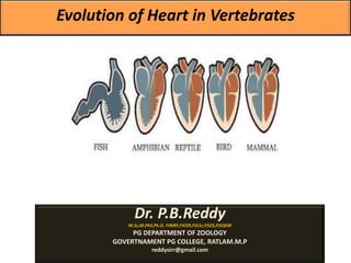
5. Evolutionary change in heart of vertebrates
Heart is situated ventral to the oseophagus in the pericardial
section of the coelom.
Heart is a highly muscular pumping organ that pumps blood into
arteries and sucks it back through the veins.
In vertebrates it has undergone transformation by twisting from a
straight tube to a complex multi-chambered organ.
. There has been an increase in the number of chambers in heart
during evolution of vertebrates.
The heart is covered by a transparent protective covering, called
pericardium. It is a single layer in fish.
Within pericardium there is a pericardial fluid, protects the heart
from the external injury.
The evolution of the heart is based on the separation of
oxygenated blood from deoxygenated blood for efficient oxygen
transport.
6.
7. Single circuit circulation -
Fishes have a single circulatory system.
In this system, the blood flows through
the heart only once completing a full
circuit in the fish's body.
The blood travels from the heart to the
gills, where the exchange of gases takes
place (carbon dioxide is released and
oxygen is absorbed).
The oxygenated blood from the gills
then flows to various parts of the body
and from these parts back to the heart.
8. Double circulation is a process during which blood passes twice
through the heart during one complete cycle. This type of
circulation is found in amphibians, reptiles, birds, and mammals.
However, it is more prominent in birds and mammals as in them
the heart is completely divided into four chambers.
The movement of blood in an organism is divided into two parts:
(i) Systemic circulation
(ii) Pulmonary circulation
Systemic circulation involves the movement of oxygenated
blood from the left ventricle of the heart to the aorta. It is then
carried by blood through a network of arteries, arterioles, and
capillaries to the tissues. From the tissues, the deoxygenated
blood is collected by the venules, veins, and vena cava, and is
emptied into the left auricle.
Pulmonary circulation involves the movement of deoxygenated
blood from the right ventricle to the pulmonary artery, which then
carries blood to the lungs for oxygenation. From the lungs, the
oxygenated blood is carried by the pulmonary veins into the left
atrium.
Hence, in double circulation, blood has to pass alternately through
the lungs and the tissues.
Significance of double circulation:
The separation of oxygenated and deoxygenated blood allows a
more efficient supply of oxygen to the body cells. Blood is
circulated to the body tissues through systemic circulation and to
the lungs through the pulmonary circulation.
9. 2. Two-Chambered Heart:
In cyclostomes, there are four chambers arranged in a
linear order- a thin-walled sinus venosus, a slightly
muscular atrium (auricle), a muscular ventricle and a
muscular conus arteriosus or bulbus cordis. Out of four
chambers, only atrium and ventricle correspond to the
four chambers (paired atria and paired ventricles) of the
higher vertebrates. In the evolution of heart many
changes have taken place.
Elasmobranchs:
The circulatory system in fishes supplies only
unoxygenated blood goes to the heart, from there it is
pumped to the gills, aerated and then distributed to the
body. The heart of cartilaginous dogfish is muscular and
dorsoventrally bent S-shaped tube with four
compartments in a linear series. They are sinus venosus
and atrium for receiving venous blood, and a ventricle
and conus arteriosus for pumping this blood. The heart
is a branchial venous heart. The sinus venosus and
conus arteriosus are accessory chambers. Atrium and
ventricle are true chambers, thus, it is a 2-chambered
heart.
In protochordates, a definite heart
does not exist and major blood
vessels contract rhythmically to
maintain circulation. Amphioxus
also has no heart but have small
bulbuli to pump blood into gills.
10. Teleosts:
In teleosts, the conus is reduced and has a single pair of valves.
The proximal part of ventral aorta close to conus becomes greatly
enlarged and thick-walled, called bulbus arteriosus.
It is elastic and dilates at the time of ventricular contraction. The heart is,
thus, 2-chambered with a single circulation of blood.
3. Three-Chambered Heart:
Dipnoi:
In diphoans a septum divides the atrium into a right and left chamber.
This is correlated with the use of the swim-bladder as an organ of
respiration and represents the first step toward the development of the
double-type circulatory system whereby both oxygenated and
unoxygenated blood enter the heart and are kept separate.
Blood from right auricle of the lungfish passes into the right ventricle and
is then pumped into the primitive lung-like gas bladder by pulmonary
arteries which branch off from the sixth pair of aortic arches.
The oxygenated blood returns to the left atrium by way of pulmonary
veins like amphibians.
11.
12. Amphibia:
In amphibians, the dorsal atrium shifts anterior to ventricle.
The sinus venosus opens into right atrium dorsally and not posteriorly.
The atrium is completely divided into right and left chambers and has no
foramen ovale in the inter-auricular septum, which remains open in dipnoans.
Deep pockets develop in the ventricular cavity.
The conus arteriosus divides into systemic and pulmonary vessels by a spiral
valve. In lung less salamanders, the inter-atrial septum is incomplete and
pulmonary veins are absent.
Reptilia:
In reptiles, the heart is further advanced. The atrium is always completely
separated into a right and left chamber, and in many forms the sinus venosus
is incorporated into the wall of the right atrium.
The ventricle is also partly divided by a septum in most reptiles, and in the
alligators and crocodiles is completely two-chambered.
This means that oxygenated blood coming from the lungs to the left side of
the heart is essentially separated from the non-oxygenated blood from the
body to the right side. But in other reptiles, some mixing does occur in other
parts of the circulatory system.
13. The embryonic conus arteriosus splits into three instead of two vessels:
(i) Pulmonary arch carrying blood to the lungs from right side of the
ventricle.
(ii) Right systemic aorta carrying blood from left side of the ventricle to the
body by way of right fourth aortic arch.
(iii) Left systemic comes from the right ventricle to the left fourth aortic
arch.
At the point of contact with the systemic aorta from the left ventricle, even
in crocodilians, an opening between the two is present, called the foramen
of Panizzae where there may be some mixing of the two types of blood.
Thus, reptilian heart represents the transitional heart against amphibian
heart-2 complete auricles and 2 incomplete ventricles with a little mixing
of blood in right and left systemic.
14. Aves and Mammalia:
In birds, the ventricle is completely divided into two, so that the heart
is four chambered (2 auricles and 2 ventricles).
There is complete separation of venous and arterial blood.
The systemic aorta leaves the left ventricle and carries blood to the
head and body. While the pulmonary artery leaves the right ventricle
and carries blood to the lungs for oxygenation.
Thus, there is double circulation in which there is no mixing of blood
at any place.
The sinus venosus is completely incorporated into right auricle, which
receives two precavals and a postcaval.
The left auricle receives oxygenated blood through pulmonary veins,
conus arteriosus is absent, the pulmonary aorta arises from the right
ventricle, and single systemic aorta arises from the left ventricle, and
both have valves at their bases.