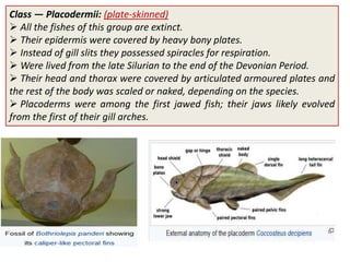
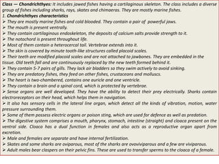
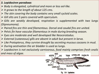
Download to read offline
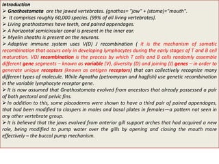


Gnathostomata, the jawed vertebrates, encompass around 60,000 species and are characterized by features such as teeth, paired appendages, and an adaptive immune system. The document discusses the evolution, classification, and anatomy of gnathostomes, particularly focusing on classes like chondrichthyes (cartilaginous fishes) and osteichthyes (bony fishes), while emphasizing the unique characteristics of early jawed vertebrates. It details the subclasses, specific groups like dipnoi (lungfishes), and their evolutionary significance as potential ancestors to amphibians.


![Structure and Types of Scales in Fishes[1].pptx](https://cdn.slidesharecdn.com/ss_thumbnails/structureandtypesofscalesinfishes1-250325121125-2f63aca3-thumbnail.jpg?width=640&height=640&fit=bounds)


![Polymer [ बहुलक ] Chemistry Notes PDF - Irfanullah Mehar - JJ Sir Chemistry.pdf](https://cdn.slidesharecdn.com/ss_thumbnails/polymerchemistrynotespdf-irfanullahmehar-jjsirchemistry-260210172118-3f9b37f7-thumbnail.jpg?width=640&height=640&fit=bounds)


