In physiology, respiration is the movement of oxygen from the outside environment to the cells within tissues, and the removal of carbon dioxide in the opposite direction. In these slides you will get to know about Physiology of Respiration in Invertibrates.
Why do animals need to breathe?
Breathing is important to organisms because cells require energy (oxygen) to move, reproduce and function. Breath also expels carbon dioxide, which is a by-product of cellular processes within the bodies of animals.
Respiration is the process of releasing energy from food and this takes place inside the cells of the body.
The process of respiration involves taking in oxygen (of air) into cells, using it for releasing energy by burning food, and then eliminating the waste products (carbon dioxide and water) from the body.
Respiration is essential for life because it provides energy for carrying out all the life processes which are necessary to keep the organisms alive.
The energy produced during respiration is stored in the form of ATP (Adenosine Tri- Phosphate) molecules in the cells of the body and used by the organism as when required.
KEY POINTS
Life started in an anaerobic environment in the so called ‘primodial broth’ (a mixture of organic molecules.
Subsequently, oxygen strangely enough became an crucial factor for aerobic metabolism especially in the higher life forms.
The rise of an oxygenic environment was an important event in the diversification of life.
It evoked a dramatic shift from inefficient to sophisticated oxygen dependent oxidizing ecosystems.
Anaerobic fermentation, the metabolic process that prevailed for the first about 2 billion years of the evolution of life, was a very inefficient way of extracting energy from organic molecules. Ex: A molecule of glucose, e.g., produces only two molecules of ATP (≈ 15 kCal) compared with 36 ATP molecules (≈ 263 kCal) in oxygenic respiration.
Aerobic metabolism must have developed at a critical point when the partial pressure of oxygen rose from an initial level to one adequately high to drive it passively across the cell membrane.
Respiration is a complex and highly integrated biomechanical, physiological, and behavioral processes.
The transfer of O2 occurs through a flow of tissue barriers and compartments by diffusion down a partial pressure gradient, which drops to about zero at the mitochondrial level.
Acquisition of molecular oxygen (O2) from the external fluid media (water and air) and the discharge of carbon dioxide (CO2) into the same milieu is the primary role of respiration.
The respiratory system is a biological system consisting of specific organs and structures.
Origin of the Lateral Line System
Lateral line is a canal along the side of a fish containing pores that open into tubes supplied with sense organs sensitive to low vibrations.
Robert H. Denison explained the origin of the lateral line system. He explained that early vertebrates had a pore-canal system in the dermis which functioned as a primitive sensory system in detecting water movement.
Through the evidences from fossils, embryology and comparative anatomy, Denison (1966) established that the inner ear is closely related to the lateral line system. He found a distinct relationship between the pore canal system and the lateral line in Osteotraci.
The inner ear and the lateral line are developed from ectodermal thickenings, called dorso-lateral placodes. These have a number of similarities, including receptors with sensory hairs, and are both innervated by fibers in the acoustico-lateral area of the brain.
The pore canal system is present and developed in Osteostraci (ostracoderm).
It is also present in Heterostraci which is another group of ostracoderms and includes early vertebrates such as lungfishes and crossopterygians.
As its presence is extensive, it is reasonable to suggest that the pore canal system was a primitive character in early vertebrates .
In transverse sections also , it is very difficult to differentiate the pore canal system from a lateral line canal.
Structure of the Lateral Line System
Epidermal structures called neuromasts form the peripheral area of the lateral line.
Neuromasts consist of two types of cells, hair cells and supporting cells.
Hair cells have an epidermal origin and each hair cell has one high kynocyle (5-10 μm) and 30 to 150 short stereocilia (2-3 μm).
The number of hair cells in each neuromast depends on its size, and they can range from dozens to thousands.
Hair cells can be oriented in two opposite directions with each hair cell surrounded by supporting cells.
At the basal part of each hair cell, there are synaptic contacts with afferent and efferent nerve fibers. Afferent fibers, transmit signals to the neural centres of the lateral line and expand at the neuromast base. The regulation of hair cells is achieved by the action of efferent fibers.
Stereocilia and kinocilium of hair cells are immersed into a cupula and are located above the surface of the sensory epithelium.
The cupula is created by a gel-like media, which is secreted by non-receptor cells of the neuromast.
osmoregulation in invertebrates- it is a processes by which any organisms maintains the fluid and salt balance of its body, which is important for proper functioning of organs .
Taxonomic Collections, Preservation and Curating of InsectsKamlesh Patel
Taxonomy: Taxonomy is the science of defining and naming groups of biological organisms on the basis of shared characteristics.
The classification of organisms is according to hierarchal system or in taxonomic ranks (eg; domain, kingdom, phylum class, order, family, genus and species) based on phylogenetic relationship established by genetic analysis.
Taxonomic Collection : Biological collection are typically preserved plant or animals specimens along with specimen documentations such as labels and notations.
Dry Collection - Dry collections consist of those specimens that are preserved in a dry state.
Wet Collection - Wet collections are specimens kept in a liquid preservative to prevent their deterioration.
INTRODUCTION
The jaw (Upper and lower) is any opposable articulated structure at the entrance of the mouth.
It is typically used for grasping and manipulating food.
Jaw suspension means the fusion of upper jaw and lower jaw or skull for efficient biting.
There are different ways in which these attachments are attained depending upon the modifications in visceral arches in vertebrates.
In most vertebrates, the jaws are bony or cartilaginous and oppose vertically.
The vertebrate jaw is derived from the most anterior two pharyngeal arches supporting the gills, and usually bears numerous teeth.
The vertebrate jaw probably originally evolved in the Silurian period and appeared in the Placoderm fish which further diversified in the Devonian.
It is believed that the hyoid system suspends the jaw from the brain case of the skull, permitting great mobility of the jaws.
The original selective advantage offered by the jaw may not be related to feeding, but rather to increased respiration efficiency.
The jaws were used in the buccal pump (observable in modern fish and amphibians) that pumps water across the gills of fish or air into the lungs in the case of amphibians.
Over evolutionary time the more familiar use of jaws (to humans), in feeding, was selected for and became a very important function in vertebrates. Many teleost fish have substantially modified jaws for suction feeding and jaw protrusion, resulting in highly complex jaws with dozens of bones involved.
Jaw Suspension or Suspensoria:
The method by which the upper and lower jaws are suspended or attached from the chondrocranium is known as jaw suspension or suspensorium.
Amongst the visceral arches, the first (mandibular) arch consists of
= a dorsal palato pterygoquadrate bar forming the upper jaw,
= and ventral Meckel’s cartilage forms the lower jaw.
The second (hyoid) arch consists of = a dorsal hyomandibular supporting and suspending the jaws with the cranium, and a ventral hyoid.
The remaining visceral arches support the gills and are, hence, called branchial arches. Thus, splanchnocranium forms the jaws and suspends them with the chondrocranium.
Iczn(The International Commission on Zoological Nomenclature )Al Nahian Avro
The International Commission on Zoological Nomenclature (ICZN) acts as adviser and arbiter for the zoological community by generating and disseminating information on the correct use of the scientific names of animals. The ICZN is responsible for producing the International Code of Zoological Nomenclature - a set of rules for the naming of animals and the resolution of nomenclatural problems.
ORIGIN OF CHORDATES
Animal kingdom is basically divided into two sub kingdoms:
Non-chordata- including animals without notochord.
Chordata- This comprising animals having notochord or chorda dorsalis.
Chordates were evolved sometime 500 million years ago during Cambrian period (invertebrates were also began to evolve in this period) .
Chamberlain (1900) pointed out that all modern chordates possess glomerular kidneys that are designed to remove excess water from body.
It is believed that Chordates have originated from invertebrates.
It is difficult to determine from which invertebrate group the chordates were developed.
Chordate ancestors were soft bodied animals. Hence they were not preserved as Fossils.
However, early fossils of chordates have all been recovered from marine sediments and even modern protochordates are all marine forms.
Also glomerular kidneys are also found in some marine forms such as myxinoids and sharks. That makes the marine origin of chordates more believable.
Chordates evolved from some deuterostome ancestor (echinoderms, hemichordates, pogonophorans etc.) as they have similarities in embryonic development, type of coelom and larval stages.
Many theories infers origin of chordates, hemichordates and echinoderms from a common ancestor.
The integumentary system comprises the skin and its appendages. Skin + derivatives= Integument.
It aims to protect the body from various kinds of damage, such as loss of water or damages from outside.
The integumentary system in chordates includes hair, scales, feathers, hooves, and nails.
It may serve to water proof, and protect the deeper tissues.
Excrete wastes, and regulate body temperature.
It is the attachment site for sensory receptors to detect pain, sensation, pressure, and temperature.
INTRODUCTION
The term urogenital refers to something that has both urinary and genital origins. The word urogenital is used because the urinary and reproductive systems in males merge.
These are grouped together because of their proximity to each other, their common embryological origin and the use of common pathways (ex. urethra).
Kidneys and urinary ducts form the urinary system.
The Urinary system performs two important homeostatic processes like excretion and osmoregulation. This system is intimately associated both anatomically, and in terms of embryonic origin with the genital system.
The genital system includes the gonads which generate gametes and the genital ducts that serve as passages for the gametes.
Though functionally different the two organ systems the urinary and the genital system are treated together as the urino- genital system, since both develop from the same segmental blocks of trunk mesoderm or adjacent tissues and share many of the ducts.
Thus although the two systems have nothing common functionally they are closely associated in their use of common ducts and are studied under the broad heading of urinogenital system.
The function of the excretory system is crucial in considering the possible environment of the ‘vertebrate life ’. Several main functions can be attributed to all vertebrate excretory systems:
Excretion of nitrogenous waste products.
Maintaining homeostasis with regard to ions (i.e. salt balance).
Regaining valuable substances (glucose, salts, amino acids, etc.)
Maintaining a physiological osmotic value (i.e. water balance).
The excretory system is formed by a series of paired, segmental nephrons that begin with a nephrostome opening into the coelomic cavity.
A pair of glomeruli per segment, supplied by branches from the aorta, projects into the coelomic cavity close to these nephrostomes.
At a later stage of development, the glomerulus/nephrostome area becomes separated from the rest of the coelomic cavity by an epithelial fold.
The nephrons connect to a duct that is formed by caudal growth of the most anterior nephric tubules. These paired urinary ducts open near the anal region.
Respiration.
Types of respiration.
Various modes of respiration in animals.
Human respiratory system.
Upper respiratory tract.
Nose.
Pharynx.
Larynx.
Lower respiratory tract.
Trachea.
Bronchi and bronchioles.
Lungs.
Mechanism of respiration.
Exchange of gases.
Functions of respiratory system.
Presentation on Organ & Mechanism of Respiration in Pisces And Amphibiansvskgondia
This is Powerpoint presentation helpful for students and teachers. It includes Defination of Respiration & Function of respiratory system. Also contains mechanism of respiration and various repiratory organs of pisces and amphibians, their structures and fuctions.
Hey, this is my BSc assignment which will help you. It contains the basics about sericulture. I will provide you with a brief about sericulture as well.
Sericulture, also known as silk farming, is an ancient practice that involves the cultivation of silkworms for the production of silk. It is a labor-intensive process that requires meticulous care and attention at every stage, from selecting healthy silkworm eggs to the final processing of silk fibers. Sericulture has a rich history that spans thousands of years and has been a significant part of various cultures around the world.
The origins of sericulture can be traced back to ancient China, where it was initially kept as a closely guarded secret. The Chinese closely guarded the production techniques and methods, as silk was considered a valuable commodity and a symbol of wealth and luxury. However, the art of sericulture eventually spread to other parts of Asia and later to Europe and the rest of the world.
The sericulture process begins with the careful selection of silkworm eggs. Healthy and disease-free eggs are chosen to ensure the quality of the silkworms. These eggs are then incubated under controlled conditions until they hatch into tiny silkworm larvae. The larvae are then placed on specially prepared trays and provided with a diet consisting mainly of mulberry leaves, which are the primary food source for silkworms.
Mulberry trees, scientifically known as Morus spp., are cultivated in large quantities to sustain the silk production industry. The leaves of the mulberry trees are rich in nutrients, making them an ideal food source for the silkworms. The silkworms feed voraciously on the leaves, growing rapidly and shedding their skin multiple times in a process called molting.
After several weeks of feeding and molting, the silkworms reach their final stage, known as the cocooning stage. During this stage, the silkworms secrete a protein substance called fibroin, which is used to spin their cocoons. The silkworms create a protective covering by spinning a single continuous silk thread around themselves. This spinning process takes about two to three days, and the resulting cocoon is composed of a single thread that can measure several hundred meters in length.
To obtain the silk fibers, the cocoons are carefully harvested. However, to prevent the silkworms from breaking the silk thread, the cocoons are usually subjected to a process known as stifling. Stifling involves heating the cocoons or exposing them to steam to kill the silkworms inside. This process also makes it easier to unravel the silk thread from the cocoon.
After stifling, the silk thread is carefully unwound from the cocoon. This process is called reeling, and it requires skill and precision to ensure the quality of the silk fibers. Several strands of silk thread are combined to create a stronger and more durable silk yarn. The yarn is then cleaned to remove any impurities and twisted into a usable form.
Delhi University previous 4th semester question papers. This gonna help student to practice for the final exams. As many questions comes again and again because of limited syllabus.
Observation of Io’s Resurfacing via Plume Deposition Using Ground-based Adapt...Sérgio Sacani
Since volcanic activity was first discovered on Io from Voyager images in 1979, changes
on Io’s surface have been monitored from both spacecraft and ground-based telescopes.
Here, we present the highest spatial resolution images of Io ever obtained from a groundbased telescope. These images, acquired by the SHARK-VIS instrument on the Large
Binocular Telescope, show evidence of a major resurfacing event on Io’s trailing hemisphere. When compared to the most recent spacecraft images, the SHARK-VIS images
show that a plume deposit from a powerful eruption at Pillan Patera has covered part
of the long-lived Pele plume deposit. Although this type of resurfacing event may be common on Io, few have been detected due to the rarity of spacecraft visits and the previously low spatial resolution available from Earth-based telescopes. The SHARK-VIS instrument ushers in a new era of high resolution imaging of Io’s surface using adaptive
optics at visible wavelengths.
Cancer cell metabolism: special Reference to Lactate PathwayAADYARAJPANDEY1
Normal Cell Metabolism:
Cellular respiration describes the series of steps that cells use to break down sugar and other chemicals to get the energy we need to function.
Energy is stored in the bonds of glucose and when glucose is broken down, much of that energy is released.
Cell utilize energy in the form of ATP.
The first step of respiration is called glycolysis. In a series of steps, glycolysis breaks glucose into two smaller molecules - a chemical called pyruvate. A small amount of ATP is formed during this process.
Most healthy cells continue the breakdown in a second process, called the Kreb's cycle. The Kreb's cycle allows cells to “burn” the pyruvates made in glycolysis to get more ATP.
The last step in the breakdown of glucose is called oxidative phosphorylation (Ox-Phos).
It takes place in specialized cell structures called mitochondria. This process produces a large amount of ATP. Importantly, cells need oxygen to complete oxidative phosphorylation.
If a cell completes only glycolysis, only 2 molecules of ATP are made per glucose. However, if the cell completes the entire respiration process (glycolysis - Kreb's - oxidative phosphorylation), about 36 molecules of ATP are created, giving it much more energy to use.
IN CANCER CELL:
Unlike healthy cells that "burn" the entire molecule of sugar to capture a large amount of energy as ATP, cancer cells are wasteful.
Cancer cells only partially break down sugar molecules. They overuse the first step of respiration, glycolysis. They frequently do not complete the second step, oxidative phosphorylation.
This results in only 2 molecules of ATP per each glucose molecule instead of the 36 or so ATPs healthy cells gain. As a result, cancer cells need to use a lot more sugar molecules to get enough energy to survive.
introduction to WARBERG PHENOMENA:
WARBURG EFFECT Usually, cancer cells are highly glycolytic (glucose addiction) and take up more glucose than do normal cells from outside.
Otto Heinrich Warburg (; 8 October 1883 – 1 August 1970) In 1931 was awarded the Nobel Prize in Physiology for his "discovery of the nature and mode of action of the respiratory enzyme.
WARNBURG EFFECT : cancer cells under aerobic (well-oxygenated) conditions to metabolize glucose to lactate (aerobic glycolysis) is known as the Warburg effect. Warburg made the observation that tumor slices consume glucose and secrete lactate at a higher rate than normal tissues.
Professional air quality monitoring systems provide immediate, on-site data for analysis, compliance, and decision-making.
Monitor common gases, weather parameters, particulates.
Multi-source connectivity as the driver of solar wind variability in the heli...Sérgio Sacani
The ambient solar wind that flls the heliosphere originates from multiple
sources in the solar corona and is highly structured. It is often described
as high-speed, relatively homogeneous, plasma streams from coronal
holes and slow-speed, highly variable, streams whose source regions are
under debate. A key goal of ESA/NASA’s Solar Orbiter mission is to identify
solar wind sources and understand what drives the complexity seen in the
heliosphere. By combining magnetic feld modelling and spectroscopic
techniques with high-resolution observations and measurements, we show
that the solar wind variability detected in situ by Solar Orbiter in March
2022 is driven by spatio-temporal changes in the magnetic connectivity to
multiple sources in the solar atmosphere. The magnetic feld footpoints
connected to the spacecraft moved from the boundaries of a coronal hole
to one active region (12961) and then across to another region (12957). This
is refected in the in situ measurements, which show the transition from fast
to highly Alfvénic then to slow solar wind that is disrupted by the arrival of
a coronal mass ejection. Our results describe solar wind variability at 0.5 au
but are applicable to near-Earth observatories.
Slide 1: Title Slide
Extrachromosomal Inheritance
Slide 2: Introduction to Extrachromosomal Inheritance
Definition: Extrachromosomal inheritance refers to the transmission of genetic material that is not found within the nucleus.
Key Components: Involves genes located in mitochondria, chloroplasts, and plasmids.
Slide 3: Mitochondrial Inheritance
Mitochondria: Organelles responsible for energy production.
Mitochondrial DNA (mtDNA): Circular DNA molecule found in mitochondria.
Inheritance Pattern: Maternally inherited, meaning it is passed from mothers to all their offspring.
Diseases: Examples include Leber’s hereditary optic neuropathy (LHON) and mitochondrial myopathy.
Slide 4: Chloroplast Inheritance
Chloroplasts: Organelles responsible for photosynthesis in plants.
Chloroplast DNA (cpDNA): Circular DNA molecule found in chloroplasts.
Inheritance Pattern: Often maternally inherited in most plants, but can vary in some species.
Examples: Variegation in plants, where leaf color patterns are determined by chloroplast DNA.
Slide 5: Plasmid Inheritance
Plasmids: Small, circular DNA molecules found in bacteria and some eukaryotes.
Features: Can carry antibiotic resistance genes and can be transferred between cells through processes like conjugation.
Significance: Important in biotechnology for gene cloning and genetic engineering.
Slide 6: Mechanisms of Extrachromosomal Inheritance
Non-Mendelian Patterns: Do not follow Mendel’s laws of inheritance.
Cytoplasmic Segregation: During cell division, organelles like mitochondria and chloroplasts are randomly distributed to daughter cells.
Heteroplasmy: Presence of more than one type of organellar genome within a cell, leading to variation in expression.
Slide 7: Examples of Extrachromosomal Inheritance
Four O’clock Plant (Mirabilis jalapa): Shows variegated leaves due to different cpDNA in leaf cells.
Petite Mutants in Yeast: Result from mutations in mitochondrial DNA affecting respiration.
Slide 8: Importance of Extrachromosomal Inheritance
Evolution: Provides insight into the evolution of eukaryotic cells.
Medicine: Understanding mitochondrial inheritance helps in diagnosing and treating mitochondrial diseases.
Agriculture: Chloroplast inheritance can be used in plant breeding and genetic modification.
Slide 9: Recent Research and Advances
Gene Editing: Techniques like CRISPR-Cas9 are being used to edit mitochondrial and chloroplast DNA.
Therapies: Development of mitochondrial replacement therapy (MRT) for preventing mitochondrial diseases.
Slide 10: Conclusion
Summary: Extrachromosomal inheritance involves the transmission of genetic material outside the nucleus and plays a crucial role in genetics, medicine, and biotechnology.
Future Directions: Continued research and technological advancements hold promise for new treatments and applications.
Slide 11: Questions and Discussion
Invite Audience: Open the floor for any questions or further discussion on the topic.
The increased availability of biomedical data, particularly in the public domain, offers the opportunity to better understand human health and to develop effective therapeutics for a wide range of unmet medical needs. However, data scientists remain stymied by the fact that data remain hard to find and to productively reuse because data and their metadata i) are wholly inaccessible, ii) are in non-standard or incompatible representations, iii) do not conform to community standards, and iv) have unclear or highly restricted terms and conditions that preclude legitimate reuse. These limitations require a rethink on data can be made machine and AI-ready - the key motivation behind the FAIR Guiding Principles. Concurrently, while recent efforts have explored the use of deep learning to fuse disparate data into predictive models for a wide range of biomedical applications, these models often fail even when the correct answer is already known, and fail to explain individual predictions in terms that data scientists can appreciate. These limitations suggest that new methods to produce practical artificial intelligence are still needed.
In this talk, I will discuss our work in (1) building an integrative knowledge infrastructure to prepare FAIR and "AI-ready" data and services along with (2) neurosymbolic AI methods to improve the quality of predictions and to generate plausible explanations. Attention is given to standards, platforms, and methods to wrangle knowledge into simple, but effective semantic and latent representations, and to make these available into standards-compliant and discoverable interfaces that can be used in model building, validation, and explanation. Our work, and those of others in the field, creates a baseline for building trustworthy and easy to deploy AI models in biomedicine.
Bio
Dr. Michel Dumontier is the Distinguished Professor of Data Science at Maastricht University, founder and executive director of the Institute of Data Science, and co-founder of the FAIR (Findable, Accessible, Interoperable and Reusable) data principles. His research explores socio-technological approaches for responsible discovery science, which includes collaborative multi-modal knowledge graphs, privacy-preserving distributed data mining, and AI methods for drug discovery and personalized medicine. His work is supported through the Dutch National Research Agenda, the Netherlands Organisation for Scientific Research, Horizon Europe, the European Open Science Cloud, the US National Institutes of Health, and a Marie-Curie Innovative Training Network. He is the editor-in-chief for the journal Data Science and is internationally recognized for his contributions in bioinformatics, biomedical informatics, and semantic technologies including ontologies and linked data.
Richard's entangled aventures in wonderlandRichard Gill
Since the loophole-free Bell experiments of 2020 and the Nobel prizes in physics of 2022, critics of Bell's work have retreated to the fortress of super-determinism. Now, super-determinism is a derogatory word - it just means "determinism". Palmer, Hance and Hossenfelder argue that quantum mechanics and determinism are not incompatible, using a sophisticated mathematical construction based on a subtle thinning of allowed states and measurements in quantum mechanics, such that what is left appears to make Bell's argument fail, without altering the empirical predictions of quantum mechanics. I think however that it is a smoke screen, and the slogan "lost in math" comes to my mind. I will discuss some other recent disproofs of Bell's theorem using the language of causality based on causal graphs. Causal thinking is also central to law and justice. I will mention surprising connections to my work on serial killer nurse cases, in particular the Dutch case of Lucia de Berk and the current UK case of Lucy Letby.
Seminar of U.V. Spectroscopy by SAMIR PANDASAMIR PANDA
Spectroscopy is a branch of science dealing the study of interaction of electromagnetic radiation with matter.
Ultraviolet-visible spectroscopy refers to absorption spectroscopy or reflect spectroscopy in the UV-VIS spectral region.
Ultraviolet-visible spectroscopy is an analytical method that can measure the amount of light received by the analyte.
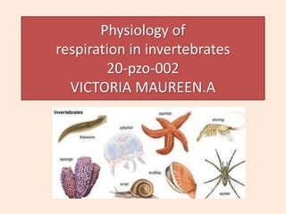
2. SYNOPSIS
Introduction
History
Respiratory organs of invertebrates
1. Trachea and spiracles
2. Gill
3. Book lung
4. Book gills
Respiration in
1. Protozoa
2. Porifera
3. Coelenterates
4. Aschelminthes and platyhelminthes
5. Annelids
6. Arthropoda
7. Mollusca
8. Echinoderm
Respiratory pigments
Functions of respiration
Videos
Question bank
Reference
3. INTRODUCTION
• The process of gas exchange in the body, called
respiration. The act of inhaling and exhaling air in
order to exchange oxygen for carbon dioxide
• The process of inhalation of oxygen and
exhalation of carbon dioxide is known
as respiration.
• There are two types of cellular respiration
aerobic and anaerobic. One occurs in the
presence of oxygen (aerobic), and one occurs in
the absence of oxygen (anaerobic).
4. HISTORY
• Marcello Malpighi (1628-1694) was an
Italian scientist who found the
anatomical basis of respiration, He
was one of the first biologists to make
use of the newly invented microscope
and is best known as the discoverer of
the pulmonary capillaries and alveoli.
• Henri Dutrochet, a French physiologist
who term 'respiration'. He also
discovered and named the process of
osmosis (passage of solvent through a
semipermeable membrane) that
occurs at cellular level in both plants
and animals.
5. Respiratory organs of invertebrates
Trachea
• This respiratory organ is a hallmark of
insects.
• It is made up of a system of branching tubes
that deliver oxygen to, and remove carbon
dioxide from, the tissues,
• The smallest tubes, tracheoles, penetrate
cells and diffuse water, oxygen, and carbon
dioxide.
• Tracheae are a system of tiny tubes that
permit passage of gases into the interior of
the body.
• Tracheal systems are highly efficient for
these small, terrestrial animals.
• The pores to the outside, called spiracles,
are typically paired structures, two in the
thorax and eight in the abdomen. Periodic
opening and closing of the spiracles prevents
water loss by evaporation,
6.
7. Gills
• Many invertebrates use gills as a major means
of gas exchange
• Gills are branching organs located on the side
of heads that have small blood vessels called
capillaries. As the organism opens its mouth,
water runs over the gills, and blood in the
capillaries picks up oxygen that’s dissolved in
the water.
• Gills consist of plate-like structures called
filaments that are covered by an array of
lamellae enclosing a capillary blood network
• Oxygen-rich water passes through the narrow
channels formed by the lamellar layers, where
oxygen diffuses into the capillaries. The
densely packed lamellar structure is
advantageous because it provides a large
surface area for oxygen transfer.
8. Book lung
• It is a form of respiratory organ found
in certain air-breathing arthropods
(scorpions and some spiders).
• Each book lung consists of a series of
thin plates that are highly vascular
(i.e., richly supplied with blood) and
are arranged in relation to each other
like the pages of a book.
• These plates extend into an internal
pouch formed by the external
skeleton that opens to the exterior by
a small slit. This provides an extensive
surface for the exchange of oxygen
and carbon dioxide with the
surrounding air. There are four pairs
in scorpions and up to two in spiders.
9.
10. Book gills
• It is believed that book lungs evolved from book gills. Although they have a
similar book-like structure,
• book gills are external, while book lungs are internal. Both are considered
appendages because book lungs develop from limb buds before the buds
flatten into segmented lamellae.
• Book gills are still present in the marine arthropod Limulus (horseshoe
crabs) which have five pairs of them,
• the flap in front of them being the genital operculum which lacks gills.
11. PROTOZOA
• Single-celled organisms,
such as bacteria and
protozoa, are in constant
contact with their external
environment. Gas
exchange occurs by
diffusion across their
membranes. The
respiratory gases may
diffuse in and diffuses out
trough the general body
surface, there are no
special organ for
respiration.
12. PORIFERA
• In sponges, the special
respiratory organs are absent
• Gaseous exchange occurs by
simple diffusion between the
cells of sponges and the
current of water
• Oxygen dissolved in water is
taken in by diffusion through
the general body surface and
carbon di oxide is given out
• Amoebocytes distributes
oxygen with in the
mesenchyme and carry away
carbon di oxide
13. COELENTERATES
• In coelenterates, the special
respiratory organs are absent
• Their body cells are ore or less
directly exposed to the
environment, both cell layers
absorb oxygen from and
expel carbon dioxide into the
surrounding water.
• the oxygen is absorbed into their
first layer of skin, called the
ectoderm.
• Then, it goes through to the second
layer, called the endoderm. The
oxygen molecules are used and
excess oxygen is released as carbon
dioxide.
14. ASCHELMINTHES AND PLATYHELMINTHES
• respiratory organs are absent The body
walls of aschelminthes are very thin and
thus it acts as their respiratory system.
• In living flatworms and roundworms, the
exchange of gases takes place through
general body surface
• In parasitic form, there is no exchange of
gases. Endo parasites lives n almost oxygen
free environment and fulfills its relatively
less energy requirements by anaerobic
respiration
• Flatworms are small, literally flat worms,
which 'breathe' through diffusion across
the outer membrane. The flat shape of
these organisms increases the surface area
for diffusion, ensuring that each cell within
the body is close to the outer membrane
surface and has access to oxygen.
15. ANNELIDA
• Respiration in annelids occurs primarily through
their moist skin, although certain species have
evolved specialized gills or use paired
projections called parapodia in gas exchange.
• In earthworms the respiration mainly occurred
or performed through skin the called as
cutaneous respiration
• The blood of earthworm contains a respiratory
pigment – Haemoglobin in a dissolved state in
its plasma
• The epidermis of the body wall acts as a
permeable membranes through which the
atmospheric oxygen diffuses in its capillaries
and combine with haemoglobin to form
oxyhaemoglobin
16. • The oxyhaemoglobin is circulated by
blood into the tissues where oxygen
tension is very low and carbon di oxide
tension is high
• The oxyhaemoglobin breaks up to release
oxygen to the tissues and haemoglobin in
a reduced state
• Now carbon di oxide from tissue diffuses
into the blood due to its high tension. The
carbon di oxide is carried by the blood
generally in a dissolved condition and
when it reacts to the epidermal capillaries
it diffuses from the blood to the
atmosphere due to low tension
17. ARTHROPODA
• Aquatic arthropods possess gills for respiration and are
covered by the exoskeleton, which is thin in this area
and not a barrier to the exchange of gases.
• Terrestrial arthropods possess tracheae and book lungs
as respiratory organs. The small, external openings
(spiracles) reduce water loss, the chitinous lining
prevents collapse
• Book lungs are chitin-lined internal pockets containing
many blood-filled plates over which air circulates. Most
spiders possess tracheae and book lungs, but large
spiders (such as tarantulas) and scorpions possess book
lungs alone.
18. • The respiratory system of cockroach
is very well developed to
compensate the absence of
respiratory pigment in the blood
• Ten pairs of spiracles or stigmata are
present on the lateral side of the
body. The largest first pair is present
on the mesothorax. The second pair
is on the metathorax and the rest
eight pairs are on the first eight
abdominal segments.
• the haemocoel contains a network
of elastic, closed air tubes or
tracheae. Three longitudinal
tracheal trunks are present on each
side of the abdominal cavity.
19. MOLLUSCA
• Basically all molluscs breathe by
gills that are called ctenidia (comb-
gills) because of their comb-like
shape.
• A ctenidium is shaped like a comb or a
feather, with a central part from
which many filaments or plate-like
structures protrude, lined up in a row
• In land snails and slugs, mantle cavity
has evolved into primitive lung
• the mantle cavity forms a pulmonary
chamber, the inner surface of which is
highly vascularised.
• Many molluscs have a siphon which
expels water and wastes
20. ECHINODERMS
• In echinoderms (starfish, sea urchins, brittle
stars), most of the respiratory exchange occurs
across tube feet (a series of suction-cup
extensions used for locomotion).
• Echinoderms typically breathe and respire by the
simple diffusion of gases like oxygen and carbon
dioxide in and out of their body cell membranes.
• However, this exchange is supplemented by
extensions of the coelomic, or body-fluid, cavity
into thin-walled “gills” or dermal branchiae
that bring the coelomic fluid into close contact
with seawater.
• Respiratory tree is the branches of cloaca just
inside the anus with the help of the drawing
water through the anus and then expelled
21. Respiratory Pigments in Invertebrates
• In order to facilitate the transport of oxygen to
different parts of the body, most animals have
developed respiratory pigments.
• In general, respiratory pigments are coloured proteins
that contain a metallic element in their constitution
and have the property of forming loose combination
with oxygen and sometimes with carbon dioxide.
• Four different (biochemically) respiratory pigments are
recognized – haemoglobin, chlorocruorin,
haemocyanin, and haemerythrin. Even in the same
phylum there may be several distinct pigments
22. • Haemoglobin:
It is the most efficient
respiratory pigment. It is
widely distributed in the
animal kingdom, starting
from some protozoa like
Paramoecium to almost all
vertebrates except eel larvae
and some Antarctic fishes.
Some invertebrate phyla viz.,
Porifera, Cnidaria and
Ctenophora, totally lack it
23. • Haemocyanin:
Among various copper-
proteins occurring in nature,
only haemo cyanin can
reversibly combine with
oxygen and thus, serves as a
transport pigment. It is
found in Chitons, some
gastropods and
cephalopods amongst the
molluscs and in crustaceans
and Limulus amongst the
arthropods. It always remain
dissolved in the plasma.
24. • Chlorocruorin:
This green coloured
metalloprotein is found in
the plasma of certain
polychaet families. It is a
metalloprotein with the
metal being iron (Fe++); the
metalloporphyrin is similar
to heme of haemoglobin
except that one vinyl (CH =
CH2) group is replaced by
formyl (0=CH) group in
Chlorocruorin. The
porphyrin is called
chlorocruoheme.
25. • Haemerythrin:
This violet coloured pigment is
found inside the corpuscles of
animals ex.polychaete worm
Magelona. It is also an iron
containing metalloprotein but
has no porphyrin.
• Pinna globin:
This brown coloured,
manganese containing
pigment is present in the
plasma of Pinna.
26. Functions of respiration
• Delivers oxygen to the cells in your body.
• Removes waste gases, including carbon
dioxide, from the body when you exhale.
• Breathing – movement of air
• Sound Production
• Olfaction, or Smelling, Is a Chemical Sensation
27.
28. Question bank
PART-A
1. Define respiration?
2. What are the types of respiration?
3. Name the Respiratory organs of invertebrates?
4. What is Trachea?
5. What are tracheoles?
6. What are Spiracles?
7. Define Gills?
8. What are gill filaments?
9. What is book lung?
10. What are book gills?
11. Define diffusion?
12. What is the role of Amoebocytes in poriferan respiration?
13. Define cutaneous respiration?
14. What is oxyhaemoglobin ?
15. What is ctenidia ?
16. What is dermal branchiae?
17. What is Respiratory tree ?
18. What is respiratory pigments?
19. What are the types of respiratory pigment?
29. PART-B
1. Explain the mechanism of respiration in insects?
2. Explain the mechanism of respiration in annelids?
3. State the functions of respiration?
4. Give an account of respiratory organ “book lung”?
PART-C
1. Explain the types of respiratory organs in invertebrates ?
2. Give a detailed account of types of respiratory pigments?
30. References
• https://byjus.com/biology/respiration-cockroach-earthworm
• https://www.notesonzoology.com/cockroaches/process-of-
respiration-in-cockroaches-invertebrates/1989
• https://www.britannica.com/science/respiratory-system/Basic-
types-of-respiratory-
• https://www.webmd.com/lung/picture-of-the-trachea
• https://www.toppr.com/guides/science/respiration-in-
organism/respiration-in-other-animals/
• https://phylumrespiratioryexamin.weebly.com/porifera.html
• https://www.vedantu.com/question-answer/respiration-occurs-
in-annelids-by-a-skin-b-gills-class-11-biology-cbse-
6010e70fdfcfb40cf08fd05c
• Book: Manual of zoology part 1 Invertebrata
Volume 1 and 2
BY- M. Ekambaranatha Ayyar T.N Ananthakrishnan