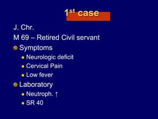
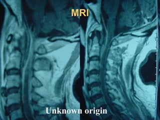
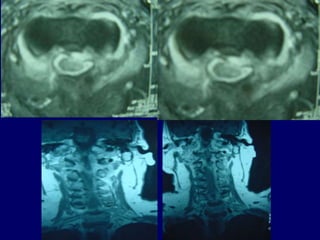
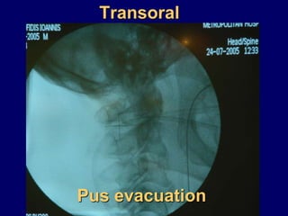
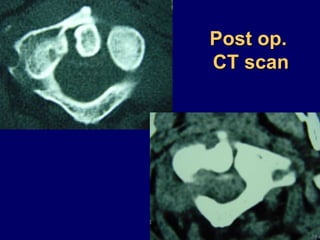
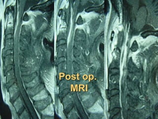
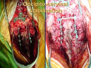
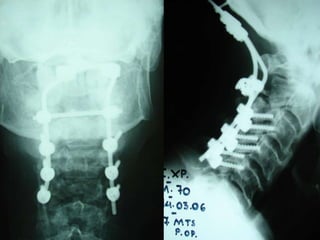
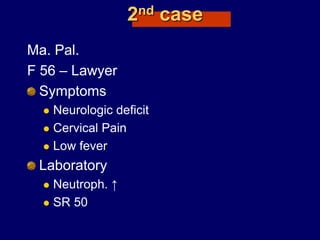
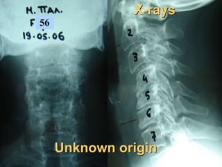
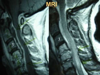
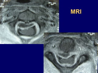
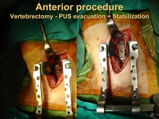
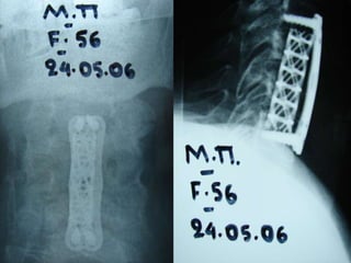
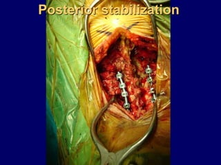
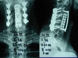
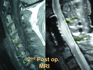
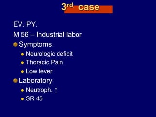
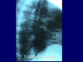
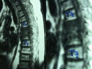
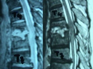
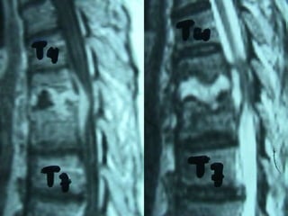
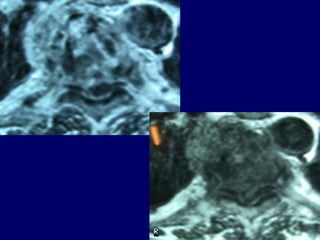
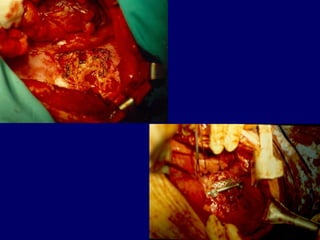
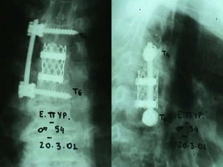
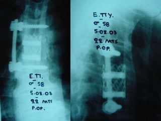
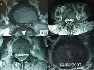
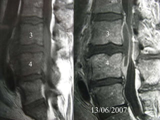
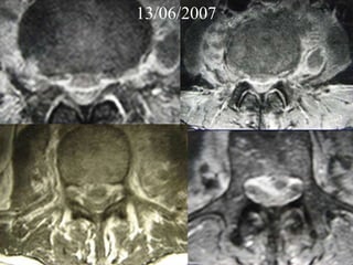
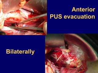
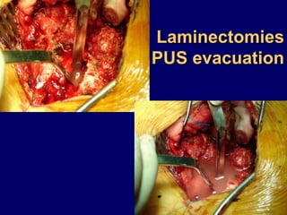
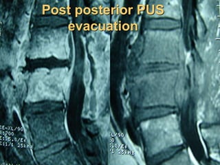
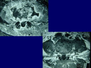
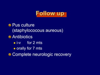
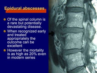
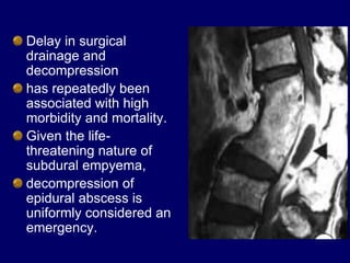
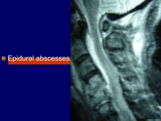
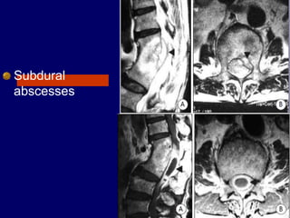
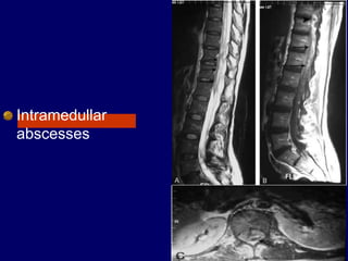
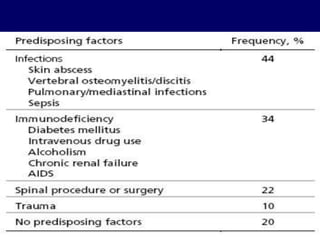
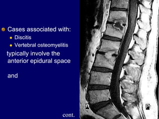
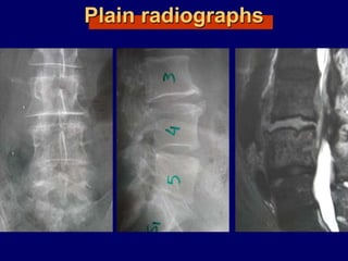
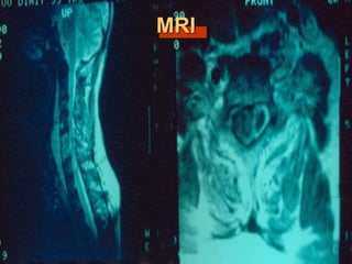
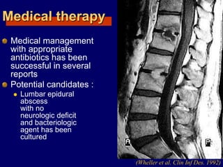
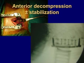
Vertebral Osteomyelitis
complicated with
Epidural Absceses
GEORGE SAPKAS
PROFESSOR AT ORTHOPAEDICS
Metropolitan Hospital
Athens Greece
Cases
1st case
J. Chr.
M 69 – Retired Civil servant
Symptoms
 Neurologic deficit
 Cervical Pain
 Low fever
Laboratory
 Neutroph. ↑
 SR 40
MRI
Unknown origin
Treatment
Transoral
Pus evacuation
Post op.
CT scan
Post op.
MRI
Occipito-cervical
stabilization
2nd Post op.
CT scan
2nd Post op.
MRI (6mts)
Post op.
3D scan (3yrs)
Follow up
Culture
 staphylococous aureous
Antibiotics
i-v for 2 mts
orally for 4 mts
Full neurologic recovery
2nd case
Ma. Pal.
F 56 – Lawyer
Symptoms
 Neurologic deficit
 Cervical Pain
 Low fever
Laboratory
 Neutroph. ↑
 SR 50
56
X-rays
Unknown origin
MRI
3
3
MRI
Anterior procedure
Vertebrectomy - PUS evacuation + Stabilization
Posterior stabilization
2nd Post op.
MRI
Follow up
Pus culture
(staphylococous aureous)
Antibiotics
 i-v for 2 mts
 orally for 6 mts
Complete neurologic recovery
3rd case
EV. PY.
M 56 – Industrial labor
Symptoms
 Neurologic deficit
 Thoracic Pain
 Low fever
Laboratory
 Neutroph. ↑
 SR 45
Unknown Origin
Follow up
Pus culture
(staphylococous aureous)
Antibiotics
 i-v for 2 mts
 orally for 6 mts
Complete neurologic recovery
4th case
D.N.
M 61– Cardiologist
Symptoms
 Severe Neurologic deficit
 Intensive Thoracic-lumbar Pain
 High fever
Laboratory
 Neutroph. ↑
 SR 60
PMH
 Heavy smoker
 Diabetes melitus
 Recent Elbow Furuncle (untreated)
Elbow furuncle
3
3
4 4
04/06/2007
04/06/2007
04/06/2007
13/06/2007
3
4
3
4
13/06/2007
Anterior
PUS evacuation
Bilaterally
Laminectomies
PUS evacuation
Post posterior PUS
evacuation
Follow up
Pus culture
(staphylococous aureous)
Antibiotics
 i-v for 2 mts
 orally for 7 mts
Complete neurologic recovery
Conclusions
Epidural abscesses
Of the spinal column is
a rare but potentially
devastating disease.
When recognized early
and treated
appropriately the
outcome can be
excellent
However the mortality
is as high as 20% even
in modern series
Surgical therapy
Decompression of the neural elements
and
drainage of purulent material or
debridement of granulation tissue
is recognized as the best method to
prevent neurologic deficits
and if performed quickly after the onset
of deficits, may allow full recovery.
Delay in surgical
drainage and
decompression
has repeatedly been
associated with high
morbidity and mortality.
Given the life-
threatening nature of
subdural empyema,
decompression of
epidural abscess is
uniformly considered an
emergency.
SPINAL EPIDURAL,
AND
SUBDURAL - INTRAMEDULAR
ABSCESSES
GEORGE SAPKAS
PROFESSOR AT ORHTOPAEDICS
Metropolitan Hospital
Athens Greece
Epidural abscesses
Subdural
abscesses
Intramedullar
abscesses
Historical review
1761 Morgagni
first to allude pyogenic infection in the spinal
epidural space
(Feldenzer et al. Neurosurgery 1987)
1820 Bergamaschi
first description
(Hlavin et al. Neurosurgery 1990)
1892 (Unknown)
first surgical drainage
1901 Barth
first successful drainage
(Hlavin et al. Neurosurgery 1990)
Epidemiology
2 cases per 10.000
hospital admissions per year
(Hlavin et al. Neurosurgery 1990)
The peak age incidence is in the sixth
and seventh decade of life
(Danner et al. Rev infection disease 1987)
(Wheeler et al. Clin Infect, disease 1992)
Rare in the pediatric population
(Rubin et al. Pediatric infect disease 1993)
Comorbid conditions
Diabetes mellitus
Intravenous drug use
Chronic renal failure
Alcoholism
Cancer
(Redekop et al. Can J. Neurol. Sci 1992)
Source of infection
Skin and soft tissue 25%
Previous spinal surgery
Osteomyelitis
Spinal trauma
Urinary tracts
Respiratory tracts
(Redekop et al. Can J. Neurol. Sci 1992)
Unknown and not indentified 16% - 40%
(Hlavin et al. Neurosurgery 1990)
(Redekop et al. Can J. Neurol. Sci 1992)
Etiologic agent
(Hlavin et al. Neurosurgery 1990)
(Redekop et al. Can J. Neurol. Sci 1992)
Pathophysiology
The spinal epidural
space is a metameric
segmental structure in
which some areas are
filled with fat and veins
and other areas the
dura is in direct contact
with bone or ligament
In addition individual
metameres are septated
preventing free
communication between
the anterior and
posterior epidural space (Redekop et al. Can J. Neurol. Sci 1992)
The majority of
epidural abscesses
are from
hematogenous
spread and are
localized posteriorly
(Redekop et al. Can J. Neurol. Sci 1992)
Cases associated with:
 Discitis
 Vertebral osteomyelitis
typically involve the
anterior epidural space
and
cont.
In few cases that
are commonly
post-surgical, the
abscess may be
circumferential
because of
disruption of the
normal anatomic
septations
The extend of the
abscess is usually
limited with an average
of 3 to 4 vertebral
segments
(Del Curling et al. Neurosurgery 1990)
(Hlavin et al. Neurosurgery 1990)
Location
Cervical 15%
Thoracic 50%
Lumbar 34%
Posterior 80%
Anterior 20%
The precise pathophysiologic cause of
the neurological impairment is not
known
 Rapid and irreversible deterioration
prompted several authors to postulate an
ischemic mechanism either from arterial
occlusion or venous stasis
(Baker et al. N. Engl J Med 1975)
Recent studies indicate
that the progressive
neurologic deficits were
secondary to
compression
(Feldenzer et al. Neurosurgery 1987)
(Feldenzer et al. Neurosurgery 1988)
It is most likely that the
cause of neurological
deficit is multi factorial
with compression been
the major component
Diagnosis
Onset of symptoms usually occurs
within hours to days
but may be more chronic in nature,
presenting with weeks to months
of symptoms.
The microbiology often dictates the
pace of progression.
If left untreated, the progression of
symptoms is usually sequential:
 back pain (70-100%),
 radicular irritation (50%),
 motor weakness (30-40%)
 sphincter incontinence (30-40%)
 sensory changes (12%),
 then paralysis (6-48%)
 fever is frequently present especially in acute
phases
(Redekop et al. Can J. Neurol. Sci 1992)
(Maslen et al. Arch inten Med 1993)
Tuberculous abscesses
The clinical presentation is slightly
different
 Back pain ~ 100%
 The prodrome is longer
 Leukocytosis frequently absent
 Fever - // -
 The patients are usually younger than
patients with bacterial abscesses
Differential diagnosis
Spondylosis or disk syndromes
Epidural Hematoma
Leptomeningeal Carcinomatosis
Metastatic Disease to the Spine
Spinal Cord Hemorrhage or Infarction
Subdural Hematoma or Empyema
HIV-1 Associated Vacuolar Myelopathy
Tropical Myeloneuropathies
Vitamin B-12 Associated Neurological Diseases
Alcohol (Ethanol) Related Neuropathy
Laboratory studies
Leukocytosis,
(left shift),
anemia.
In one retrospective analysis,
leukocytosis was present in only
60% of patients.
Blood cultures positive in 60%.
ESR and CRP elevated.
Lumbar puncture relatively contraindicated
(risk of spreading the bacteria into the
subarachnoid space).
Usually reveals inflammation, cultures positive
in 25% of cases.
Imaging studies
Plain radiographs occasionally demonstrate
osteomyelitis but are of almost no utility.
Spinal MRI is the procedure of choice
(sensitivity 95%, specificity 92%).
Gadolinium enhancement increases sensitivity
and enables better differentiation between
abscess and surrounding neurological
structures.
CT-guided needle aspiration may be used to
obtain material for analysis.
Plain radiographs
MRI
Treatment
Medical therapy
Medical management
with appropriate
antibiotics has been
successful in several
reports
Potential candidates :
 Lumbar epidural
abscess
with no
neurologic deficit
and bacteriologic
agent has been
cultured
(Wheller et al. Clin Inf Des. 1992)
Proposed criteria for exclusive medical
treatment in spinal epidural abscesses
Poor surgical candidates because of sever
concomitant medical problems
Cases in which the abscess involves a
considerable length of spinal canal and who
have epiduritis from the cervical to the lumbar
level
Patients not suffering from severe loss of spinal
cord or cauda equina function
Patients with complete paralysis for more 3
days
The length of suggested medical
therapy is at least 8 to 12 weeks
of intravenous antibiotics
followed by oral agents
(Leys et al. Ann Neurol. 1985)
Management
Initiate empirical therapy; must continue for 3 – 4 weeks
Ceftriaxone (ROCEPHIN) 2g x 2
Ceftazidime (SOLVETAN) 2g x 3
Cefazolin (BIOZOLIN) 2g x3
+
Meropenem (MERONEM) 1g x 3 (antipseudomonal)
±
Metronidazole (FLAGYL) 500mg x 3
±
Gentamycin (GARAMYCIN) 80mg x3 in D/W 5%
(if post – op or IV drug user or endocarditis)
±
Vancomycin (VONCON) 1g x 2 (MRSA)
Operative treatment
Posterior decompression
and stabilization
Anterior decompression
± stabilization
Vertebral Osteomyelitis complicated with Epidural Absceses

Vertebral Osteomyelitis complicated with Epidural Absceses

  • 1.
    Vertebral Osteomyelitis complicated with EpiduralAbsceses GEORGE SAPKAS PROFESSOR AT ORTHOPAEDICS Metropolitan Hospital Athens Greece
  • 2.
  • 3.
    1st case J. Chr. M69 – Retired Civil servant Symptoms  Neurologic deficit  Cervical Pain  Low fever Laboratory  Neutroph. ↑  SR 40
  • 4.
  • 6.
  • 7.
  • 8.
  • 9.
  • 10.
  • 12.
  • 13.
  • 14.
  • 15.
    Follow up Culture  staphylococousaureous Antibiotics i-v for 2 mts orally for 4 mts Full neurologic recovery
  • 16.
    2nd case Ma. Pal. F56 – Lawyer Symptoms  Neurologic deficit  Cervical Pain  Low fever Laboratory  Neutroph. ↑  SR 50
  • 17.
  • 18.
  • 19.
  • 20.
    Anterior procedure Vertebrectomy -PUS evacuation + Stabilization
  • 22.
  • 24.
  • 25.
    Follow up Pus culture (staphylococousaureous) Antibiotics  i-v for 2 mts  orally for 6 mts Complete neurologic recovery
  • 26.
    3rd case EV. PY. M56 – Industrial labor Symptoms  Neurologic deficit  Thoracic Pain  Low fever Laboratory  Neutroph. ↑  SR 45
  • 27.
  • 34.
    Follow up Pus culture (staphylococousaureous) Antibiotics  i-v for 2 mts  orally for 6 mts Complete neurologic recovery
  • 36.
    4th case D.N. M 61–Cardiologist Symptoms  Severe Neurologic deficit  Intensive Thoracic-lumbar Pain  High fever Laboratory  Neutroph. ↑  SR 60 PMH  Heavy smoker  Diabetes melitus  Recent Elbow Furuncle (untreated)
  • 37.
  • 38.
  • 39.
  • 40.
  • 41.
  • 42.
  • 43.
  • 44.
  • 45.
  • 47.
    Follow up Pus culture (staphylococousaureous) Antibiotics  i-v for 2 mts  orally for 7 mts Complete neurologic recovery
  • 48.
  • 49.
    Epidural abscesses Of thespinal column is a rare but potentially devastating disease. When recognized early and treated appropriately the outcome can be excellent However the mortality is as high as 20% even in modern series
  • 50.
    Surgical therapy Decompression ofthe neural elements and drainage of purulent material or debridement of granulation tissue is recognized as the best method to prevent neurologic deficits and if performed quickly after the onset of deficits, may allow full recovery.
  • 51.
    Delay in surgical drainageand decompression has repeatedly been associated with high morbidity and mortality. Given the life- threatening nature of subdural empyema, decompression of epidural abscess is uniformly considered an emergency.
  • 52.
    SPINAL EPIDURAL, AND SUBDURAL -INTRAMEDULAR ABSCESSES GEORGE SAPKAS PROFESSOR AT ORHTOPAEDICS Metropolitan Hospital Athens Greece
  • 53.
  • 54.
  • 55.
  • 56.
    Historical review 1761 Morgagni firstto allude pyogenic infection in the spinal epidural space (Feldenzer et al. Neurosurgery 1987) 1820 Bergamaschi first description (Hlavin et al. Neurosurgery 1990) 1892 (Unknown) first surgical drainage 1901 Barth first successful drainage (Hlavin et al. Neurosurgery 1990)
  • 57.
    Epidemiology 2 cases per10.000 hospital admissions per year (Hlavin et al. Neurosurgery 1990) The peak age incidence is in the sixth and seventh decade of life (Danner et al. Rev infection disease 1987) (Wheeler et al. Clin Infect, disease 1992) Rare in the pediatric population (Rubin et al. Pediatric infect disease 1993)
  • 58.
    Comorbid conditions Diabetes mellitus Intravenousdrug use Chronic renal failure Alcoholism Cancer (Redekop et al. Can J. Neurol. Sci 1992)
  • 60.
    Source of infection Skinand soft tissue 25% Previous spinal surgery Osteomyelitis Spinal trauma Urinary tracts Respiratory tracts (Redekop et al. Can J. Neurol. Sci 1992) Unknown and not indentified 16% - 40% (Hlavin et al. Neurosurgery 1990) (Redekop et al. Can J. Neurol. Sci 1992)
  • 61.
    Etiologic agent (Hlavin etal. Neurosurgery 1990) (Redekop et al. Can J. Neurol. Sci 1992)
  • 62.
    Pathophysiology The spinal epidural spaceis a metameric segmental structure in which some areas are filled with fat and veins and other areas the dura is in direct contact with bone or ligament In addition individual metameres are septated preventing free communication between the anterior and posterior epidural space (Redekop et al. Can J. Neurol. Sci 1992)
  • 63.
    The majority of epiduralabscesses are from hematogenous spread and are localized posteriorly (Redekop et al. Can J. Neurol. Sci 1992)
  • 64.
    Cases associated with: Discitis  Vertebral osteomyelitis typically involve the anterior epidural space and cont.
  • 65.
    In few casesthat are commonly post-surgical, the abscess may be circumferential because of disruption of the normal anatomic septations
  • 66.
    The extend ofthe abscess is usually limited with an average of 3 to 4 vertebral segments (Del Curling et al. Neurosurgery 1990) (Hlavin et al. Neurosurgery 1990)
  • 67.
    Location Cervical 15% Thoracic 50% Lumbar34% Posterior 80% Anterior 20%
  • 69.
    The precise pathophysiologiccause of the neurological impairment is not known  Rapid and irreversible deterioration prompted several authors to postulate an ischemic mechanism either from arterial occlusion or venous stasis (Baker et al. N. Engl J Med 1975)
  • 70.
    Recent studies indicate thatthe progressive neurologic deficits were secondary to compression (Feldenzer et al. Neurosurgery 1987) (Feldenzer et al. Neurosurgery 1988) It is most likely that the cause of neurological deficit is multi factorial with compression been the major component
  • 71.
    Diagnosis Onset of symptomsusually occurs within hours to days but may be more chronic in nature, presenting with weeks to months of symptoms. The microbiology often dictates the pace of progression.
  • 72.
    If left untreated,the progression of symptoms is usually sequential:  back pain (70-100%),  radicular irritation (50%),  motor weakness (30-40%)  sphincter incontinence (30-40%)  sensory changes (12%),  then paralysis (6-48%)  fever is frequently present especially in acute phases (Redekop et al. Can J. Neurol. Sci 1992) (Maslen et al. Arch inten Med 1993)
  • 73.
    Tuberculous abscesses The clinicalpresentation is slightly different  Back pain ~ 100%  The prodrome is longer  Leukocytosis frequently absent  Fever - // -  The patients are usually younger than patients with bacterial abscesses
  • 74.
    Differential diagnosis Spondylosis ordisk syndromes Epidural Hematoma Leptomeningeal Carcinomatosis Metastatic Disease to the Spine Spinal Cord Hemorrhage or Infarction Subdural Hematoma or Empyema HIV-1 Associated Vacuolar Myelopathy Tropical Myeloneuropathies Vitamin B-12 Associated Neurological Diseases Alcohol (Ethanol) Related Neuropathy
  • 75.
    Laboratory studies Leukocytosis, (left shift), anemia. Inone retrospective analysis, leukocytosis was present in only 60% of patients. Blood cultures positive in 60%. ESR and CRP elevated. Lumbar puncture relatively contraindicated (risk of spreading the bacteria into the subarachnoid space). Usually reveals inflammation, cultures positive in 25% of cases.
  • 76.
    Imaging studies Plain radiographsoccasionally demonstrate osteomyelitis but are of almost no utility. Spinal MRI is the procedure of choice (sensitivity 95%, specificity 92%). Gadolinium enhancement increases sensitivity and enables better differentiation between abscess and surrounding neurological structures. CT-guided needle aspiration may be used to obtain material for analysis.
  • 77.
  • 78.
  • 79.
  • 80.
    Medical therapy Medical management withappropriate antibiotics has been successful in several reports Potential candidates :  Lumbar epidural abscess with no neurologic deficit and bacteriologic agent has been cultured (Wheller et al. Clin Inf Des. 1992)
  • 81.
    Proposed criteria forexclusive medical treatment in spinal epidural abscesses Poor surgical candidates because of sever concomitant medical problems Cases in which the abscess involves a considerable length of spinal canal and who have epiduritis from the cervical to the lumbar level Patients not suffering from severe loss of spinal cord or cauda equina function Patients with complete paralysis for more 3 days
  • 82.
    The length ofsuggested medical therapy is at least 8 to 12 weeks of intravenous antibiotics followed by oral agents (Leys et al. Ann Neurol. 1985)
  • 83.
    Management Initiate empirical therapy;must continue for 3 – 4 weeks Ceftriaxone (ROCEPHIN) 2g x 2 Ceftazidime (SOLVETAN) 2g x 3 Cefazolin (BIOZOLIN) 2g x3 + Meropenem (MERONEM) 1g x 3 (antipseudomonal) ± Metronidazole (FLAGYL) 500mg x 3 ± Gentamycin (GARAMYCIN) 80mg x3 in D/W 5% (if post – op or IV drug user or endocarditis) ± Vancomycin (VONCON) 1g x 2 (MRSA)
  • 84.
  • 86.
  • 87.
  • 88.