SlideShare a Scribd company logo
1 of 35
Dr/ ABD ALLAH NAZEER. MD.
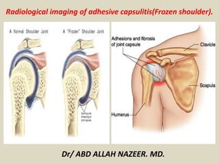
Radiological imaging of adhesive capsulitis(Frozen shoulder).
Adhesive capsulitis of the shoulder is a clinical
condition characterized by progressive limitation of active
and passive mobility of the glenohumeral joint, generally
associated with high levels of pain. Although the diagnosis
of adhesive capsulitis is based mainly on clinical
examination, different imaging modalities including
arthrography, ultrasound, magnetic resonance, and
magnetic resonance arthrography may help to confirm the
diagnosis, detecting a number of findings such as capsular
and coracohumeral ligament thickening, poor capsular
distension, extracapsular contrast leakage, and synovial
hypertrophy and scar tissue formation at the rotator
interval. Ultrasound can also be used to guide intra- and
periarticular procedures for treating patients with
adhesive capsulitis.
Clinical presentation
Adhesive capsulitis presentation can be broken into three distinct stages:
freezing: painful stage
patients may not present during this stage because they think that
eventually, the pain will resolve if self-treated
as the symptoms progress, pain worsens and both active and passive range
of motion (ROM) becomes more restricted
this can eventually result in the patient seeking medical consultation
typically lasts between 3 and 9 months and is characterised by an acute
synovitis of the glenohumeral joint
frozen: transitional stage
most patients will progress to the second stage
during this stage, shoulder pain does not necessarily worsen
because of pain at the end of the range of motion, arm movement may be
limited, causing muscular disuse
can last between 4 to 12 months
the common capsular pattern of limitation has historically been described as
diminishing motions with external shoulder rotation being the most limited,
followed closely by shoulder flexion, and internal rotation
a point is eventually reached in the frozen stage where pain does not occur
at the end of the range of motion
thawing stage(Resolution stage).
begins when the range of motion starts to improve
lasts anywhere from 12 to 42 months and is
defined by a gradual return of shoulder mobility
Pathology
Adhesive capsulitis is divided into two main types:
primary or idiopathic
absence of preceding trauma
secondary
major or minor repetitive trauma
shoulder or thoracic surgery
endocrine, e.g. diabetes, hyperthyroidism
rheumatological conditions
Radiographic features
Fluoroscopic arthrography
Described features include:
limited injectable fluid capacity of the glenohumeral joint
small dependent axillary fold
small subscapularis bursa
irregularity of the anterior capsular insertion at the
anatomic neck of the humerus
lymphatic filling may be present
Ultrasound
Limitation of movement of the supraspinatus is
considered a sensitive feature. A thickened coracohumeral
ligament (CHL) can also be a suggestive feature of
adhesive capsulitis.
MRI/MR arthrography
normal inferior glenohumeral ligament measures <4 mm
and is best seen on coronal oblique images at the mid
glenoid level; in adhesive capsulitis, the axillary recess
may show thickening ≥1.3 cm
joint capsule thickening
abnormal soft tissue thickening within the rotator
interval with signal alteration
abnormal soft tissue encasing the biceps anchor
variable enhancement of the capsule and synovium within
the axillary recess and rotator interval
Other MR arthrography features include:
thickening of the coracohumeral ligament (CHL)
subcoracoid triangle sign
Conventional arthrography, anteroposterior view. (a) Normal distension of the axillary
recess (black arrow) and the subscapular recess (thick arrow). (b) Reduced distension
of the axillary recess (black arrow) and subscapular recess associated with medial
leakage of contrast (white arrow) in a patient with adhesive capsulitis.
Axillary long-axis view of the inferior glenohumeral ligament with arm in
abduction. (a) Thickening of the inferior capsular profile (calipers, 3.3 mm)
in a shoulder affected by adhesive capsulitis. H humerus (b) In the
contralateral shoulder, the capsule has normal thickness (calipers, 1.5 mm)
Coronal oblique T2-
weighted fat-saturated
(a, b) and axial proton
density fat-saturated
(c, d) images. In a healthy
subject (a, c), the capsular
recess has normal signal
intensity (arrows), while
in a patient with adhesive
capsulitis (b, d), clear
signal hyperintensity can
be seen
(arrows). H humerus,
G glenoid, S supraspinatu
s tendon
Coronal oblique proton density image in a patient with adhesive capsulitis.
The axillary pouch (arrows) is thickened. G glenoid, H humerus.
MR arthrography, coronal oblique T1-weighted fat-saturated image. (a) In a healthy subject,
the axillary pouch is normally distensible (arrow). H humerus, G glenoid. (b). In a patient
with adhesive capsulitis, the axillary pouch is contracted and poorly distended (arrow)
MRI scan showing thickened (5mm) inferior capsule (normally 1mm).
Left Shoulder. Coronal T2 FS image of a 33-years-old male patient.
Shows thickened GHLI (5,58mm) synovial intraarticular liquid increase.
T2-fatsat MRI: Thickening of the capsule in the axillary
recess, and intermediate signal in adjacent soft tissues.
Coronal T1 (left) and PD-fat sat (right) MRI. Thickening (left) and intermediate signal
(right) of the joint capsule in the axillary recess in a patient with adhesive capsulitis.
MR arthrography, (a) coronal oblique and (b) axial T1-weighted fat-saturated
image in a patient with adhesive capsulitis. Leakage of contrast agent can be seen
on the anterior inferior margin of the scapula (arrow). H humerus, G glenoid
Oblique coronal PDFS image of a 45-year-
old woman after recent trauma. There is
thickening and edema of the axillary
recess(white arrow).
Oblique coronal T1 weighted image of a 57-year-
old man with clinically proven adhesive capsulitis.
There is thickening of the axillary recess, which is
only mildly hyperintense (white arrows).
Adhesive capsulitis of the shoulder.
Adhesive Capsulitis.
Adhesive capsulitis.
MRI findings are subtle in frozen shoulder. To be certain, frozen shoulder is a
clinical diagnosis, and there are no specific direct signs that are pathognomonic
for frozen shoulder. Described direct signs suggestive of frozen shoulder include:
Thickening of the glenohumeral joint capsule along the axillary pouch
Thickening of the coracohumeral ligament
Obliteration of the subcoracoid fat triangle
Rotator interval synovitis.
MRI shows- poor definition of coracohumeral ligament with abnormal intermediate
amorphous signal (with hypointense foci on all sequences) surrounding the ligament
with ill definition of superior glenohumeral ligament & rotator interval with mild free
fluid in the shoulder joint – likely suggesting adhesive capsulitis.
Inferior glenohumeral ligament and the
inferior joint capsule (yellow block arrow) in
the axillary recess shows diffuse thickening.
Diffuse thickening of the coracohumeral ligament
(white arrow) extending up to the rotator cuff
interval and is hyperintense on the T2 images.
(2a) The sagittal T2-weighted image demonstrates a thickened coracohumeral ligament (arrow)
and soft tissue thickening in the rotator interval (arrowheads). The short arrow indicates the
adjacent long head of the biceps tendon.
(2b) The coronal T2-weighted fat-suppressed image demonstrates an abnormally thickened
inferior glenohumeral ligament (arrow).
(2a)
(2b)
Adhesive capsulitis is a self-limited clinical syndrome characterized
by painful, gradual loss of active and passive glenohumeral motion.
Sagittal oblique T1-weighted
image (TR/TE = 550 ms/15 ms) (1A)
shows thickened CHL (arrows).
C = inferior margin for the coracoid
process. Sagittal oblique (1B),
oblique coronal (1C), and
transverse (1D) fat-suppressed,
proton density weighted, spin-echo
image (TR/TE = 3000 ms/34 ms)
show high-signal intensity soft
tissue in the rotator cuff interval
for the same patient (arrows).
Coronal oblique (1E) and
transverse (1F) fat-suppressed,
proton density, weighted spin-echo
image (TR/TE = 3000 ms/34 ms)
demonstrate a thickened inferior
glenohumeral ligament (axillary
recess) for the same patient
(arrows).
Magnetic Resonance Imaging, Arthrography, and Ultrasonography Reflect Clinical
Impairment in Patients With Idiopathic Adhesive Capsulitis of the Shoulder.
Oblique sagittal PDFS image of a 69-year-
old man. There is thickening of the CHL,
and fibrovascular tissue with mild edema
in the rotator cuff interval(white arrow).
Fibrovascular changes are visible around
CHL and around SGHL. Axial PDFS images
(A,B) through the rotator cuff interval
shows extensive fibrovascular tissue
around the biceps tendon (white arrow).
Sagittal oblique T2-weighted image in a patient
with adhesive capsulitis. The coracohumeral
ligament (arrows) is markedly thickened.
MR arthrography, sagittal oblique T1-
weighted image. In a patient with adhesive
capsulitis for 15 weeks, the fat triangle
(arrowheads) signal is considerably reduced.
Oblique sagittal PD weighted image shows
obliteration and fibrosis in subcoracoid space.
Oblique sagittal oblique PD weighted
image shows thickened CHL (arrow) in a
57-year-old man with adhesive capsulitis.
A coronal T2-weighted fat suppressed image through the
anterior aspect of the shoulder demonstrates increased
signal intensity (asterisk) in the rotator interval region,
obscuring the normal fat, suspicious for adhesive capsulitis.
Post IV gadolinium enhanced sagittal T1-weighted
image with fat-suppression demonstrates
enhancement in the rotator interval region (arrows),
confirming the diagnosis of adhesive capsulitis.
Sagittal oblique T1 MRI. Thickened coracohumeral
ligament (arrows) in a patient with frozen shoulder.
T1 sagittal oblique MRI (left): Almost complete obliteration of subcoracoid fat (arrow);
T2 fat-sat sagittal oblique MRI (right): intermediate signal scar in the rotator interval.
Adhesive capsulitis with frozen Shoulder.
Thank You.

More Related Content

What's hot

Presentation1.pptx, ultrasound examination of the elbow joint.
Presentation1.pptx, ultrasound examination of the elbow joint.Presentation1.pptx, ultrasound examination of the elbow joint.
Presentation1.pptx, ultrasound examination of the elbow joint.Abdellah Nazeer
 
Presentation1.pptx, ultrasound examination of the knee joint.
Presentation1.pptx, ultrasound examination of the knee joint.Presentation1.pptx, ultrasound examination of the knee joint.
Presentation1.pptx, ultrasound examination of the knee joint.Abdellah Nazeer
 
Presentation1.pptx, radiological imaging of bursae.
Presentation1.pptx, radiological imaging of bursae.Presentation1.pptx, radiological imaging of bursae.
Presentation1.pptx, radiological imaging of bursae.Abdellah Nazeer
 
HOW TO READ SHOULDER MRI
HOW TO READ SHOULDER MRI HOW TO READ SHOULDER MRI
HOW TO READ SHOULDER MRI Arjun Kouloth
 
Radiology of the Elbow Joint. Dr. Sumit Sharma
Radiology of the Elbow Joint. Dr. Sumit SharmaRadiology of the Elbow Joint. Dr. Sumit Sharma
Radiology of the Elbow Joint. Dr. Sumit SharmaSumit Sharma
 
Imaging in arthritis
Imaging in arthritisImaging in arthritis
Imaging in arthritisNavni Garg
 
Presentation1, radiological film reading of the sholder joint.
Presentation1,  radiological film reading of the sholder joint.Presentation1,  radiological film reading of the sholder joint.
Presentation1, radiological film reading of the sholder joint.Abdellah Nazeer
 
Post-graduate Certifcate Musculoskeletal Ultrasound - The Shoulder
Post-graduate Certifcate Musculoskeletal Ultrasound - The ShoulderPost-graduate Certifcate Musculoskeletal Ultrasound - The Shoulder
Post-graduate Certifcate Musculoskeletal Ultrasound - The ShoulderDr. Peter Resteghini
 
Presentation1.pptx. shoulder joint.
Presentation1.pptx. shoulder joint.Presentation1.pptx. shoulder joint.
Presentation1.pptx. shoulder joint.Abdellah Nazeer
 
Muscloskeletal Ultrasound of the shoulder (basic level)
Muscloskeletal Ultrasound of the shoulder (basic level)Muscloskeletal Ultrasound of the shoulder (basic level)
Muscloskeletal Ultrasound of the shoulder (basic level)Samar Tharwat
 
Avascular necrosis Radiology
Avascular necrosis RadiologyAvascular necrosis Radiology
Avascular necrosis Radiologyrajss007
 
Mri shoulder joint with common pathologies
Mri shoulder joint with common pathologiesMri shoulder joint with common pathologies
Mri shoulder joint with common pathologiesGobardhan Thapa
 
Imaging in hip disorders
Imaging in hip disordersImaging in hip disorders
Imaging in hip disordersVikram Patil
 
Presentation1.pptx, radiological anatomy of the thigh and leg.
Presentation1.pptx, radiological anatomy of the thigh and leg.Presentation1.pptx, radiological anatomy of the thigh and leg.
Presentation1.pptx, radiological anatomy of the thigh and leg.Abdellah Nazeer
 

What's hot (20)

Imaging of knee by mr and usg
Imaging of knee by mr and usgImaging of knee by mr and usg
Imaging of knee by mr and usg
 
Presentation1.pptx, ultrasound examination of the elbow joint.
Presentation1.pptx, ultrasound examination of the elbow joint.Presentation1.pptx, ultrasound examination of the elbow joint.
Presentation1.pptx, ultrasound examination of the elbow joint.
 
Presentation1.pptx, ultrasound examination of the knee joint.
Presentation1.pptx, ultrasound examination of the knee joint.Presentation1.pptx, ultrasound examination of the knee joint.
Presentation1.pptx, ultrasound examination of the knee joint.
 
Presentation1.pptx, radiological imaging of bursae.
Presentation1.pptx, radiological imaging of bursae.Presentation1.pptx, radiological imaging of bursae.
Presentation1.pptx, radiological imaging of bursae.
 
Mri knee trauma
Mri knee traumaMri knee trauma
Mri knee trauma
 
MRI Knee trauma
MRI Knee traumaMRI Knee trauma
MRI Knee trauma
 
HOW TO READ SHOULDER MRI
HOW TO READ SHOULDER MRI HOW TO READ SHOULDER MRI
HOW TO READ SHOULDER MRI
 
Radiology of the Elbow Joint. Dr. Sumit Sharma
Radiology of the Elbow Joint. Dr. Sumit SharmaRadiology of the Elbow Joint. Dr. Sumit Sharma
Radiology of the Elbow Joint. Dr. Sumit Sharma
 
Imaging in arthritis
Imaging in arthritisImaging in arthritis
Imaging in arthritis
 
Presentation1, radiological film reading of the sholder joint.
Presentation1,  radiological film reading of the sholder joint.Presentation1,  radiological film reading of the sholder joint.
Presentation1, radiological film reading of the sholder joint.
 
Post-graduate Certifcate Musculoskeletal Ultrasound - The Shoulder
Post-graduate Certifcate Musculoskeletal Ultrasound - The ShoulderPost-graduate Certifcate Musculoskeletal Ultrasound - The Shoulder
Post-graduate Certifcate Musculoskeletal Ultrasound - The Shoulder
 
Hip joint
Hip jointHip joint
Hip joint
 
Presentation1.pptx. shoulder joint.
Presentation1.pptx. shoulder joint.Presentation1.pptx. shoulder joint.
Presentation1.pptx. shoulder joint.
 
Muscloskeletal Ultrasound of the shoulder (basic level)
Muscloskeletal Ultrasound of the shoulder (basic level)Muscloskeletal Ultrasound of the shoulder (basic level)
Muscloskeletal Ultrasound of the shoulder (basic level)
 
Avascular necrosis Radiology
Avascular necrosis RadiologyAvascular necrosis Radiology
Avascular necrosis Radiology
 
Mri shoulder joint with common pathologies
Mri shoulder joint with common pathologiesMri shoulder joint with common pathologies
Mri shoulder joint with common pathologies
 
Imaging in hip disorders
Imaging in hip disordersImaging in hip disorders
Imaging in hip disorders
 
Mr imaging of spine
Mr imaging of spineMr imaging of spine
Mr imaging of spine
 
Presentation1.pptx, radiological anatomy of the thigh and leg.
Presentation1.pptx, radiological anatomy of the thigh and leg.Presentation1.pptx, radiological anatomy of the thigh and leg.
Presentation1.pptx, radiological anatomy of the thigh and leg.
 
MRI OF SHOULDER INJURY
MRI OF SHOULDER INJURYMRI OF SHOULDER INJURY
MRI OF SHOULDER INJURY
 

Viewers also liked

Chest radiology in intensive care
Chest radiology in intensive careChest radiology in intensive care
Chest radiology in intensive careAndrew Ferguson
 
Presentation2.pptx, radiological imaging of gastric lesions.
Presentation2.pptx, radiological imaging of gastric lesions.Presentation2.pptx, radiological imaging of gastric lesions.
Presentation2.pptx, radiological imaging of gastric lesions.Abdellah Nazeer
 
Radiology of digestive system
Radiology of digestive systemRadiology of digestive system
Radiology of digestive systemghalan
 
Presentation2.pptx. radilogical imaging of intestinal obstruction.
Presentation2.pptx. radilogical imaging of intestinal obstruction.Presentation2.pptx. radilogical imaging of intestinal obstruction.
Presentation2.pptx. radilogical imaging of intestinal obstruction.Abdellah Nazeer
 
Presentation1.pptx, ultrasound study of the spleen and pancreas.
Presentation1.pptx, ultrasound study of the spleen and pancreas.Presentation1.pptx, ultrasound study of the spleen and pancreas.
Presentation1.pptx, ultrasound study of the spleen and pancreas.Abdellah Nazeer
 
Presentation1.pptx pancreatic disease.
Presentation1.pptx pancreatic disease.Presentation1.pptx pancreatic disease.
Presentation1.pptx pancreatic disease.Abdellah Nazeer
 
Git and hepatobiliary radiology mocks fcps
Git and hepatobiliary radiology mocks fcpsGit and hepatobiliary radiology mocks fcps
Git and hepatobiliary radiology mocks fcpsdrneelammalik
 
Radiology signs and symptoms
Radiology signs and symptomsRadiology signs and symptoms
Radiology signs and symptomsakifab93
 
Presentation1, film readiing for barium studies.
Presentation1, film readiing for barium studies.Presentation1, film readiing for barium studies.
Presentation1, film readiing for barium studies.Abdellah Nazeer
 
Presentation1.pptx, radiological imaging of the larngeal diseases.
Presentation1.pptx, radiological imaging of the larngeal diseases.Presentation1.pptx, radiological imaging of the larngeal diseases.
Presentation1.pptx, radiological imaging of the larngeal diseases.Abdellah Nazeer
 
Presentation1, mri quiz cases.
Presentation1, mri quiz cases.Presentation1, mri quiz cases.
Presentation1, mri quiz cases.Abdellah Nazeer
 
Chronic obstructive pulmonary disease
Chronic obstructive pulmonary diseaseChronic obstructive pulmonary disease
Chronic obstructive pulmonary diseaseEdson Mutandwa
 
Presentation1.pptx, radiological imaging of chronic obstructive airway disease.
Presentation1.pptx, radiological imaging of chronic obstructive airway disease.Presentation1.pptx, radiological imaging of chronic obstructive airway disease.
Presentation1.pptx, radiological imaging of chronic obstructive airway disease.Abdellah Nazeer
 
Presentation1.pptx, radiological imaging of copd.
Presentation1.pptx, radiological imaging of copd.Presentation1.pptx, radiological imaging of copd.
Presentation1.pptx, radiological imaging of copd.Abdellah Nazeer
 
Hrct in diagnosis of diffuse lung diseases
Hrct in diagnosis of  diffuse lung diseasesHrct in diagnosis of  diffuse lung diseases
Hrct in diagnosis of diffuse lung diseasesAhmed Bahnassy
 
Presentation2.pptx, radiological imaging of the rectal diseases.
Presentation2.pptx, radiological imaging of the rectal diseases.Presentation2.pptx, radiological imaging of the rectal diseases.
Presentation2.pptx, radiological imaging of the rectal diseases.Abdellah Nazeer
 
Presentation1.pptx, radiological imaging of large bowel diseases
Presentation1.pptx, radiological imaging of large bowel diseasesPresentation1.pptx, radiological imaging of large bowel diseases
Presentation1.pptx, radiological imaging of large bowel diseasesAbdellah Nazeer
 

Viewers also liked (20)

Chest radiology in intensive care
Chest radiology in intensive careChest radiology in intensive care
Chest radiology in intensive care
 
Presentation2.pptx, radiological imaging of gastric lesions.
Presentation2.pptx, radiological imaging of gastric lesions.Presentation2.pptx, radiological imaging of gastric lesions.
Presentation2.pptx, radiological imaging of gastric lesions.
 
Radiology of digestive system
Radiology of digestive systemRadiology of digestive system
Radiology of digestive system
 
Presentation2.pptx. radilogical imaging of intestinal obstruction.
Presentation2.pptx. radilogical imaging of intestinal obstruction.Presentation2.pptx. radilogical imaging of intestinal obstruction.
Presentation2.pptx. radilogical imaging of intestinal obstruction.
 
Presentation1.pptx, ultrasound study of the spleen and pancreas.
Presentation1.pptx, ultrasound study of the spleen and pancreas.Presentation1.pptx, ultrasound study of the spleen and pancreas.
Presentation1.pptx, ultrasound study of the spleen and pancreas.
 
Presentation1.pptx pancreatic disease.
Presentation1.pptx pancreatic disease.Presentation1.pptx pancreatic disease.
Presentation1.pptx pancreatic disease.
 
Git and hepatobiliary radiology mocks fcps
Git and hepatobiliary radiology mocks fcpsGit and hepatobiliary radiology mocks fcps
Git and hepatobiliary radiology mocks fcps
 
Radiology signs and symptoms
Radiology signs and symptomsRadiology signs and symptoms
Radiology signs and symptoms
 
Emphysema
EmphysemaEmphysema
Emphysema
 
Presentation1, film readiing for barium studies.
Presentation1, film readiing for barium studies.Presentation1, film readiing for barium studies.
Presentation1, film readiing for barium studies.
 
Presentation1.pptx, radiological imaging of the larngeal diseases.
Presentation1.pptx, radiological imaging of the larngeal diseases.Presentation1.pptx, radiological imaging of the larngeal diseases.
Presentation1.pptx, radiological imaging of the larngeal diseases.
 
Presentation1, mri quiz cases.
Presentation1, mri quiz cases.Presentation1, mri quiz cases.
Presentation1, mri quiz cases.
 
Chronic obstructive pulmonary disease
Chronic obstructive pulmonary diseaseChronic obstructive pulmonary disease
Chronic obstructive pulmonary disease
 
Presentation1.pptx, radiological imaging of chronic obstructive airway disease.
Presentation1.pptx, radiological imaging of chronic obstructive airway disease.Presentation1.pptx, radiological imaging of chronic obstructive airway disease.
Presentation1.pptx, radiological imaging of chronic obstructive airway disease.
 
Presentation1.pptx, radiological imaging of copd.
Presentation1.pptx, radiological imaging of copd.Presentation1.pptx, radiological imaging of copd.
Presentation1.pptx, radiological imaging of copd.
 
Hepatic imaging
Hepatic imagingHepatic imaging
Hepatic imaging
 
Hrct in diagnosis of diffuse lung diseases
Hrct in diagnosis of  diffuse lung diseasesHrct in diagnosis of  diffuse lung diseases
Hrct in diagnosis of diffuse lung diseases
 
Laryngeal Cancer
Laryngeal CancerLaryngeal Cancer
Laryngeal Cancer
 
Presentation2.pptx, radiological imaging of the rectal diseases.
Presentation2.pptx, radiological imaging of the rectal diseases.Presentation2.pptx, radiological imaging of the rectal diseases.
Presentation2.pptx, radiological imaging of the rectal diseases.
 
Presentation1.pptx, radiological imaging of large bowel diseases
Presentation1.pptx, radiological imaging of large bowel diseasesPresentation1.pptx, radiological imaging of large bowel diseases
Presentation1.pptx, radiological imaging of large bowel diseases
 

Similar to Presentation1, radiological imaging of adhesive capsulitis(frozen shoulder).

Regional conditions of upper limb
Regional conditions of upper limbRegional conditions of upper limb
Regional conditions of upper limbAaishwaryaa Rai
 
Regional conditions of upper limb BY MIN^ED ACADEMY
Regional conditions of upper limb BY MIN^ED ACADEMYRegional conditions of upper limb BY MIN^ED ACADEMY
Regional conditions of upper limb BY MIN^ED ACADEMYMINED ACADEMY
 
Shoulder impingement syndrome
Shoulder impingement syndromeShoulder impingement syndrome
Shoulder impingement syndromeAaishwaryaa Rai
 
Frozen shoulder BY MIN^ED ACADEMY
Frozen shoulder BY MIN^ED ACADEMYFrozen shoulder BY MIN^ED ACADEMY
Frozen shoulder BY MIN^ED ACADEMYMINED ACADEMY
 
SHOULDER USG RAJ.pptx
SHOULDER USG RAJ.pptxSHOULDER USG RAJ.pptx
SHOULDER USG RAJ.pptxRajVaghasia
 
Ankylosing spondylitis clinical feature and diagnosis
Ankylosing spondylitis clinical feature and diagnosisAnkylosing spondylitis clinical feature and diagnosis
Ankylosing spondylitis clinical feature and diagnosisdattasrisaila
 
Presentation1, radiological imaging of shoulder dislocation.
Presentation1, radiological imaging of shoulder dislocation.Presentation1, radiological imaging of shoulder dislocation.
Presentation1, radiological imaging of shoulder dislocation.Abdellah Nazeer
 
Presentation1.pptx, ultrasound examination of the wrist joint.
Presentation1.pptx, ultrasound examination of the wrist joint.Presentation1.pptx, ultrasound examination of the wrist joint.
Presentation1.pptx, ultrasound examination of the wrist joint.Abdellah Nazeer
 
The Rotator Interval A Link Between Anatomy and Ultrasound.pdf
The Rotator Interval A Link Between Anatomy and Ultrasound.pdfThe Rotator Interval A Link Between Anatomy and Ultrasound.pdf
The Rotator Interval A Link Between Anatomy and Ultrasound.pdfThoiPham12
 
ULTRASONOGRAPHY OF DIAPHRAGM AND ITS CLINICAL IMPLICATION
ULTRASONOGRAPHY OF DIAPHRAGM AND ITS CLINICAL IMPLICATIONULTRASONOGRAPHY OF DIAPHRAGM AND ITS CLINICAL IMPLICATION
ULTRASONOGRAPHY OF DIAPHRAGM AND ITS CLINICAL IMPLICATIONAbhilasha Singh
 
Cervical Arthritis, Cervical Spondylotic Myelopathy by Pablo Pazmino MD
Cervical Arthritis, Cervical Spondylotic Myelopathy by Pablo Pazmino MDCervical Arthritis, Cervical Spondylotic Myelopathy by Pablo Pazmino MD
Cervical Arthritis, Cervical Spondylotic Myelopathy by Pablo Pazmino MDPablo Pazmino
 
Cervical Arthritis / Cervical Spondylotic Myelopathy / Cervical Stenosis by P...
Cervical Arthritis / Cervical Spondylotic Myelopathy / Cervical Stenosis by P...Cervical Arthritis / Cervical Spondylotic Myelopathy / Cervical Stenosis by P...
Cervical Arthritis / Cervical Spondylotic Myelopathy / Cervical Stenosis by P...Pablo Pazmino
 
usg shoulder by dr. kanishka.pptx
usg shoulder by dr. kanishka.pptxusg shoulder by dr. kanishka.pptx
usg shoulder by dr. kanishka.pptxRajVaghasia
 
Presentation1, radiological imaging of popliteal fossa masses.
Presentation1, radiological imaging of popliteal fossa masses.Presentation1, radiological imaging of popliteal fossa masses.
Presentation1, radiological imaging of popliteal fossa masses.Abdellah Nazeer
 
Imaging of lumbar spondylosis
Imaging of lumbar spondylosisImaging of lumbar spondylosis
Imaging of lumbar spondylosishazem youssef
 

Similar to Presentation1, radiological imaging of adhesive capsulitis(frozen shoulder). (20)

Regional conditions of upper limb
Regional conditions of upper limbRegional conditions of upper limb
Regional conditions of upper limb
 
Regional conditions of upper limb BY MIN^ED ACADEMY
Regional conditions of upper limb BY MIN^ED ACADEMYRegional conditions of upper limb BY MIN^ED ACADEMY
Regional conditions of upper limb BY MIN^ED ACADEMY
 
MRI sholdure
MRI sholdureMRI sholdure
MRI sholdure
 
Shoulder impingement syndrome
Shoulder impingement syndromeShoulder impingement syndrome
Shoulder impingement syndrome
 
Frozen shoulder BY MIN^ED ACADEMY
Frozen shoulder BY MIN^ED ACADEMYFrozen shoulder BY MIN^ED ACADEMY
Frozen shoulder BY MIN^ED ACADEMY
 
SHOULDER USG RAJ.pptx
SHOULDER USG RAJ.pptxSHOULDER USG RAJ.pptx
SHOULDER USG RAJ.pptx
 
Ankylosing spondylitis clinical feature and diagnosis
Ankylosing spondylitis clinical feature and diagnosisAnkylosing spondylitis clinical feature and diagnosis
Ankylosing spondylitis clinical feature and diagnosis
 
Rotator cuff tears
Rotator cuff tearsRotator cuff tears
Rotator cuff tears
 
Presentation1, radiological imaging of shoulder dislocation.
Presentation1, radiological imaging of shoulder dislocation.Presentation1, radiological imaging of shoulder dislocation.
Presentation1, radiological imaging of shoulder dislocation.
 
Picture Medicine
Picture MedicinePicture Medicine
Picture Medicine
 
Presentation1.pptx, ultrasound examination of the wrist joint.
Presentation1.pptx, ultrasound examination of the wrist joint.Presentation1.pptx, ultrasound examination of the wrist joint.
Presentation1.pptx, ultrasound examination of the wrist joint.
 
The Rotator Interval A Link Between Anatomy and Ultrasound.pdf
The Rotator Interval A Link Between Anatomy and Ultrasound.pdfThe Rotator Interval A Link Between Anatomy and Ultrasound.pdf
The Rotator Interval A Link Between Anatomy and Ultrasound.pdf
 
KUMMEL DISEASE.pptx
KUMMEL DISEASE.pptxKUMMEL DISEASE.pptx
KUMMEL DISEASE.pptx
 
ULTRASONOGRAPHY OF DIAPHRAGM AND ITS CLINICAL IMPLICATION
ULTRASONOGRAPHY OF DIAPHRAGM AND ITS CLINICAL IMPLICATIONULTRASONOGRAPHY OF DIAPHRAGM AND ITS CLINICAL IMPLICATION
ULTRASONOGRAPHY OF DIAPHRAGM AND ITS CLINICAL IMPLICATION
 
Cervical Arthritis, Cervical Spondylotic Myelopathy by Pablo Pazmino MD
Cervical Arthritis, Cervical Spondylotic Myelopathy by Pablo Pazmino MDCervical Arthritis, Cervical Spondylotic Myelopathy by Pablo Pazmino MD
Cervical Arthritis, Cervical Spondylotic Myelopathy by Pablo Pazmino MD
 
Cervical Arthritis / Cervical Spondylotic Myelopathy / Cervical Stenosis by P...
Cervical Arthritis / Cervical Spondylotic Myelopathy / Cervical Stenosis by P...Cervical Arthritis / Cervical Spondylotic Myelopathy / Cervical Stenosis by P...
Cervical Arthritis / Cervical Spondylotic Myelopathy / Cervical Stenosis by P...
 
usg shoulder by dr. kanishka.pptx
usg shoulder by dr. kanishka.pptxusg shoulder by dr. kanishka.pptx
usg shoulder by dr. kanishka.pptx
 
Presentation1, radiological imaging of popliteal fossa masses.
Presentation1, radiological imaging of popliteal fossa masses.Presentation1, radiological imaging of popliteal fossa masses.
Presentation1, radiological imaging of popliteal fossa masses.
 
Ddd rem rai2
Ddd rem rai2Ddd rem rai2
Ddd rem rai2
 
Imaging of lumbar spondylosis
Imaging of lumbar spondylosisImaging of lumbar spondylosis
Imaging of lumbar spondylosis
 

More from Abdellah Nazeer

Muculoskeletal Pediatic Imaging..pptx
Muculoskeletal Pediatic Imaging..pptxMuculoskeletal Pediatic Imaging..pptx
Muculoskeletal Pediatic Imaging..pptxAbdellah Nazeer
 
Presentation1, Ultrasound of the bowel loops and the lymph nodes..pptx
Presentation1, Ultrasound of the bowel loops and the lymph nodes..pptxPresentation1, Ultrasound of the bowel loops and the lymph nodes..pptx
Presentation1, Ultrasound of the bowel loops and the lymph nodes..pptxAbdellah Nazeer
 
Presentation1 Short cases MD..pptx
Presentation1 Short cases MD..pptxPresentation1 Short cases MD..pptx
Presentation1 Short cases MD..pptxAbdellah Nazeer
 
Presentation1, MD MCQ Cases..pptx
Presentation1, MD MCQ Cases..pptxPresentation1, MD MCQ Cases..pptx
Presentation1, MD MCQ Cases..pptxAbdellah Nazeer
 
Presentation1, Short Cases Quiz..pptx
Presentation1, Short Cases Quiz..pptxPresentation1, Short Cases Quiz..pptx
Presentation1, Short Cases Quiz..pptxAbdellah Nazeer
 
Presentation1, radiological imaging of lateral hindfoot impingement.
Presentation1, radiological imaging of lateral hindfoot impingement.Presentation1, radiological imaging of lateral hindfoot impingement.
Presentation1, radiological imaging of lateral hindfoot impingement.Abdellah Nazeer
 
Presentation2, radiological anatomy of the liver and spleen.
Presentation2, radiological anatomy of the liver and spleen.Presentation2, radiological anatomy of the liver and spleen.
Presentation2, radiological anatomy of the liver and spleen.Abdellah Nazeer
 
Presentation1, artifacts and pitfalls of the wrist and elbow joints.
Presentation1, artifacts and pitfalls of the wrist and elbow joints.Presentation1, artifacts and pitfalls of the wrist and elbow joints.
Presentation1, artifacts and pitfalls of the wrist and elbow joints.Abdellah Nazeer
 
Presentation1, artifact and pitfalls of the knee, hip and ankle joints.
Presentation1, artifact and pitfalls of the knee, hip and ankle joints.Presentation1, artifact and pitfalls of the knee, hip and ankle joints.
Presentation1, artifact and pitfalls of the knee, hip and ankle joints.Abdellah Nazeer
 
Presentation1, radiological imaging of artifact and pitfalls in shoulder join...
Presentation1, radiological imaging of artifact and pitfalls in shoulder join...Presentation1, radiological imaging of artifact and pitfalls in shoulder join...
Presentation1, radiological imaging of artifact and pitfalls in shoulder join...Abdellah Nazeer
 
Presentation1, radiological imaging of internal abdominal hernia.
Presentation1, radiological imaging of internal abdominal hernia.Presentation1, radiological imaging of internal abdominal hernia.
Presentation1, radiological imaging of internal abdominal hernia.Abdellah Nazeer
 
Presentation11, radiological imaging of ovarian torsion.
Presentation11, radiological imaging of ovarian torsion.Presentation11, radiological imaging of ovarian torsion.
Presentation11, radiological imaging of ovarian torsion.Abdellah Nazeer
 
Presentation1, musculoskeletal anatomy.
Presentation1, musculoskeletal anatomy.Presentation1, musculoskeletal anatomy.
Presentation1, musculoskeletal anatomy.Abdellah Nazeer
 
Presentation1, new mri techniques in the diagnosis and monitoring of multiple...
Presentation1, new mri techniques in the diagnosis and monitoring of multiple...Presentation1, new mri techniques in the diagnosis and monitoring of multiple...
Presentation1, new mri techniques in the diagnosis and monitoring of multiple...Abdellah Nazeer
 
Presentation1, radiological application of diffusion weighted mri in neck mas...
Presentation1, radiological application of diffusion weighted mri in neck mas...Presentation1, radiological application of diffusion weighted mri in neck mas...
Presentation1, radiological application of diffusion weighted mri in neck mas...Abdellah Nazeer
 
Presentation1, radiological application of diffusion weighted images in breas...
Presentation1, radiological application of diffusion weighted images in breas...Presentation1, radiological application of diffusion weighted images in breas...
Presentation1, radiological application of diffusion weighted images in breas...Abdellah Nazeer
 
Presentation1, radiological application of diffusion weighted images in abdom...
Presentation1, radiological application of diffusion weighted images in abdom...Presentation1, radiological application of diffusion weighted images in abdom...
Presentation1, radiological application of diffusion weighted images in abdom...Abdellah Nazeer
 
Presentation1, radiological application of diffusion weighted imges in neuror...
Presentation1, radiological application of diffusion weighted imges in neuror...Presentation1, radiological application of diffusion weighted imges in neuror...
Presentation1, radiological application of diffusion weighted imges in neuror...Abdellah Nazeer
 
Presentation1, mr physics.
Presentation1, mr physics.Presentation1, mr physics.
Presentation1, mr physics.Abdellah Nazeer
 
Presentation1. ct physics.
Presentation1. ct physics.Presentation1. ct physics.
Presentation1. ct physics.Abdellah Nazeer
 

More from Abdellah Nazeer (20)

Muculoskeletal Pediatic Imaging..pptx
Muculoskeletal Pediatic Imaging..pptxMuculoskeletal Pediatic Imaging..pptx
Muculoskeletal Pediatic Imaging..pptx
 
Presentation1, Ultrasound of the bowel loops and the lymph nodes..pptx
Presentation1, Ultrasound of the bowel loops and the lymph nodes..pptxPresentation1, Ultrasound of the bowel loops and the lymph nodes..pptx
Presentation1, Ultrasound of the bowel loops and the lymph nodes..pptx
 
Presentation1 Short cases MD..pptx
Presentation1 Short cases MD..pptxPresentation1 Short cases MD..pptx
Presentation1 Short cases MD..pptx
 
Presentation1, MD MCQ Cases..pptx
Presentation1, MD MCQ Cases..pptxPresentation1, MD MCQ Cases..pptx
Presentation1, MD MCQ Cases..pptx
 
Presentation1, Short Cases Quiz..pptx
Presentation1, Short Cases Quiz..pptxPresentation1, Short Cases Quiz..pptx
Presentation1, Short Cases Quiz..pptx
 
Presentation1, radiological imaging of lateral hindfoot impingement.
Presentation1, radiological imaging of lateral hindfoot impingement.Presentation1, radiological imaging of lateral hindfoot impingement.
Presentation1, radiological imaging of lateral hindfoot impingement.
 
Presentation2, radiological anatomy of the liver and spleen.
Presentation2, radiological anatomy of the liver and spleen.Presentation2, radiological anatomy of the liver and spleen.
Presentation2, radiological anatomy of the liver and spleen.
 
Presentation1, artifacts and pitfalls of the wrist and elbow joints.
Presentation1, artifacts and pitfalls of the wrist and elbow joints.Presentation1, artifacts and pitfalls of the wrist and elbow joints.
Presentation1, artifacts and pitfalls of the wrist and elbow joints.
 
Presentation1, artifact and pitfalls of the knee, hip and ankle joints.
Presentation1, artifact and pitfalls of the knee, hip and ankle joints.Presentation1, artifact and pitfalls of the knee, hip and ankle joints.
Presentation1, artifact and pitfalls of the knee, hip and ankle joints.
 
Presentation1, radiological imaging of artifact and pitfalls in shoulder join...
Presentation1, radiological imaging of artifact and pitfalls in shoulder join...Presentation1, radiological imaging of artifact and pitfalls in shoulder join...
Presentation1, radiological imaging of artifact and pitfalls in shoulder join...
 
Presentation1, radiological imaging of internal abdominal hernia.
Presentation1, radiological imaging of internal abdominal hernia.Presentation1, radiological imaging of internal abdominal hernia.
Presentation1, radiological imaging of internal abdominal hernia.
 
Presentation11, radiological imaging of ovarian torsion.
Presentation11, radiological imaging of ovarian torsion.Presentation11, radiological imaging of ovarian torsion.
Presentation11, radiological imaging of ovarian torsion.
 
Presentation1, musculoskeletal anatomy.
Presentation1, musculoskeletal anatomy.Presentation1, musculoskeletal anatomy.
Presentation1, musculoskeletal anatomy.
 
Presentation1, new mri techniques in the diagnosis and monitoring of multiple...
Presentation1, new mri techniques in the diagnosis and monitoring of multiple...Presentation1, new mri techniques in the diagnosis and monitoring of multiple...
Presentation1, new mri techniques in the diagnosis and monitoring of multiple...
 
Presentation1, radiological application of diffusion weighted mri in neck mas...
Presentation1, radiological application of diffusion weighted mri in neck mas...Presentation1, radiological application of diffusion weighted mri in neck mas...
Presentation1, radiological application of diffusion weighted mri in neck mas...
 
Presentation1, radiological application of diffusion weighted images in breas...
Presentation1, radiological application of diffusion weighted images in breas...Presentation1, radiological application of diffusion weighted images in breas...
Presentation1, radiological application of diffusion weighted images in breas...
 
Presentation1, radiological application of diffusion weighted images in abdom...
Presentation1, radiological application of diffusion weighted images in abdom...Presentation1, radiological application of diffusion weighted images in abdom...
Presentation1, radiological application of diffusion weighted images in abdom...
 
Presentation1, radiological application of diffusion weighted imges in neuror...
Presentation1, radiological application of diffusion weighted imges in neuror...Presentation1, radiological application of diffusion weighted imges in neuror...
Presentation1, radiological application of diffusion weighted imges in neuror...
 
Presentation1, mr physics.
Presentation1, mr physics.Presentation1, mr physics.
Presentation1, mr physics.
 
Presentation1. ct physics.
Presentation1. ct physics.Presentation1. ct physics.
Presentation1. ct physics.
 

Recently uploaded

College Call Girls Pune Mira 9907093804 Short 1500 Night 6000 Best call girls...
College Call Girls Pune Mira 9907093804 Short 1500 Night 6000 Best call girls...College Call Girls Pune Mira 9907093804 Short 1500 Night 6000 Best call girls...
College Call Girls Pune Mira 9907093804 Short 1500 Night 6000 Best call girls...Miss joya
 
Call Girls Darjeeling Just Call 9907093804 Top Class Call Girl Service Available
Call Girls Darjeeling Just Call 9907093804 Top Class Call Girl Service AvailableCall Girls Darjeeling Just Call 9907093804 Top Class Call Girl Service Available
Call Girls Darjeeling Just Call 9907093804 Top Class Call Girl Service AvailableDipal Arora
 
Kesar Bagh Call Girl Price 9548273370 , Lucknow Call Girls Service
Kesar Bagh Call Girl Price 9548273370 , Lucknow Call Girls ServiceKesar Bagh Call Girl Price 9548273370 , Lucknow Call Girls Service
Kesar Bagh Call Girl Price 9548273370 , Lucknow Call Girls Servicemakika9823
 
Book Paid Powai Call Girls Mumbai 𖠋 9930245274 𖠋Low Budget Full Independent H...
Book Paid Powai Call Girls Mumbai 𖠋 9930245274 𖠋Low Budget Full Independent H...Book Paid Powai Call Girls Mumbai 𖠋 9930245274 𖠋Low Budget Full Independent H...
Book Paid Powai Call Girls Mumbai 𖠋 9930245274 𖠋Low Budget Full Independent H...Call Girls in Nagpur High Profile
 
Call Girls Horamavu WhatsApp Number 7001035870 Meeting With Bangalore Escorts
Call Girls Horamavu WhatsApp Number 7001035870 Meeting With Bangalore EscortsCall Girls Horamavu WhatsApp Number 7001035870 Meeting With Bangalore Escorts
Call Girls Horamavu WhatsApp Number 7001035870 Meeting With Bangalore Escortsvidya singh
 
VIP Call Girls Pune Vrinda 9907093804 Short 1500 Night 6000 Best call girls S...
VIP Call Girls Pune Vrinda 9907093804 Short 1500 Night 6000 Best call girls S...VIP Call Girls Pune Vrinda 9907093804 Short 1500 Night 6000 Best call girls S...
VIP Call Girls Pune Vrinda 9907093804 Short 1500 Night 6000 Best call girls S...Miss joya
 
Call Girls Service In Shyam Nagar Whatsapp 8445551418 Independent Escort Service
Call Girls Service In Shyam Nagar Whatsapp 8445551418 Independent Escort ServiceCall Girls Service In Shyam Nagar Whatsapp 8445551418 Independent Escort Service
Call Girls Service In Shyam Nagar Whatsapp 8445551418 Independent Escort Serviceparulsinha
 
CALL ON ➥9907093804 🔝 Call Girls Baramati ( Pune) Girls Service
CALL ON ➥9907093804 🔝 Call Girls Baramati ( Pune)  Girls ServiceCALL ON ➥9907093804 🔝 Call Girls Baramati ( Pune)  Girls Service
CALL ON ➥9907093804 🔝 Call Girls Baramati ( Pune) Girls ServiceMiss joya
 
♛VVIP Hyderabad Call Girls Chintalkunta🖕7001035870🖕Riya Kappor Top Call Girl ...
♛VVIP Hyderabad Call Girls Chintalkunta🖕7001035870🖕Riya Kappor Top Call Girl ...♛VVIP Hyderabad Call Girls Chintalkunta🖕7001035870🖕Riya Kappor Top Call Girl ...
♛VVIP Hyderabad Call Girls Chintalkunta🖕7001035870🖕Riya Kappor Top Call Girl ...astropune
 
Call Girls Ludhiana Just Call 9907093804 Top Class Call Girl Service Available
Call Girls Ludhiana Just Call 9907093804 Top Class Call Girl Service AvailableCall Girls Ludhiana Just Call 9907093804 Top Class Call Girl Service Available
Call Girls Ludhiana Just Call 9907093804 Top Class Call Girl Service AvailableDipal Arora
 
Bangalore Call Girls Nelamangala Number 7001035870 Meetin With Bangalore Esc...
Bangalore Call Girls Nelamangala Number 7001035870  Meetin With Bangalore Esc...Bangalore Call Girls Nelamangala Number 7001035870  Meetin With Bangalore Esc...
Bangalore Call Girls Nelamangala Number 7001035870 Meetin With Bangalore Esc...narwatsonia7
 
Call Girl Coimbatore Prisha☎️ 8250192130 Independent Escort Service Coimbatore
Call Girl Coimbatore Prisha☎️  8250192130 Independent Escort Service CoimbatoreCall Girl Coimbatore Prisha☎️  8250192130 Independent Escort Service Coimbatore
Call Girl Coimbatore Prisha☎️ 8250192130 Independent Escort Service Coimbatorenarwatsonia7
 
Premium Call Girls Cottonpet Whatsapp 7001035870 Independent Escort Service
Premium Call Girls Cottonpet Whatsapp 7001035870 Independent Escort ServicePremium Call Girls Cottonpet Whatsapp 7001035870 Independent Escort Service
Premium Call Girls Cottonpet Whatsapp 7001035870 Independent Escort Servicevidya singh
 
VIP Call Girls Pune Vani 9907093804 Short 1500 Night 6000 Best call girls Ser...
VIP Call Girls Pune Vani 9907093804 Short 1500 Night 6000 Best call girls Ser...VIP Call Girls Pune Vani 9907093804 Short 1500 Night 6000 Best call girls Ser...
VIP Call Girls Pune Vani 9907093804 Short 1500 Night 6000 Best call girls Ser...Miss joya
 
Call Girls Service Pune Vaishnavi 9907093804 Short 1500 Night 6000 Best call ...
Call Girls Service Pune Vaishnavi 9907093804 Short 1500 Night 6000 Best call ...Call Girls Service Pune Vaishnavi 9907093804 Short 1500 Night 6000 Best call ...
Call Girls Service Pune Vaishnavi 9907093804 Short 1500 Night 6000 Best call ...Miss joya
 
Call Girls Colaba Mumbai ❤️ 9920874524 👈 Cash on Delivery
Call Girls Colaba Mumbai ❤️ 9920874524 👈 Cash on DeliveryCall Girls Colaba Mumbai ❤️ 9920874524 👈 Cash on Delivery
Call Girls Colaba Mumbai ❤️ 9920874524 👈 Cash on Deliverynehamumbai
 
Best Rate (Hyderabad) Call Girls Jahanuma ⟟ 8250192130 ⟟ High Class Call Girl...
Best Rate (Hyderabad) Call Girls Jahanuma ⟟ 8250192130 ⟟ High Class Call Girl...Best Rate (Hyderabad) Call Girls Jahanuma ⟟ 8250192130 ⟟ High Class Call Girl...
Best Rate (Hyderabad) Call Girls Jahanuma ⟟ 8250192130 ⟟ High Class Call Girl...astropune
 
VIP Russian Call Girls in Varanasi Samaira 8250192130 Independent Escort Serv...
VIP Russian Call Girls in Varanasi Samaira 8250192130 Independent Escort Serv...VIP Russian Call Girls in Varanasi Samaira 8250192130 Independent Escort Serv...
VIP Russian Call Girls in Varanasi Samaira 8250192130 Independent Escort Serv...Neha Kaur
 

Recently uploaded (20)

College Call Girls Pune Mira 9907093804 Short 1500 Night 6000 Best call girls...
College Call Girls Pune Mira 9907093804 Short 1500 Night 6000 Best call girls...College Call Girls Pune Mira 9907093804 Short 1500 Night 6000 Best call girls...
College Call Girls Pune Mira 9907093804 Short 1500 Night 6000 Best call girls...
 
Call Girls Darjeeling Just Call 9907093804 Top Class Call Girl Service Available
Call Girls Darjeeling Just Call 9907093804 Top Class Call Girl Service AvailableCall Girls Darjeeling Just Call 9907093804 Top Class Call Girl Service Available
Call Girls Darjeeling Just Call 9907093804 Top Class Call Girl Service Available
 
Escort Service Call Girls In Sarita Vihar,, 99530°56974 Delhi NCR
Escort Service Call Girls In Sarita Vihar,, 99530°56974 Delhi NCREscort Service Call Girls In Sarita Vihar,, 99530°56974 Delhi NCR
Escort Service Call Girls In Sarita Vihar,, 99530°56974 Delhi NCR
 
Kesar Bagh Call Girl Price 9548273370 , Lucknow Call Girls Service
Kesar Bagh Call Girl Price 9548273370 , Lucknow Call Girls ServiceKesar Bagh Call Girl Price 9548273370 , Lucknow Call Girls Service
Kesar Bagh Call Girl Price 9548273370 , Lucknow Call Girls Service
 
Book Paid Powai Call Girls Mumbai 𖠋 9930245274 𖠋Low Budget Full Independent H...
Book Paid Powai Call Girls Mumbai 𖠋 9930245274 𖠋Low Budget Full Independent H...Book Paid Powai Call Girls Mumbai 𖠋 9930245274 𖠋Low Budget Full Independent H...
Book Paid Powai Call Girls Mumbai 𖠋 9930245274 𖠋Low Budget Full Independent H...
 
Call Girls Horamavu WhatsApp Number 7001035870 Meeting With Bangalore Escorts
Call Girls Horamavu WhatsApp Number 7001035870 Meeting With Bangalore EscortsCall Girls Horamavu WhatsApp Number 7001035870 Meeting With Bangalore Escorts
Call Girls Horamavu WhatsApp Number 7001035870 Meeting With Bangalore Escorts
 
VIP Call Girls Pune Vrinda 9907093804 Short 1500 Night 6000 Best call girls S...
VIP Call Girls Pune Vrinda 9907093804 Short 1500 Night 6000 Best call girls S...VIP Call Girls Pune Vrinda 9907093804 Short 1500 Night 6000 Best call girls S...
VIP Call Girls Pune Vrinda 9907093804 Short 1500 Night 6000 Best call girls S...
 
Call Girls Service In Shyam Nagar Whatsapp 8445551418 Independent Escort Service
Call Girls Service In Shyam Nagar Whatsapp 8445551418 Independent Escort ServiceCall Girls Service In Shyam Nagar Whatsapp 8445551418 Independent Escort Service
Call Girls Service In Shyam Nagar Whatsapp 8445551418 Independent Escort Service
 
CALL ON ➥9907093804 🔝 Call Girls Baramati ( Pune) Girls Service
CALL ON ➥9907093804 🔝 Call Girls Baramati ( Pune)  Girls ServiceCALL ON ➥9907093804 🔝 Call Girls Baramati ( Pune)  Girls Service
CALL ON ➥9907093804 🔝 Call Girls Baramati ( Pune) Girls Service
 
♛VVIP Hyderabad Call Girls Chintalkunta🖕7001035870🖕Riya Kappor Top Call Girl ...
♛VVIP Hyderabad Call Girls Chintalkunta🖕7001035870🖕Riya Kappor Top Call Girl ...♛VVIP Hyderabad Call Girls Chintalkunta🖕7001035870🖕Riya Kappor Top Call Girl ...
♛VVIP Hyderabad Call Girls Chintalkunta🖕7001035870🖕Riya Kappor Top Call Girl ...
 
Call Girls Ludhiana Just Call 9907093804 Top Class Call Girl Service Available
Call Girls Ludhiana Just Call 9907093804 Top Class Call Girl Service AvailableCall Girls Ludhiana Just Call 9907093804 Top Class Call Girl Service Available
Call Girls Ludhiana Just Call 9907093804 Top Class Call Girl Service Available
 
Bangalore Call Girls Nelamangala Number 7001035870 Meetin With Bangalore Esc...
Bangalore Call Girls Nelamangala Number 7001035870  Meetin With Bangalore Esc...Bangalore Call Girls Nelamangala Number 7001035870  Meetin With Bangalore Esc...
Bangalore Call Girls Nelamangala Number 7001035870 Meetin With Bangalore Esc...
 
Call Girl Coimbatore Prisha☎️ 8250192130 Independent Escort Service Coimbatore
Call Girl Coimbatore Prisha☎️  8250192130 Independent Escort Service CoimbatoreCall Girl Coimbatore Prisha☎️  8250192130 Independent Escort Service Coimbatore
Call Girl Coimbatore Prisha☎️ 8250192130 Independent Escort Service Coimbatore
 
Premium Call Girls Cottonpet Whatsapp 7001035870 Independent Escort Service
Premium Call Girls Cottonpet Whatsapp 7001035870 Independent Escort ServicePremium Call Girls Cottonpet Whatsapp 7001035870 Independent Escort Service
Premium Call Girls Cottonpet Whatsapp 7001035870 Independent Escort Service
 
VIP Call Girls Pune Vani 9907093804 Short 1500 Night 6000 Best call girls Ser...
VIP Call Girls Pune Vani 9907093804 Short 1500 Night 6000 Best call girls Ser...VIP Call Girls Pune Vani 9907093804 Short 1500 Night 6000 Best call girls Ser...
VIP Call Girls Pune Vani 9907093804 Short 1500 Night 6000 Best call girls Ser...
 
Call Girls Service Pune Vaishnavi 9907093804 Short 1500 Night 6000 Best call ...
Call Girls Service Pune Vaishnavi 9907093804 Short 1500 Night 6000 Best call ...Call Girls Service Pune Vaishnavi 9907093804 Short 1500 Night 6000 Best call ...
Call Girls Service Pune Vaishnavi 9907093804 Short 1500 Night 6000 Best call ...
 
sauth delhi call girls in Bhajanpura 🔝 9953056974 🔝 escort Service
sauth delhi call girls in Bhajanpura 🔝 9953056974 🔝 escort Servicesauth delhi call girls in Bhajanpura 🔝 9953056974 🔝 escort Service
sauth delhi call girls in Bhajanpura 🔝 9953056974 🔝 escort Service
 
Call Girls Colaba Mumbai ❤️ 9920874524 👈 Cash on Delivery
Call Girls Colaba Mumbai ❤️ 9920874524 👈 Cash on DeliveryCall Girls Colaba Mumbai ❤️ 9920874524 👈 Cash on Delivery
Call Girls Colaba Mumbai ❤️ 9920874524 👈 Cash on Delivery
 
Best Rate (Hyderabad) Call Girls Jahanuma ⟟ 8250192130 ⟟ High Class Call Girl...
Best Rate (Hyderabad) Call Girls Jahanuma ⟟ 8250192130 ⟟ High Class Call Girl...Best Rate (Hyderabad) Call Girls Jahanuma ⟟ 8250192130 ⟟ High Class Call Girl...
Best Rate (Hyderabad) Call Girls Jahanuma ⟟ 8250192130 ⟟ High Class Call Girl...
 
VIP Russian Call Girls in Varanasi Samaira 8250192130 Independent Escort Serv...
VIP Russian Call Girls in Varanasi Samaira 8250192130 Independent Escort Serv...VIP Russian Call Girls in Varanasi Samaira 8250192130 Independent Escort Serv...
VIP Russian Call Girls in Varanasi Samaira 8250192130 Independent Escort Serv...
 

Presentation1, radiological imaging of adhesive capsulitis(frozen shoulder).

  • 1. Dr/ ABD ALLAH NAZEER. MD. Radiological imaging of adhesive capsulitis(Frozen shoulder).
  • 2. Adhesive capsulitis of the shoulder is a clinical condition characterized by progressive limitation of active and passive mobility of the glenohumeral joint, generally associated with high levels of pain. Although the diagnosis of adhesive capsulitis is based mainly on clinical examination, different imaging modalities including arthrography, ultrasound, magnetic resonance, and magnetic resonance arthrography may help to confirm the diagnosis, detecting a number of findings such as capsular and coracohumeral ligament thickening, poor capsular distension, extracapsular contrast leakage, and synovial hypertrophy and scar tissue formation at the rotator interval. Ultrasound can also be used to guide intra- and periarticular procedures for treating patients with adhesive capsulitis.
  • 3. Clinical presentation Adhesive capsulitis presentation can be broken into three distinct stages: freezing: painful stage patients may not present during this stage because they think that eventually, the pain will resolve if self-treated as the symptoms progress, pain worsens and both active and passive range of motion (ROM) becomes more restricted this can eventually result in the patient seeking medical consultation typically lasts between 3 and 9 months and is characterised by an acute synovitis of the glenohumeral joint frozen: transitional stage most patients will progress to the second stage during this stage, shoulder pain does not necessarily worsen because of pain at the end of the range of motion, arm movement may be limited, causing muscular disuse can last between 4 to 12 months the common capsular pattern of limitation has historically been described as diminishing motions with external shoulder rotation being the most limited, followed closely by shoulder flexion, and internal rotation a point is eventually reached in the frozen stage where pain does not occur at the end of the range of motion
  • 4. thawing stage(Resolution stage). begins when the range of motion starts to improve lasts anywhere from 12 to 42 months and is defined by a gradual return of shoulder mobility Pathology Adhesive capsulitis is divided into two main types: primary or idiopathic absence of preceding trauma secondary major or minor repetitive trauma shoulder or thoracic surgery endocrine, e.g. diabetes, hyperthyroidism rheumatological conditions
  • 5. Radiographic features Fluoroscopic arthrography Described features include: limited injectable fluid capacity of the glenohumeral joint small dependent axillary fold small subscapularis bursa irregularity of the anterior capsular insertion at the anatomic neck of the humerus lymphatic filling may be present Ultrasound Limitation of movement of the supraspinatus is considered a sensitive feature. A thickened coracohumeral ligament (CHL) can also be a suggestive feature of adhesive capsulitis.
  • 6. MRI/MR arthrography normal inferior glenohumeral ligament measures <4 mm and is best seen on coronal oblique images at the mid glenoid level; in adhesive capsulitis, the axillary recess may show thickening ≥1.3 cm joint capsule thickening abnormal soft tissue thickening within the rotator interval with signal alteration abnormal soft tissue encasing the biceps anchor variable enhancement of the capsule and synovium within the axillary recess and rotator interval Other MR arthrography features include: thickening of the coracohumeral ligament (CHL) subcoracoid triangle sign
  • 7. Conventional arthrography, anteroposterior view. (a) Normal distension of the axillary recess (black arrow) and the subscapular recess (thick arrow). (b) Reduced distension of the axillary recess (black arrow) and subscapular recess associated with medial leakage of contrast (white arrow) in a patient with adhesive capsulitis.
  • 8. Axillary long-axis view of the inferior glenohumeral ligament with arm in abduction. (a) Thickening of the inferior capsular profile (calipers, 3.3 mm) in a shoulder affected by adhesive capsulitis. H humerus (b) In the contralateral shoulder, the capsule has normal thickness (calipers, 1.5 mm)
  • 9. Coronal oblique T2- weighted fat-saturated (a, b) and axial proton density fat-saturated (c, d) images. In a healthy subject (a, c), the capsular recess has normal signal intensity (arrows), while in a patient with adhesive capsulitis (b, d), clear signal hyperintensity can be seen (arrows). H humerus, G glenoid, S supraspinatu s tendon
  • 10. Coronal oblique proton density image in a patient with adhesive capsulitis. The axillary pouch (arrows) is thickened. G glenoid, H humerus.
  • 11. MR arthrography, coronal oblique T1-weighted fat-saturated image. (a) In a healthy subject, the axillary pouch is normally distensible (arrow). H humerus, G glenoid. (b). In a patient with adhesive capsulitis, the axillary pouch is contracted and poorly distended (arrow)
  • 12. MRI scan showing thickened (5mm) inferior capsule (normally 1mm).
  • 13. Left Shoulder. Coronal T2 FS image of a 33-years-old male patient. Shows thickened GHLI (5,58mm) synovial intraarticular liquid increase.
  • 14. T2-fatsat MRI: Thickening of the capsule in the axillary recess, and intermediate signal in adjacent soft tissues.
  • 15. Coronal T1 (left) and PD-fat sat (right) MRI. Thickening (left) and intermediate signal (right) of the joint capsule in the axillary recess in a patient with adhesive capsulitis.
  • 16. MR arthrography, (a) coronal oblique and (b) axial T1-weighted fat-saturated image in a patient with adhesive capsulitis. Leakage of contrast agent can be seen on the anterior inferior margin of the scapula (arrow). H humerus, G glenoid
  • 17. Oblique coronal PDFS image of a 45-year- old woman after recent trauma. There is thickening and edema of the axillary recess(white arrow). Oblique coronal T1 weighted image of a 57-year- old man with clinically proven adhesive capsulitis. There is thickening of the axillary recess, which is only mildly hyperintense (white arrows).
  • 18. Adhesive capsulitis of the shoulder.
  • 21. MRI findings are subtle in frozen shoulder. To be certain, frozen shoulder is a clinical diagnosis, and there are no specific direct signs that are pathognomonic for frozen shoulder. Described direct signs suggestive of frozen shoulder include: Thickening of the glenohumeral joint capsule along the axillary pouch Thickening of the coracohumeral ligament Obliteration of the subcoracoid fat triangle Rotator interval synovitis.
  • 22. MRI shows- poor definition of coracohumeral ligament with abnormal intermediate amorphous signal (with hypointense foci on all sequences) surrounding the ligament with ill definition of superior glenohumeral ligament & rotator interval with mild free fluid in the shoulder joint – likely suggesting adhesive capsulitis.
  • 23. Inferior glenohumeral ligament and the inferior joint capsule (yellow block arrow) in the axillary recess shows diffuse thickening. Diffuse thickening of the coracohumeral ligament (white arrow) extending up to the rotator cuff interval and is hyperintense on the T2 images.
  • 24. (2a) The sagittal T2-weighted image demonstrates a thickened coracohumeral ligament (arrow) and soft tissue thickening in the rotator interval (arrowheads). The short arrow indicates the adjacent long head of the biceps tendon. (2b) The coronal T2-weighted fat-suppressed image demonstrates an abnormally thickened inferior glenohumeral ligament (arrow). (2a) (2b)
  • 25. Adhesive capsulitis is a self-limited clinical syndrome characterized by painful, gradual loss of active and passive glenohumeral motion.
  • 26. Sagittal oblique T1-weighted image (TR/TE = 550 ms/15 ms) (1A) shows thickened CHL (arrows). C = inferior margin for the coracoid process. Sagittal oblique (1B), oblique coronal (1C), and transverse (1D) fat-suppressed, proton density weighted, spin-echo image (TR/TE = 3000 ms/34 ms) show high-signal intensity soft tissue in the rotator cuff interval for the same patient (arrows). Coronal oblique (1E) and transverse (1F) fat-suppressed, proton density, weighted spin-echo image (TR/TE = 3000 ms/34 ms) demonstrate a thickened inferior glenohumeral ligament (axillary recess) for the same patient (arrows).
  • 27. Magnetic Resonance Imaging, Arthrography, and Ultrasonography Reflect Clinical Impairment in Patients With Idiopathic Adhesive Capsulitis of the Shoulder.
  • 28. Oblique sagittal PDFS image of a 69-year- old man. There is thickening of the CHL, and fibrovascular tissue with mild edema in the rotator cuff interval(white arrow). Fibrovascular changes are visible around CHL and around SGHL. Axial PDFS images (A,B) through the rotator cuff interval shows extensive fibrovascular tissue around the biceps tendon (white arrow).
  • 29. Sagittal oblique T2-weighted image in a patient with adhesive capsulitis. The coracohumeral ligament (arrows) is markedly thickened. MR arthrography, sagittal oblique T1- weighted image. In a patient with adhesive capsulitis for 15 weeks, the fat triangle (arrowheads) signal is considerably reduced.
  • 30. Oblique sagittal PD weighted image shows obliteration and fibrosis in subcoracoid space. Oblique sagittal oblique PD weighted image shows thickened CHL (arrow) in a 57-year-old man with adhesive capsulitis.
  • 31. A coronal T2-weighted fat suppressed image through the anterior aspect of the shoulder demonstrates increased signal intensity (asterisk) in the rotator interval region, obscuring the normal fat, suspicious for adhesive capsulitis. Post IV gadolinium enhanced sagittal T1-weighted image with fat-suppression demonstrates enhancement in the rotator interval region (arrows), confirming the diagnosis of adhesive capsulitis.
  • 32. Sagittal oblique T1 MRI. Thickened coracohumeral ligament (arrows) in a patient with frozen shoulder.
  • 33. T1 sagittal oblique MRI (left): Almost complete obliteration of subcoracoid fat (arrow); T2 fat-sat sagittal oblique MRI (right): intermediate signal scar in the rotator interval.
  • 34. Adhesive capsulitis with frozen Shoulder.