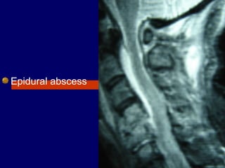
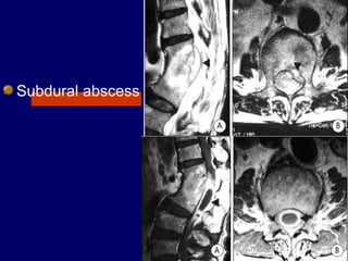
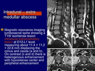
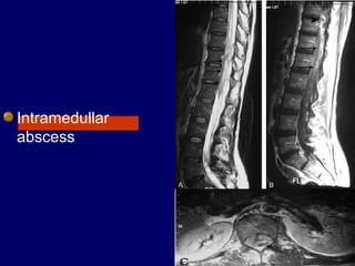
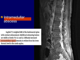
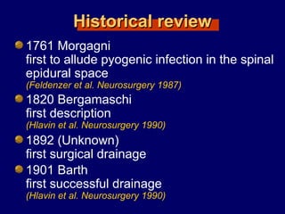
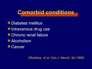
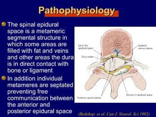
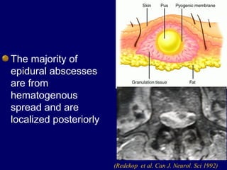
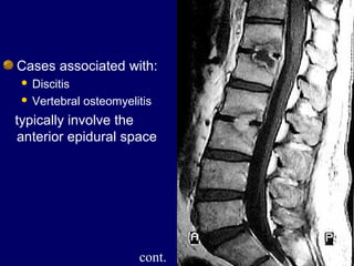
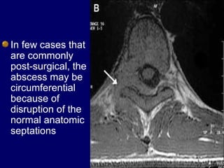
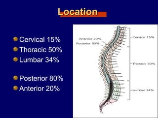
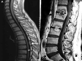
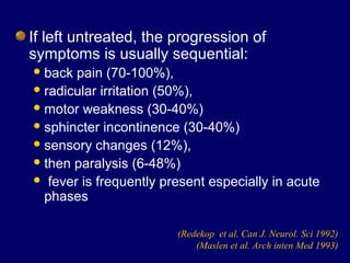
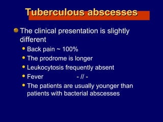
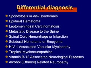
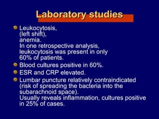
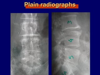
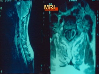
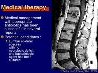
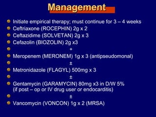
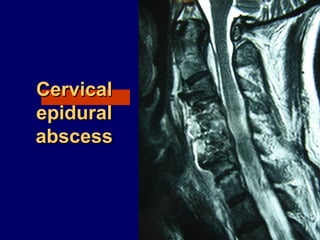
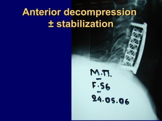
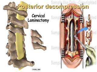
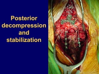
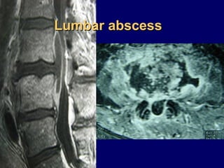
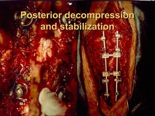
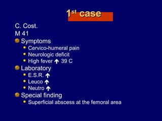
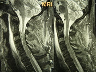
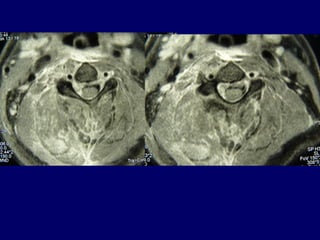
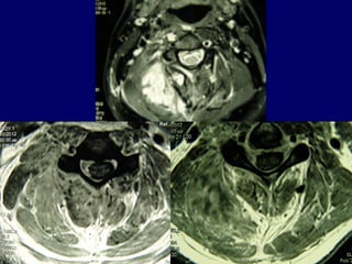
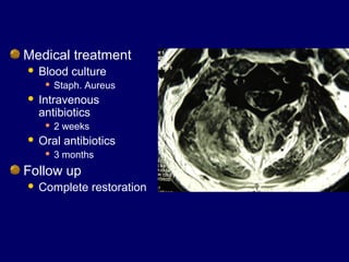
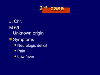
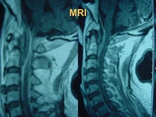
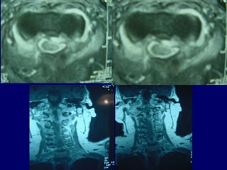
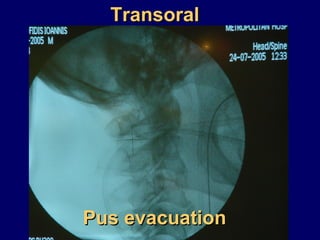
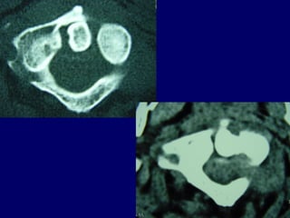
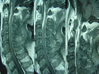
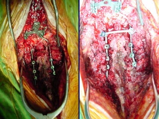
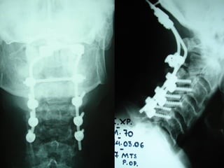
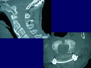
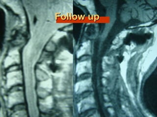
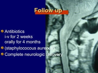
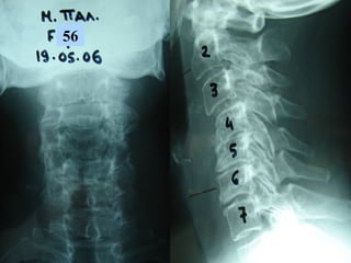
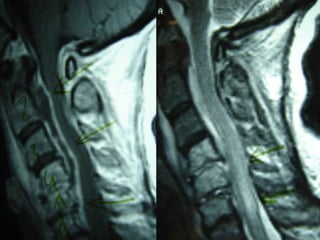
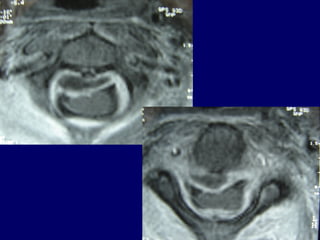
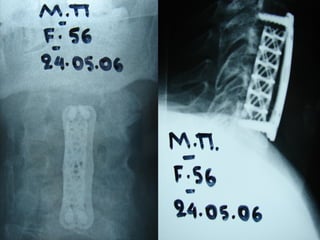
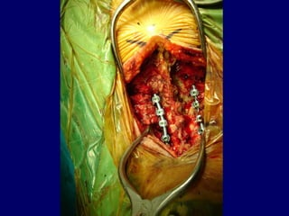
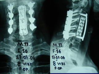
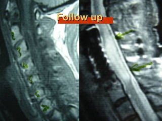
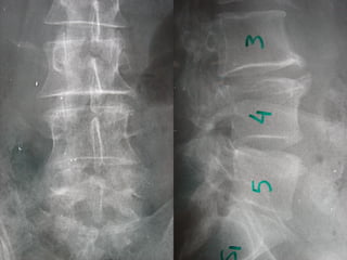
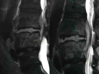
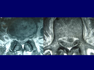
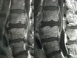
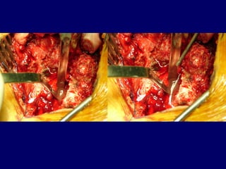
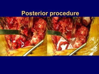
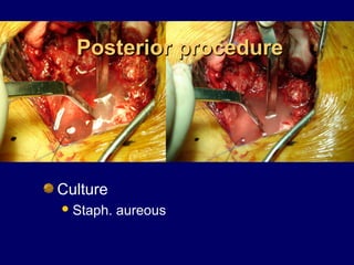
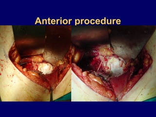
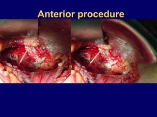
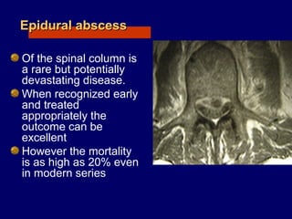
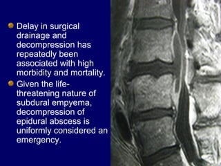
SPINAL EPIDURAL,SPINAL EPIDURAL,
ANDAND
SUBDURAL - INTRAMEDULLARSUBDURAL - INTRAMEDULLAR
ABSCESSESABSCESSES
GEORGE SAPKAS
ASC. PROFESSOR
1st Orthopaedic Department
Medical School-Athens University
Attikon Hospital
Metropolitan Hospital
Athens Greece
Epidural abscess
Subdural abscess
Intradural – extraIntradural – extra
medullar abscessmedullar abscess
Magnetic resonance imaging
lumbosacral spine showing a
T1W isointense lesion
intradural extramedullary
lesion at D12-L1 level
measuring about 11.4 × 11.2
× 22.8 mm displacing the
conus and cauda (a and b).
On contrast (c and d) there is
heterogenous enhancement
with hypointense center and
peripheral enhancement
Intramedullar
abscess
Intramedullar
abscess
Historical reviewHistorical review
1761 Morgagni
first to allude pyogenic infection in the spinal
epidural space
(Feldenzer et al. Neurosurgery 1987)
1820 Bergamaschi
first description
(Hlavin et al. Neurosurgery 1990)
1892 (Unknown)
first surgical drainage
1901 Barth
first successful drainage
(Hlavin et al. Neurosurgery 1990)
EpidemiologyEpidemiology
2 cases per 10.000
hospital admissions per year
(Hlavin et al. Neurosurgery 1990)
The peak age incidence is in the sixth
and seventh decade of life
(Danner et al. Rev infection disease 1987)
(Wheeler et al. Clin Infect, disease 1992)
Rare in the pediatric population
(Rubin et al. Pediatric infect disease 1993)
Comorbid conditionsComorbid conditions
Diabetes mellitus
Intravenous drug use
Chronic renal failure
Alcoholism
Cancer
(Redekop et al. Can J. Neurol. Sci 1992)
Source of infectionSource of infection
Skin and soft tissue 25%
Previous spinal surgery
Osteomyelitis
Spinal trauma
Urinary tracts
Respiratory tracts
(Redekop et al. Can J. Neurol. Sci 1992)
Unknown and not indentified 16% - 40%
(Hlavin et al. Neurosurgery 1990)
(Redekop et al. Can J. Neurol. Sci 1992)
Etiologic agentEtiologic agent
(Hlavin et al. Neurosurgery 1990)
(Redekop et al. Can J. Neurol. Sci 1992)
PathophysiologyPathophysiology
The spinal epidural
space is a metameric
segmental structure in
which some areas are
filled with fat and veins
and other areas the dura
is in direct contact with
bone or ligament
In addition individual
metameres are septated
preventing free
communication between
the anterior and
posterior epidural space (Redekop et al. Can J. Neurol. Sci 1992)
The majority of
epidural abscesses
are from
hematogenous
spread and are
localized posteriorly
(Redekop et al. Can J. Neurol. Sci 1992)
Cases associated with:
 Discitis
 Vertebral osteomyelitis
typically involve the
anterior epidural space
cont.
In few cases that
are commonly
post-surgical, the
abscess may be
circumferential
because of
disruption of the
normal anatomic
septations
The extend of the
abscess is usually
limited with an average
of 3 to 4 vertebral
segments
(Del Curling et al. Neurosurgery 1990)
(Hlavin et al. Neurosurgery 1990)
LocationLocation
Cervical 15%
Thoracic 50%
Lumbar 34%
Posterior 80%
Anterior 20%
The precise pathophysiologic cause of
the neurological impairment is not
known
Rapid and irreversible deterioration
prompted several authors to postulate an
ischemic mechanism either from arterial
occlusion or venous stasis
(Baker et al. N. Engl J Med 1975)
Recent studies indicate
that the progressive
neurologic deficits were
secondary to
compression
(Feldenzer et al. Neurosurgery 1987)
(Feldenzer et al. Neurosurgery 1988)
It is most likely that the
cause of neurological
deficit is multi factorial
with compression been
the major component
DiagnosisDiagnosis
Onset of symptoms usually occurs
within hours to days
but may be more chronic in nature,
presenting with weeks to months
of symptoms.
The microbiology often dictates the
pace of progression.
If left untreated, the progression of
symptoms is usually sequential:
back pain (70-100%),
radicular irritation (50%),
motor weakness (30-40%)
sphincter incontinence (30-40%)
sensory changes (12%),
then paralysis (6-48%)
 fever is frequently present especially in acute
phases
(Redekop et al. Can J. Neurol. Sci 1992)
(Maslen et al. Arch inten Med 1993)
Tuberculous abscessesTuberculous abscesses
The clinical presentation is slightly
different
Back pain ~ 100%
The prodrome is longer
Leukocytosis frequently absent
Fever - // -
The patients are usually younger than
patients with bacterial abscesses
Differential diagnosisDifferential diagnosis
Spondylosis or disk syndromes
Epidural Hematoma
Leptomeningeal Carcinomatosis
Metastatic Disease to the Spine
Spinal Cord Hemorrhage or Infarction
Subdural Hematoma or Empyema
HIV-1 Associated Vacuolar Myelopathy
Tropical Myeloneuropathies
Vitamin B-12 Associated Neurological Diseases
Alcohol (Ethanol) Related Neuropathy
Laboratory studiesLaboratory studies
Leukocytosis,
(left shift),
anemia.
In one retrospective analysis,
leukocytosis was present in only
60% of patients.
Blood cultures positive in 60%.
ESR and CRP elevated.
Lumbar puncture relatively contraindicated
(risk of spreading the bacteria into the
subarachnoid space).
Usually reveals inflammation, cultures positive
in 25% of cases.
Imaging studiesImaging studies
Plain radiographs occasionally demonstrate
osteomyelitis but are of almost no utility.
Spinal MRI is the procedure of choice
(sensitivity 95%, specificity 92%).
Gadolinium enhancement increases sensitivity
and enables better differentiation between
abscess and surrounding neurological
structures.
CT-guided needle aspiration may be used to
obtain material for analysis.
Plain radiographsPlain radiographs
MRIMRI
TreatmentTreatment
Medical therapyMedical therapy
Medical management
with appropriate
antibiotics has been
successful in several
reports
Potential candidates :
 Lumbar epidural
abscess
with no
neurologic deficit
and bacteriologic
agent has been
cultured
(Wheller et al. Clin Inf Des. 1992)
Proposed criteria for exclusive medicalProposed criteria for exclusive medical
treatment in spinal epidural abscessestreatment in spinal epidural abscesses
Poor surgical candidates because of severe
concomitant medical problems
Cases in which the abscess involves a
considerable length of spinal canal and who
have epiduritis from the cervical to the lumbar
level
Patients not suffering from severe loss of spinal
cord or cauda equina function
Patients with complete paralysis for more 3
days
The length of suggested medical
therapy is at least 8 to 12 weeks
of intravenous antibiotics
followed by oral agents
(Leys et al. Ann Neurol. 1985)
ManagementManagement
Initiate empirical therapy; must continue for 3 – 4 weeks
Ceftriaxone (ROCEPHIN) 2g x 2
Ceftazidime (SOLVETAN) 2g x 3
Cefazolin (BIOZOLIN) 2g x3
+
Meropenem (MERONEM) 1g x 3 (antipseudomonal)
±
Metronidazole (FLAGYL) 500mg x 3
±
Gentamycin (GARAMYCIN) 80mg x3 in D/W 5%
(if post – op or IV drug user or endocarditis)
±
Vancomycin (VONCON) 1g x 2 (MRSA)
Operative treatmentOperative treatment
CervicalCervical
epiduralepidural
abscessabscess
Anterior decompressionAnterior decompression
±± stabilizationstabilization
Posterior decompressionPosterior decompression
PosteriorPosterior
decompressiondecompression
andand
stabilizationstabilization
Lumbar abscessLumbar abscess
Posterior decompressionPosterior decompression
and stabilizationand stabilization
CasesCases
11stst
casecase
C. Cost.
M 41
Symptoms
 Cervico-humeral pain
 Neurologic deficit
 High fever  39 C
Laboratory
 E.S.R. 
 Leuco 
 Neutro 
Special finding
 Superficial abscess at the femoral area
MRIMRI
Medical treatment
 Blood culture
 Staph. Aureus
 Intravenous
antibiotics
 2 weeks
 Oral antibiotics
 3 months
Follow up
 Complete restoration
22ndnd
casecase
J. Chr.
M 69
Unknown origin
Symptoms
Neurologic deficit
Pain
Low fever
MRIMRI
TransoralTransoral
Pus evacuationPus evacuation
Follow upFollow up
Follow upFollow up
Antibiotics
i-v for 2 weeks
orally for 4 months
(staphylococous aureous)
Complete neurologic recovery
33rdrd
casecase
Ma. Pal.
F 56
Unknown origin
Symptoms
Neurologic deficit
Pain
Low fever
56
Follow upFollow up
Follow upFollow up
Pus culture
(staphylococous aureous)
Antibiotics
i-v for 2 mts
orally for 6 mts
Complete neurologic recovery
44thth
casecase
Diak. Nick.
M 61
possible origin
superficial skin infection at the elbow area
Symptoms
 High fever
 Back Pain
Laboratory
 E.S.R. 
 Leucocitosis 
 Neutro 
Posterior procedurePosterior procedure
Culture
Staph. aureous
Anterior procedureAnterior procedure
Follow upFollow up
Antibiotic treatment
Various schemes
 I.V. – Oral
for 6 months
Sufficient recovery
ConclusionsConclusions
Epidural abscessEpidural abscess
Of the spinal column is
a rare but potentially
devastating disease.
When recognized early
and treated
appropriately the
outcome can be
excellent
However the mortality
is as high as 20% even
in modern series
Surgical therapySurgical therapy
Decompression of the neural elements
and drainage of purulent material or
debridement of granulation tissue is
recognized as the best method to
prevent neurologic deficits and if
performed quickly after the onset of
deficits, may allow full recovery.
Delay in surgical
drainage and
decompression has
repeatedly been
associated with high
morbidity and mortality.
Given the life-
threatening nature of
subdural empyema,
decompression of
epidural abscess is
uniformly considered an
emergency.
University Hospital “ATTIKON”

SPINAL EPIDURAL, AND SUBDURAL - INTRAMEDULLAR ABSCESSES