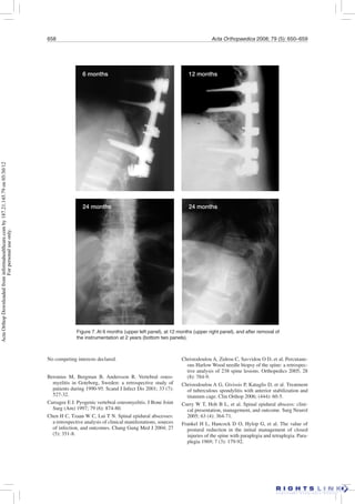
650 Acta Orthopaedica 2008; 79 (5): 650–659
Spondylodiscitis. A retrospective study of 163
patients
Efthimios J Karadimas1, Cody Bunger1, Bend Erling Lindblad2,
Ebbe Stender Hansen1, Kristian Høy1, Peter Helmig1, Anne Sofie Kannerup2, and
Bent Niedermann1
1Spinal Unit and 2Department of Orthopedics, Aarhus University Hospital, Aarhus, Denmark
Correspondence EJK: eikaradimas@yahoo.gr
Submitted 07-04-26. Accepted 08-03-02
Copyright © Taylor & Francis 2008. ISSN 1745–3674. Printed in Sweden – all rights reserved.
DOI 10.1080/17453670810016678
Background and purpose Spondylodiscitis may be a
serious disease due to diagnostic delay and inadequate
treatment. There is no consensus on when and how to
operate. We therefore retrospectively analyzed the out-
come of a large series of patients treated either nonop-
eratively or surgically.
Patients and methods Between 1992 and 2000, 163
patients (101 males) were hospitalized due to spondy-
lodiscitis. The mean age was 56 (1–83) years. The infec-
tion was located in the cervical spine in 13 patients
(8%), in the thoracic spine in 62 patients (38%), at
the thoracolumbar junction in 10 patients (6%), and
in the lumbo-sacral spine in 78 patients (48%). In 67
patients (41%), no microorganisms were detected. Most
of the other patients had Staphylococcus aureus infec-
tion (53 patients) and/or Mycobacterium tuberculosis
(22 patients). The patients were divided into 3 groups:
(A) 70 patients who had nonoperative treatment, (B) 56
patients who underwent posterior decompression alone,
and (C) 37 patients who underwent decompression and
stabilization.
Results At 12-month follow-up, nonoperative treat-
ment (A) had failed in 8/70 patients, who had subse-
quently been operated. 24/56 and 6/37 had been reoper-
ated in groups B and C, respectively. Group A patients
had no neurological symptoms. In group B, 11 had
neurological deficits and surgery was beneficial for 5 of
them; 4 remained unchanged and 2 deteriorated (1 due
to cerebral abscess). 11 patients in group C had altered
neurogical deficits, which improved in 9 of them. 20
patients had died during the 1-year follow-up, 3 in hos-
pital, directly related with infection.
Interpretation Nonoperative treatment was effective
in nine-tenths of the patients. Decompression alone had
high a reoperation rate compared to decompression and
internal stabilization.
■
Spondylodiscitis is a rare disease with a highly
variable outcome. Mild cases can be treated nonop-
eratively. Severe cases have a substantial morbidity
(back pain, deformity, and neurological deficits)
and a high mortality rate regardless of advanced
drug therapy and surgical intervention (Carragee
1997, Soehle and Wallenfang 2002).
Diabetes mellitus, malnutrition, substance abuse
(alcohol/drug users), sickle cell anemia, immu-
nosuppressive conditions (HIV, steroid users),
tumors, renal failure, and septicemia may aggra-
vate the final outcome of spondylodiscitis (Sam-
path and Rigamonti 1999, Reihsaus et al. 2000,
Soehle and Wallenfang 2002). The most common
symptoms are pain, fever, night sweats, anemia,
loss of weight, malaise, and neurological symp-
toms due to ischemia or mechanical compression
(abscess) (Reihsaus et al. 2000) Late diagnosis can
result in general multiple organ failure and epidu-
ral abscesses (Carragee 1997, Hadjipavlou et al.
2000a, Rhee and Heller 2004).
The most common infectious organism is Staph-
ylococcus aureus, followed by Streptococcus viri-
dans, Echerichia coli, Staphylococcus epidermis,
Brucella mellitensis and Proteus spp. Pseudomo-
ActaOrthopDownloadedfrominformahealthcare.comby187.21.145.79on05/30/12
Forpersonaluseonly.
Acta Orthopaedica 2008; 79 (5): 650–659 651
The location of the infection is mainly in the
lumbar spine, followed by the thoracic and the cer-
vical spine (Rhee et al. 2004). Neurocompression
is commonest in the thoracic and cervical region,
due to the close relation of the spinal canal and
dural sac (Hadjipavlou et al. 2000b).
Improved techniques for early diagnosis (MRI/
STIR sequence), appropriate antibiotic therapy,
and surgery in selected cases have resulted both in
reduced mortality and morbidity. However, there
have been few studies reporting the outcome of
treatment of spinal infection. The aim of our study
was to analyze the effectiveness of conservative
or surgical treatment in patients with spinal infec-
tion and determine the pathway of optimal surgical
treatment.
Patients and methods
163 patients (101 males) with spondylodiscitis,
treated in our hospital between 1992 and 2000,
were studied retrospectively. The mean age of
patients was 56 (1–83) years. Only 12 patients
were younger than 20 years and 60% of the patients
were older than 50 years. 143 patients were native
Danish citizens, 16 were immigrants from Africa,
and 4 were from other countries. Patients with bone
metastases or primary tumors were excluded.
Evaluation prior to admission to the spinal
unit
All patients had initially consulted either their GP
or a district hospital. For most patients, the diagno-
sis of the infection before arrival at the spinal unit
had been based on plain radiographs, often com-
bined with CT or MRI. Although ideally antibiotic
therapy should not be started until the organism
has been identified (Grados et al. 2007), 100 of the
163 patients had already received antibiotic treat-
ment for an average duration of 48 (5–194) days
before being referred to us. In most of the cases,
the spinal infection was related to infection else-
where (skin trauma, upper respiratory tract, urinary
tract, or gastrointestinal tract).
Evaluation after admission to the spinal unit
Our department is a referral center for spinal
infections, and it covers a population of almost
1.5 million people. A spinal specialist evaluated
the patients at admission. The Frankel classifica-
tion was used in order to evaluate the neurological
status (Frankel et al. 1969). The kyphotic angle and
Cobb angles were measured radiographically. All
operations were performed in our hospital.
The location of the infection was in the cervical
spine in 13 patients (8%), the thoracic spine in 62
patients (38%), at the thoracolumbar junction in 10
patients (6%) and in the lumbo-sacral spine in 78
patients (48%). Plain radiographs with MRI and
CT revealed 355 infected levels. In 15 cases, the
infection was localized at a single level, in 117 it
was at 2 levels, in 18 it was at 3 levels, in 6 it was at
multiple levels. 7 cases were multiple-focus (Table
1). 54% of the patients with cervical spine infec-
tion and 65% of those with thoracic spine infection
were operated on.
Concomitant disease was common: diabetes
mellitus in 16 patients, chronic obstructive lung
disease in 14 patients, cancer in 12 patients, drug
addiction in 20 patients, and other diseases (poor
nutrition, chronic steroid use, alcohol addiction,
lupus erythematosus, rheumatoid arthritis, anky-
losing spondylitis, immunosuppressive conditions,
HIV infection) in 33 patients. 26 patients had 2 or
more concomitant diseases. 9 cases were classified
Table 1. Level distribution in 163 patients with spinal infection
1 2 3 Multiple Multiple Total
Location level levels levels levels foci
Cervical 4 6 – 2 1 13
Thoracic 7 40 9 4 2 62
Thorac-lumbar – 5 1 – 4 10
Lumbo-sacral 4 66 8 78
Total 15 117 18 6 7 163
nas aeruginosa, Candida albicans,
and viral and fungal organisms (e.g.
Aspergillus fumigatus) can cause
spinal infections in immunosup-
pressed patients and intravenous
drug users (Carragee 1997, Rhee
and Heller 2004). In isolated cases,
Serratia marcescens and Echino-
coccus spp. can also be detected
(Kaufman et al. 1980, Hadjipavlou
et al. 2004).
ActaOrthopDownloadedfrominformahealthcare.comby187.21.145.79on05/30/12
Forpersonaluseonly.
652 Acta Orthopaedica 2008; 79 (5): 650–659
as iatrogenic infections; 4 of these were related to
epidural catheters/spinal punctures and 5 occurred
postoperatively, 2 of them following discectomy.
A CT-guided bone biopsy (Christodoulou et al.
2005, Hadjipavlou et al. 2004) was performed in
31 patients but the culture was positive for micro-
organisms in only 16 patients, perhaps because of
previous use of antibiotics. In all patients who were
operated, bone samples for histology and culture
were taken during surgery. Overall, bacteria were
isolated in 74 patients and tuberculosis was found
in 22 other patients (Table 2).
Nonoperative treatment (group A; 70 patients)
(Figures 1 and 2)
Criteria for nonoperative treatment were: no neu-
rology, no clinical manifestation of unstable spine,
no kyphosis, and good heath status. In 15 patients,
the infection was located at one level. The patients
received intravenous antibiotic therapy for 4 weeks
followed by oral antibiotics until clinical and labo-
ratory findings had normalized (usually for 1–6
months).
The choice of the antibiotic was based on the
results of culture. The patients with negative bac-
teriological findings followed a protocol consisting
of dicloxacillin sodium and gentamycin (initially
intravenously) for at least 1 month.
The patients with tuberculosis were given iso-
niazid 300 mg × 1 per os, rifampicin 450 mg × 1
per os, ethambutol 1,200 mg × 1 per os, and pyra-
zinamid 2,000 mg × 1 per os for 1 year.
All patients treated conservatively used a cast
brace for mean 13 (11–17) weeks in order to sup-
port the infected spine and to prevent deformity.
Surgical treatment (93 patients) (Figures 3–7)
The indications for surgical treatment were: exten-
sive bone destruction (kyphosis of > 15 degrees),
the presence of an epidural abscess, neurological
deterioration, and failure of nonoperative treatment
(persistent pain and fever, and persistent abnormal
Table 2. Microorganisms identified
Microorganism Number Percentage
of patients
None 67 41
Staphylococcus aureus 53 33
Escherichia coli 5 3
Pseudomonas aeruginosa 3 2
Mycobacterium tuberculosis 22 13
Coagulation-negative
staphylococci 3 2
Candida albicans 1 0.5
Salmonella sp. 1 0.5
A Streptococcus 3 2
Pneumococcus 2 1
Hemophilus aphrophilus 1 0.5
Clostridium perfringens 2 1
Figure 1. MRI image (left) and a CT scan (right) from a patient treated nonoperatively.
ActaOrthopDownloadedfrominformahealthcare.comby187.21.145.79on05/30/12
Forpersonaluseonly.
Acta Orthopaedica 2008; 79 (5): 650–659 653
C-reactive protein and erythrocyte sedimentation
rate). None of the patients was enrolled for two
stage operation.
In group B (56 patients) (Figures 3 and 4), a pos-
terior decompression was performed. The infection
was located at 2 levels in 45 cases. 1 patient died in
hospital and 6 died during follow-up. The location
of the infection was lumbo-sacral in 32 patients
and thoracic in 21 patients.
The extension of the decompression depended
on the extent of the destruction/abscess. In mul-
tiple-level infections, bilateral extensive laminec-
tomies were preferred. Wide decompression was
used for extensive single-level infections in order
to allow a clear view and complete removal of the
foci of infection.
In group C (37 patients) (Figures 5–7), the
decompression was followed by stabilization. The
location was thoracic in 17 patients, lumbo-sacral
in 11, and cervical in 5. In 29 cases, the infection
was located at 2 levels. 1 patient died in hospital
and 4 died during the follow-up.
Results
The patients were reviewed on a regular basis at 3,
6, and 12 months. 20 patients died during the first
year: 3 in hospital from complications related to the
spinal infection, 8 within 6 months, and 9 within 1
year. The latter 15 patients died from causes not
directly related to the infection.
One-quarter (38/163) of the patients required
further operative treatment during the first year
(Table 3). A reoperation was performed in one-
third (30/93) of the patients who underwent sur-
gery. 8/70 patients in group A required reoperation
to control infection and stabilize the spine. All had
normal neurology. 24/56 group B patients were
reoperated with debridement and stabilization. In
contrast, only 6/37 patients in group C were reop-
erated.
During treatment, in the patients without com-
plications the markers of infection (WBC, CRP,
and ESR) gradually normalized earlier in group A
than in groups B and C.
Figure 2. Pretreatment radiograph (left) and CT-guided biopsy (right) from a
patient treated nonoperatively.
A posterior approach for both
decompression and stabiliza-
tion was used in 31 patients. 4
patients underwent an anterior
decompression and stabilization
and 2 patients were decompressed
through both an anterior and a
posterior approach, followed by
circumferential stabilization.
Pus was drained and the infected
bone masses were debrided, fol-
lowed by decompression and
bone grafts were used in all cases
to increase the percentage of
fusion.
We did use cages in the primary
treatment; these were used solely
in cases of revision surgery.
In 33 cases, a posterior instru-
mentation with posterolateral
bone graft was used and in 4 an
anterior Kaneda stabilization
system was used. A postoperative
brace was used in 17 cases.
ActaOrthopDownloadedfrominformahealthcare.comby187.21.145.79on05/30/12
Forpersonaluseonly.
654 Acta Orthopaedica 2008; 79 (5): 650–659
Neurological status (Table 4)
All 70 patients in groupA had normal neurology. In
group B, 33/44 patients had normal neurology and
11 had altered neurology (incomplete data in 12
patients) after the decompression which was ben-
eficial in only 5 cases. In group C, 20/31 patients
had normal neurology and 11 had neurological
impairment (incomplete data in 6 patients). Their
status improved postoperatively in all but 2 cases.
Complications
3 patients died during hospitalization due to mul-
tiorgan failure. Other complications during hospi-
talization were: pulmonary embolism (2 patients)
atelectasis (2), postoperative hematoma (2), paral-
ysis of the diaphragm (1), cardiological problems
(4), and cerebral thrombosis (1). In addition, 3
patients had residual psoas abscess, 2 had pancre-
atic abscess, 1 had cerebellar abscess (altered neu-
rology), and 3 had pulmonary infection. Figure 4. MRI from a patient treated surgically.
Figure 3. Three-month, 6-month
and 12-month radiographs from
a patient treated nonoperatively.
3 months 6 months 6 months
12 months 12 months
ActaOrthopDownloadedfrominformahealthcare.comby187.21.145.79on05/30/12
Forpersonaluseonly.
Acta Orthopaedica 2008; 79 (5): 650–659 655
After the 1-year study period, 15 patients were
re-admitted to hospital and had further surgery
related to their spinal infection. Of the 4 patients
in group A, 3 underwent posterior stabilization due
to progressive kyphosis and 1 patient underwent
posterior decompression due to spinal stenosis. Of
the 7 patients in group B, 4 underwent posterior
stabilization due to progressive kyphosis, 2 under-
went posterior decompression due to spinal steno-
sis, and 1 underwent anterior stabilization with a
cage due to progressive kyphosis. Of the 4 patients
in group C, 1 underwent revision of the metalwork
due to hardware failure; in 1 patient the posterior
fusion was converted to a circumferential (360°)
fusion; 1 patient had vertebral traumatic fracture at
a level adjacent to the instrumentation, which was
treated with an extension of the stabilization; and 1
patient developed a reinfection at an adjacent level,
requiring an extension of the decompression and
the posterior instrumentation.
Discussion
In our series, spinal infections were more often
located in the lumbar spine, neurological compli-
cations were commoner in the thoracic and cervi-
cal spine, many patients were old and had other
diseases or drug addiction, the infection was com-
moner in men, and Staphylococcus aureus was the
most commonly identified microorganism, thus
confirming previous findings (Carragee 1997, Had-
jipavlou et al. 2000b, Reihsaus et al. 2000, Chen et
al. 2004, Hadjipavlou et al. 2004, Rhee and Heller
2004, Curry et al. 2005, Lohr et al. 2005, Pereira
and Lynch 2005).
Table 3. Revision of the initial treatment
Location, No. Further operative
treatment treatment
Cervical 13 patients
Group A nonop. 7 2
Group B op. 1 –
Group C op. 5 2
Thoracic 62 patients
Group A nonop. 24 2
Group B op. 21 10
Group C op. 17 3
Thoraco-lumbar 10 patients
Group A nonop. 4 1
Group B op. 2 –
Group C op. 4 –
Lumbo-sacral 78 patients
Group A nonop. 35 3
Group B op. 32 13
Group C op. 11 2
Nonoperative
Group A 70 8
Operative
Group B 56 24
Group C 37 6
Total 163 38
A: nonoperative treatment;
B: treatment with posterior decompression only;
C: treatment with anterior debridement and posterior or
anterior internal stabilization
Table 4. Breakdown of neurological status
Nonoperative group A
No neurological symptoms 70
Further operation (all Frankel E) 8
Operative group
Incomplete information 12
Deaths 6
Altered neurology 22
cervical 2
thoracic 12
thoraco-lumbar 1
lumbo-sacral 7
Group B (incomplete data in 12 patients)
Classified as Frankel E 33
Altered neurology (Frankel’s classification) 11
improved by 2 grades
A to C 1
improved by 1 grade
C to D 1
D to E 3
unchanged conditions
A 1
D 3
deterioration
E to C (due to cerebral abscess) 1
E to D 1
Overall improvement: 46%
Group C (incomplete data in 6 patients)
Classified as Frankel E 20
Altered neurology (Frankel’s classification) 11
improved by 2 grades
A to C 1
improved by 1 grade
B to C 1
C to D 1
D to E 2
unchanged conditions
A 1
D 1
Radicular pain a 4
Overall improvement: 82%
a all with complete recovery after the operation
ActaOrthopDownloadedfrominformahealthcare.comby187.21.145.79on05/30/12
Forpersonaluseonly.
656 Acta Orthopaedica 2008; 79 (5): 650–659
The incidence of staphylococcal infections in
our series (33%) is similar to that in a previous
study (34%) (Beronius et al. 2001), but is lower
than in most of the previously mentioned studies.
We believe that this is related to the fact that in
our series, only 47% of bone biopsies were posi-
tive for microorganisms—probably because many
of the patients had been on antibiotic therapy prior
to referral to our unit.
The second most common microorganism in our
study was Mycobacterium tuberculosis (22/163
patients). Although this finding contrasts with the
results of a recent literature review (Reihsaus et al.
2000), other studies have shown that tuberculosis
is still causing spinal infections (predominately
thoracic) despite extensive global vaccination pro-
grams (Turgut 2001, Curry et al. 2005, Pereira and
Lynch 2005, Christodoulou et al. 2006).
Of the 163 patients, 38 (23.3%) required further
treatment, which is less than a recently reported
figure of 48% (Curry et al. 2005). It is interesting
that none of these patients was younger than 20
years of age.
In cases with mild clinical symptoms and
absence of neurological impairment (group A),
nonoperative management was adequate to control
the infection, as reported by other authors (Reih-
saus et al. 2000, Rhee and Heller 2004, Nene and
Bhojraj 2005). We reoperated 8/70 patients in this
group, a rate in-between what has been reported in
other series (Curry et al. 2005, Nene and Bhojraj
2005). In retrospect, we miscalculated the severity
Figure 5. Preoperatively, with evidence of kyphosis. Postoperatively during hospitalization, at
6 months, and B at 12 months
Preoperative Postoperative
6 months 12 months
ActaOrthopDownloadedfrominformahealthcare.comby187.21.145.79on05/30/12
Forpersonaluseonly.
Acta Orthopaedica 2008; 79 (5): 650–659 657
of the infection in 7 cases, which should have been
stabilized from the beginning.
Of the 93 patients who were operated, 30
required revision surgery. This was mainly related
to the fact that single posterior decompression,
although sufficient in order to control the infection,
did not prevent progression of deformity. A similar
reoperation rate was presented in a paper by Lohr
et al. (2005) involving 27 patients.
Posterior laminectomy with the purpose of
decompressing the spinal canal or wide decom-
pression may destabilize the spine. In our series,
decompression alone failed in nearly half of the
patients and for that reason we abandoned it as a
stand-alone operation. However, for isolated small
abscesses or in cases with high co-morbidity, per-
cutaneous techniques and transpendicular discec-
tomy are promising for the control of infection and
vertebral destruction (Hadjipavlou et al. 2004).
In 37 patients with vertebral collapse and exten-
sive abscess formation, we combined decompres-
sion with anterior debridement and reconstruction
with structural bone graft (from ribs or iliac crest)
and posterior or anterior stabilization. The use of
implants carries risks, but it supports the instru-
mentation and accelerates bone fusion (Carragee
1997, Curry et al. 2005, Lohr et al. 2005). We
found no complications with this technique.
Early diagnosis was related to better outcome,
specifically in patients with neurological deficits.
Figure 6. Preoperative and postoperative radiographs taken during hospitalization.
Our policy was to offer patients with deteriorat-
ing neurology emergency surgery within 24 h of
admission. The overall neurological outcome of
our study was similar to that reported in a review
paper (Reihsaus et al. 2000), and it is also com-
parable with other published series (Hadjipavlou
et al. 2000b, Lohr et al. 2005). Other studies have
shown better results (Schinkel et al. 2003, Pereira
and Lynch 2005).
In our series, 20 patients (12%) died during the
1-year follow-up, which is similar to what has
been reported in many other studies (Carragee
1997, Reihsaus et al. 2000, Tang et al. 2002). Other
authors have reported lower or negligible mortality
rates (Schinkel et al. 2003, Lohr et al. 2005, Pereira
and Lynch 2005).
Contributions of authors
CB, BEL, ESH, KH, PH, and BN were the surgeons who
recruited the patients, decided on the treatment, and per-
formed the operations. PH also retrieved the images from the
X-ray files. ASK and BEL prepared the initial database. EJK
updated it and analyzed the data; he also wrote the manu-
script. CB and KH revised the manuscript.
Mr George Drosos and Mrs Linda Marie Nygaard made the
appropriate correction as linguistic personnel.
ActaOrthopDownloadedfrominformahealthcare.comby187.21.145.79on05/30/12
Forpersonaluseonly.
658 Acta Orthopaedica 2008; 79 (5): 650–659
Christodoulou A, Zidrou C, Savvidou O D, et al. Percutane-
ous Harlow Wood needle biopsy of the spine: a retrospec-
tive analysis of 238 spine lesions. Orthopedics 2005; 28
(8): 784-9.
Christodoulou A G, Givissis P, Kataglis D, et al. Treatment
of tuberculous spondylitis with anterior stabilization and
titanium cage. Clin Orthop 2006; (444): 60-5.
Curry W T, Hoh B L, et al. Spinal epidural abscess: clini-
cal presentation, management, and outcome. Surg Neurol
2005; 63 (4): 364-71.
Frankel H L, Hancock D O, Hylop G, et al. The value of
postural reduction in the initial management of closed
injuries of the spine with paraplegia and tetraplegia. Para-
plegia 1969; 7 (3): 179-92.
Figure 7. At 6 months (upper left panel), at 12 months (upper right panel), and after removal of
the instrumentation at 2 years (bottom two panels).
6 months 12 months
24 months 24 months
No competing interests declared.
Beronius M, Bergman B, Andersson R. Vertebral osteo-
myelitis in Goteborg, Sweden: a retrospective study of
patients during 1990-95. Scand J Infect Dis 2001; 33 (7):
527-32.
Carragee E J. Pyogenic vertebral osteomyelitis. J Bone Joint
Surg (Am) 1997; 79 (6): 874-80.
Chen H C, Tzaan W C, Lui T N. Spinal epidural abscesses:
a retrospective analysis of clinical manifestations, sources
of infection, and outcomes. Chang Gung Med J 2004; 27
(5): 351-8.
ActaOrthopDownloadedfrominformahealthcare.comby187.21.145.79on05/30/12
Forpersonaluseonly.
Acta Orthopaedica 2008; 79 (5): 650–659 659
Grados F, Lescure F X, Senneville E, et al. Suggestions for
managing pyogenic (non tuberculous) discitis in adults.
Joint Bone Spine 2007; 74: 133-9.
Hadjipavlou A G, Bergquist S C, Chen J W, et al. Vertebral
Osteomyelitis. Current Treatment Options in Infect Dis
2000a; 2: 226-37.
Hadjipavlou A G, Mader J T, Necessary J T, et al. Hematog-
enous pyogenic spinal infections and their surgical man-
agement. Spine 2000b 1; 25 (13): 1668-79.
Hadjipavlou A G, Katonis P K, Gaitanis I N, et al. Percu-
taneous transpedicular discectomy and drainage in pyo-
genic spondylodiscitis. Eur Spine J 2004; 13 (8): 707-13.
Kaufman D M, Kaplan J G, Litman N. Infectious agents in
spinal epidural abscesses. Neurology 1980; 30: 844-50.
Lohr M, Reithmeier T, et al. Spinal epidural abscess: prog-
nostic factors and comparison of different surgical treat-
ment strategies. Acta Neurochir (Wien) 2005; 147 (2):
159-66.
Nene A, Bhojraj S. Results of nonsurgical treatment of tho-
racic spinal tuberculosis in adults. Spine J 2005; 5 (1):
79-84.
Pereira C E, Lynch J C. Spinal epidural abscess: an analysis
of 24 cases. Surg Neurol (Suppl 1) 2005; 63: S26-9.
Reihsaus E, Waldbaur H, Seeling W. Spinal epidural abscess,
a meta analysis of 915 patients. Neurosurg Rev 2000; 23:
175-204.
Rhee J M, Heller J G. Spinal infections. In: The adult and
pediatric spine 3rd edition (eds. J W Frymoyer and S W
Wiesel), Lippincott Williams and Wilkins, 2004, chapter
10: 165-89.
Sampath P, Rigamonti D. Spinal epidural abscess, a review
of the epidemiology, diagnosis and treatment. J Spinal
Disord Tech 1999; 12: 185-92.
Schinkel C, Gotward M, Adress H J. Surgical treatment of
spondylodiscitis. Surg Infect (Larchmt) 2003; 4 (4): 387-
91.
Soehle M, Wallenfang T. Spinal epidural abscesses, clinical
manifestation, prognostic factors and outcomes. Neuro-
surgery 2002; 51 (1): 79-85.
Tang H J, Lin H J, Liu Y C. Spinal epidural abscess-experi-
ence with 46 patients and evaluation of prognostic factors.
J Infect 2002; 45 (2): 76-81.
Turgut M. Spinal tuberculosis (Pott’s disease): its clini-
cal presentation, surgical management, and outcome. A
survey study on 694 patients. Neurosurg Rev 2001; 24
(1): 8-13.
ActaOrthopDownloadedfrominformahealthcare.comby187.21.145.79on05/30/12
Forpersonaluseonly.

Espondilodiscite[1]

  • 1.
    650 Acta Orthopaedica2008; 79 (5): 650–659 Spondylodiscitis. A retrospective study of 163 patients Efthimios J Karadimas1, Cody Bunger1, Bend Erling Lindblad2, Ebbe Stender Hansen1, Kristian Høy1, Peter Helmig1, Anne Sofie Kannerup2, and Bent Niedermann1 1Spinal Unit and 2Department of Orthopedics, Aarhus University Hospital, Aarhus, Denmark Correspondence EJK: eikaradimas@yahoo.gr Submitted 07-04-26. Accepted 08-03-02 Copyright © Taylor & Francis 2008. ISSN 1745–3674. Printed in Sweden – all rights reserved. DOI 10.1080/17453670810016678 Background and purpose Spondylodiscitis may be a serious disease due to diagnostic delay and inadequate treatment. There is no consensus on when and how to operate. We therefore retrospectively analyzed the out- come of a large series of patients treated either nonop- eratively or surgically. Patients and methods Between 1992 and 2000, 163 patients (101 males) were hospitalized due to spondy- lodiscitis. The mean age was 56 (1–83) years. The infec- tion was located in the cervical spine in 13 patients (8%), in the thoracic spine in 62 patients (38%), at the thoracolumbar junction in 10 patients (6%), and in the lumbo-sacral spine in 78 patients (48%). In 67 patients (41%), no microorganisms were detected. Most of the other patients had Staphylococcus aureus infec- tion (53 patients) and/or Mycobacterium tuberculosis (22 patients). The patients were divided into 3 groups: (A) 70 patients who had nonoperative treatment, (B) 56 patients who underwent posterior decompression alone, and (C) 37 patients who underwent decompression and stabilization. Results At 12-month follow-up, nonoperative treat- ment (A) had failed in 8/70 patients, who had subse- quently been operated. 24/56 and 6/37 had been reoper- ated in groups B and C, respectively. Group A patients had no neurological symptoms. In group B, 11 had neurological deficits and surgery was beneficial for 5 of them; 4 remained unchanged and 2 deteriorated (1 due to cerebral abscess). 11 patients in group C had altered neurogical deficits, which improved in 9 of them. 20 patients had died during the 1-year follow-up, 3 in hos- pital, directly related with infection. Interpretation Nonoperative treatment was effective in nine-tenths of the patients. Decompression alone had high a reoperation rate compared to decompression and internal stabilization. ■ Spondylodiscitis is a rare disease with a highly variable outcome. Mild cases can be treated nonop- eratively. Severe cases have a substantial morbidity (back pain, deformity, and neurological deficits) and a high mortality rate regardless of advanced drug therapy and surgical intervention (Carragee 1997, Soehle and Wallenfang 2002). Diabetes mellitus, malnutrition, substance abuse (alcohol/drug users), sickle cell anemia, immu- nosuppressive conditions (HIV, steroid users), tumors, renal failure, and septicemia may aggra- vate the final outcome of spondylodiscitis (Sam- path and Rigamonti 1999, Reihsaus et al. 2000, Soehle and Wallenfang 2002). The most common symptoms are pain, fever, night sweats, anemia, loss of weight, malaise, and neurological symp- toms due to ischemia or mechanical compression (abscess) (Reihsaus et al. 2000) Late diagnosis can result in general multiple organ failure and epidu- ral abscesses (Carragee 1997, Hadjipavlou et al. 2000a, Rhee and Heller 2004). The most common infectious organism is Staph- ylococcus aureus, followed by Streptococcus viri- dans, Echerichia coli, Staphylococcus epidermis, Brucella mellitensis and Proteus spp. Pseudomo- ActaOrthopDownloadedfrominformahealthcare.comby187.21.145.79on05/30/12 Forpersonaluseonly.
  • 2.
    Acta Orthopaedica 2008;79 (5): 650–659 651 The location of the infection is mainly in the lumbar spine, followed by the thoracic and the cer- vical spine (Rhee et al. 2004). Neurocompression is commonest in the thoracic and cervical region, due to the close relation of the spinal canal and dural sac (Hadjipavlou et al. 2000b). Improved techniques for early diagnosis (MRI/ STIR sequence), appropriate antibiotic therapy, and surgery in selected cases have resulted both in reduced mortality and morbidity. However, there have been few studies reporting the outcome of treatment of spinal infection. The aim of our study was to analyze the effectiveness of conservative or surgical treatment in patients with spinal infec- tion and determine the pathway of optimal surgical treatment. Patients and methods 163 patients (101 males) with spondylodiscitis, treated in our hospital between 1992 and 2000, were studied retrospectively. The mean age of patients was 56 (1–83) years. Only 12 patients were younger than 20 years and 60% of the patients were older than 50 years. 143 patients were native Danish citizens, 16 were immigrants from Africa, and 4 were from other countries. Patients with bone metastases or primary tumors were excluded. Evaluation prior to admission to the spinal unit All patients had initially consulted either their GP or a district hospital. For most patients, the diagno- sis of the infection before arrival at the spinal unit had been based on plain radiographs, often com- bined with CT or MRI. Although ideally antibiotic therapy should not be started until the organism has been identified (Grados et al. 2007), 100 of the 163 patients had already received antibiotic treat- ment for an average duration of 48 (5–194) days before being referred to us. In most of the cases, the spinal infection was related to infection else- where (skin trauma, upper respiratory tract, urinary tract, or gastrointestinal tract). Evaluation after admission to the spinal unit Our department is a referral center for spinal infections, and it covers a population of almost 1.5 million people. A spinal specialist evaluated the patients at admission. The Frankel classifica- tion was used in order to evaluate the neurological status (Frankel et al. 1969). The kyphotic angle and Cobb angles were measured radiographically. All operations were performed in our hospital. The location of the infection was in the cervical spine in 13 patients (8%), the thoracic spine in 62 patients (38%), at the thoracolumbar junction in 10 patients (6%) and in the lumbo-sacral spine in 78 patients (48%). Plain radiographs with MRI and CT revealed 355 infected levels. In 15 cases, the infection was localized at a single level, in 117 it was at 2 levels, in 18 it was at 3 levels, in 6 it was at multiple levels. 7 cases were multiple-focus (Table 1). 54% of the patients with cervical spine infec- tion and 65% of those with thoracic spine infection were operated on. Concomitant disease was common: diabetes mellitus in 16 patients, chronic obstructive lung disease in 14 patients, cancer in 12 patients, drug addiction in 20 patients, and other diseases (poor nutrition, chronic steroid use, alcohol addiction, lupus erythematosus, rheumatoid arthritis, anky- losing spondylitis, immunosuppressive conditions, HIV infection) in 33 patients. 26 patients had 2 or more concomitant diseases. 9 cases were classified Table 1. Level distribution in 163 patients with spinal infection 1 2 3 Multiple Multiple Total Location level levels levels levels foci Cervical 4 6 – 2 1 13 Thoracic 7 40 9 4 2 62 Thorac-lumbar – 5 1 – 4 10 Lumbo-sacral 4 66 8 78 Total 15 117 18 6 7 163 nas aeruginosa, Candida albicans, and viral and fungal organisms (e.g. Aspergillus fumigatus) can cause spinal infections in immunosup- pressed patients and intravenous drug users (Carragee 1997, Rhee and Heller 2004). In isolated cases, Serratia marcescens and Echino- coccus spp. can also be detected (Kaufman et al. 1980, Hadjipavlou et al. 2004). ActaOrthopDownloadedfrominformahealthcare.comby187.21.145.79on05/30/12 Forpersonaluseonly.
  • 3.
    652 Acta Orthopaedica2008; 79 (5): 650–659 as iatrogenic infections; 4 of these were related to epidural catheters/spinal punctures and 5 occurred postoperatively, 2 of them following discectomy. A CT-guided bone biopsy (Christodoulou et al. 2005, Hadjipavlou et al. 2004) was performed in 31 patients but the culture was positive for micro- organisms in only 16 patients, perhaps because of previous use of antibiotics. In all patients who were operated, bone samples for histology and culture were taken during surgery. Overall, bacteria were isolated in 74 patients and tuberculosis was found in 22 other patients (Table 2). Nonoperative treatment (group A; 70 patients) (Figures 1 and 2) Criteria for nonoperative treatment were: no neu- rology, no clinical manifestation of unstable spine, no kyphosis, and good heath status. In 15 patients, the infection was located at one level. The patients received intravenous antibiotic therapy for 4 weeks followed by oral antibiotics until clinical and labo- ratory findings had normalized (usually for 1–6 months). The choice of the antibiotic was based on the results of culture. The patients with negative bac- teriological findings followed a protocol consisting of dicloxacillin sodium and gentamycin (initially intravenously) for at least 1 month. The patients with tuberculosis were given iso- niazid 300 mg × 1 per os, rifampicin 450 mg × 1 per os, ethambutol 1,200 mg × 1 per os, and pyra- zinamid 2,000 mg × 1 per os for 1 year. All patients treated conservatively used a cast brace for mean 13 (11–17) weeks in order to sup- port the infected spine and to prevent deformity. Surgical treatment (93 patients) (Figures 3–7) The indications for surgical treatment were: exten- sive bone destruction (kyphosis of > 15 degrees), the presence of an epidural abscess, neurological deterioration, and failure of nonoperative treatment (persistent pain and fever, and persistent abnormal Table 2. Microorganisms identified Microorganism Number Percentage of patients None 67 41 Staphylococcus aureus 53 33 Escherichia coli 5 3 Pseudomonas aeruginosa 3 2 Mycobacterium tuberculosis 22 13 Coagulation-negative staphylococci 3 2 Candida albicans 1 0.5 Salmonella sp. 1 0.5 A Streptococcus 3 2 Pneumococcus 2 1 Hemophilus aphrophilus 1 0.5 Clostridium perfringens 2 1 Figure 1. MRI image (left) and a CT scan (right) from a patient treated nonoperatively. ActaOrthopDownloadedfrominformahealthcare.comby187.21.145.79on05/30/12 Forpersonaluseonly.
  • 4.
    Acta Orthopaedica 2008;79 (5): 650–659 653 C-reactive protein and erythrocyte sedimentation rate). None of the patients was enrolled for two stage operation. In group B (56 patients) (Figures 3 and 4), a pos- terior decompression was performed. The infection was located at 2 levels in 45 cases. 1 patient died in hospital and 6 died during follow-up. The location of the infection was lumbo-sacral in 32 patients and thoracic in 21 patients. The extension of the decompression depended on the extent of the destruction/abscess. In mul- tiple-level infections, bilateral extensive laminec- tomies were preferred. Wide decompression was used for extensive single-level infections in order to allow a clear view and complete removal of the foci of infection. In group C (37 patients) (Figures 5–7), the decompression was followed by stabilization. The location was thoracic in 17 patients, lumbo-sacral in 11, and cervical in 5. In 29 cases, the infection was located at 2 levels. 1 patient died in hospital and 4 died during the follow-up. Results The patients were reviewed on a regular basis at 3, 6, and 12 months. 20 patients died during the first year: 3 in hospital from complications related to the spinal infection, 8 within 6 months, and 9 within 1 year. The latter 15 patients died from causes not directly related to the infection. One-quarter (38/163) of the patients required further operative treatment during the first year (Table 3). A reoperation was performed in one- third (30/93) of the patients who underwent sur- gery. 8/70 patients in group A required reoperation to control infection and stabilize the spine. All had normal neurology. 24/56 group B patients were reoperated with debridement and stabilization. In contrast, only 6/37 patients in group C were reop- erated. During treatment, in the patients without com- plications the markers of infection (WBC, CRP, and ESR) gradually normalized earlier in group A than in groups B and C. Figure 2. Pretreatment radiograph (left) and CT-guided biopsy (right) from a patient treated nonoperatively. A posterior approach for both decompression and stabiliza- tion was used in 31 patients. 4 patients underwent an anterior decompression and stabilization and 2 patients were decompressed through both an anterior and a posterior approach, followed by circumferential stabilization. Pus was drained and the infected bone masses were debrided, fol- lowed by decompression and bone grafts were used in all cases to increase the percentage of fusion. We did use cages in the primary treatment; these were used solely in cases of revision surgery. In 33 cases, a posterior instru- mentation with posterolateral bone graft was used and in 4 an anterior Kaneda stabilization system was used. A postoperative brace was used in 17 cases. ActaOrthopDownloadedfrominformahealthcare.comby187.21.145.79on05/30/12 Forpersonaluseonly.
  • 5.
    654 Acta Orthopaedica2008; 79 (5): 650–659 Neurological status (Table 4) All 70 patients in groupA had normal neurology. In group B, 33/44 patients had normal neurology and 11 had altered neurology (incomplete data in 12 patients) after the decompression which was ben- eficial in only 5 cases. In group C, 20/31 patients had normal neurology and 11 had neurological impairment (incomplete data in 6 patients). Their status improved postoperatively in all but 2 cases. Complications 3 patients died during hospitalization due to mul- tiorgan failure. Other complications during hospi- talization were: pulmonary embolism (2 patients) atelectasis (2), postoperative hematoma (2), paral- ysis of the diaphragm (1), cardiological problems (4), and cerebral thrombosis (1). In addition, 3 patients had residual psoas abscess, 2 had pancre- atic abscess, 1 had cerebellar abscess (altered neu- rology), and 3 had pulmonary infection. Figure 4. MRI from a patient treated surgically. Figure 3. Three-month, 6-month and 12-month radiographs from a patient treated nonoperatively. 3 months 6 months 6 months 12 months 12 months ActaOrthopDownloadedfrominformahealthcare.comby187.21.145.79on05/30/12 Forpersonaluseonly.
  • 6.
    Acta Orthopaedica 2008;79 (5): 650–659 655 After the 1-year study period, 15 patients were re-admitted to hospital and had further surgery related to their spinal infection. Of the 4 patients in group A, 3 underwent posterior stabilization due to progressive kyphosis and 1 patient underwent posterior decompression due to spinal stenosis. Of the 7 patients in group B, 4 underwent posterior stabilization due to progressive kyphosis, 2 under- went posterior decompression due to spinal steno- sis, and 1 underwent anterior stabilization with a cage due to progressive kyphosis. Of the 4 patients in group C, 1 underwent revision of the metalwork due to hardware failure; in 1 patient the posterior fusion was converted to a circumferential (360°) fusion; 1 patient had vertebral traumatic fracture at a level adjacent to the instrumentation, which was treated with an extension of the stabilization; and 1 patient developed a reinfection at an adjacent level, requiring an extension of the decompression and the posterior instrumentation. Discussion In our series, spinal infections were more often located in the lumbar spine, neurological compli- cations were commoner in the thoracic and cervi- cal spine, many patients were old and had other diseases or drug addiction, the infection was com- moner in men, and Staphylococcus aureus was the most commonly identified microorganism, thus confirming previous findings (Carragee 1997, Had- jipavlou et al. 2000b, Reihsaus et al. 2000, Chen et al. 2004, Hadjipavlou et al. 2004, Rhee and Heller 2004, Curry et al. 2005, Lohr et al. 2005, Pereira and Lynch 2005). Table 3. Revision of the initial treatment Location, No. Further operative treatment treatment Cervical 13 patients Group A nonop. 7 2 Group B op. 1 – Group C op. 5 2 Thoracic 62 patients Group A nonop. 24 2 Group B op. 21 10 Group C op. 17 3 Thoraco-lumbar 10 patients Group A nonop. 4 1 Group B op. 2 – Group C op. 4 – Lumbo-sacral 78 patients Group A nonop. 35 3 Group B op. 32 13 Group C op. 11 2 Nonoperative Group A 70 8 Operative Group B 56 24 Group C 37 6 Total 163 38 A: nonoperative treatment; B: treatment with posterior decompression only; C: treatment with anterior debridement and posterior or anterior internal stabilization Table 4. Breakdown of neurological status Nonoperative group A No neurological symptoms 70 Further operation (all Frankel E) 8 Operative group Incomplete information 12 Deaths 6 Altered neurology 22 cervical 2 thoracic 12 thoraco-lumbar 1 lumbo-sacral 7 Group B (incomplete data in 12 patients) Classified as Frankel E 33 Altered neurology (Frankel’s classification) 11 improved by 2 grades A to C 1 improved by 1 grade C to D 1 D to E 3 unchanged conditions A 1 D 3 deterioration E to C (due to cerebral abscess) 1 E to D 1 Overall improvement: 46% Group C (incomplete data in 6 patients) Classified as Frankel E 20 Altered neurology (Frankel’s classification) 11 improved by 2 grades A to C 1 improved by 1 grade B to C 1 C to D 1 D to E 2 unchanged conditions A 1 D 1 Radicular pain a 4 Overall improvement: 82% a all with complete recovery after the operation ActaOrthopDownloadedfrominformahealthcare.comby187.21.145.79on05/30/12 Forpersonaluseonly.
  • 7.
    656 Acta Orthopaedica2008; 79 (5): 650–659 The incidence of staphylococcal infections in our series (33%) is similar to that in a previous study (34%) (Beronius et al. 2001), but is lower than in most of the previously mentioned studies. We believe that this is related to the fact that in our series, only 47% of bone biopsies were posi- tive for microorganisms—probably because many of the patients had been on antibiotic therapy prior to referral to our unit. The second most common microorganism in our study was Mycobacterium tuberculosis (22/163 patients). Although this finding contrasts with the results of a recent literature review (Reihsaus et al. 2000), other studies have shown that tuberculosis is still causing spinal infections (predominately thoracic) despite extensive global vaccination pro- grams (Turgut 2001, Curry et al. 2005, Pereira and Lynch 2005, Christodoulou et al. 2006). Of the 163 patients, 38 (23.3%) required further treatment, which is less than a recently reported figure of 48% (Curry et al. 2005). It is interesting that none of these patients was younger than 20 years of age. In cases with mild clinical symptoms and absence of neurological impairment (group A), nonoperative management was adequate to control the infection, as reported by other authors (Reih- saus et al. 2000, Rhee and Heller 2004, Nene and Bhojraj 2005). We reoperated 8/70 patients in this group, a rate in-between what has been reported in other series (Curry et al. 2005, Nene and Bhojraj 2005). In retrospect, we miscalculated the severity Figure 5. Preoperatively, with evidence of kyphosis. Postoperatively during hospitalization, at 6 months, and B at 12 months Preoperative Postoperative 6 months 12 months ActaOrthopDownloadedfrominformahealthcare.comby187.21.145.79on05/30/12 Forpersonaluseonly.
  • 8.
    Acta Orthopaedica 2008;79 (5): 650–659 657 of the infection in 7 cases, which should have been stabilized from the beginning. Of the 93 patients who were operated, 30 required revision surgery. This was mainly related to the fact that single posterior decompression, although sufficient in order to control the infection, did not prevent progression of deformity. A similar reoperation rate was presented in a paper by Lohr et al. (2005) involving 27 patients. Posterior laminectomy with the purpose of decompressing the spinal canal or wide decom- pression may destabilize the spine. In our series, decompression alone failed in nearly half of the patients and for that reason we abandoned it as a stand-alone operation. However, for isolated small abscesses or in cases with high co-morbidity, per- cutaneous techniques and transpendicular discec- tomy are promising for the control of infection and vertebral destruction (Hadjipavlou et al. 2004). In 37 patients with vertebral collapse and exten- sive abscess formation, we combined decompres- sion with anterior debridement and reconstruction with structural bone graft (from ribs or iliac crest) and posterior or anterior stabilization. The use of implants carries risks, but it supports the instru- mentation and accelerates bone fusion (Carragee 1997, Curry et al. 2005, Lohr et al. 2005). We found no complications with this technique. Early diagnosis was related to better outcome, specifically in patients with neurological deficits. Figure 6. Preoperative and postoperative radiographs taken during hospitalization. Our policy was to offer patients with deteriorat- ing neurology emergency surgery within 24 h of admission. The overall neurological outcome of our study was similar to that reported in a review paper (Reihsaus et al. 2000), and it is also com- parable with other published series (Hadjipavlou et al. 2000b, Lohr et al. 2005). Other studies have shown better results (Schinkel et al. 2003, Pereira and Lynch 2005). In our series, 20 patients (12%) died during the 1-year follow-up, which is similar to what has been reported in many other studies (Carragee 1997, Reihsaus et al. 2000, Tang et al. 2002). Other authors have reported lower or negligible mortality rates (Schinkel et al. 2003, Lohr et al. 2005, Pereira and Lynch 2005). Contributions of authors CB, BEL, ESH, KH, PH, and BN were the surgeons who recruited the patients, decided on the treatment, and per- formed the operations. PH also retrieved the images from the X-ray files. ASK and BEL prepared the initial database. EJK updated it and analyzed the data; he also wrote the manu- script. CB and KH revised the manuscript. Mr George Drosos and Mrs Linda Marie Nygaard made the appropriate correction as linguistic personnel. ActaOrthopDownloadedfrominformahealthcare.comby187.21.145.79on05/30/12 Forpersonaluseonly.
  • 9.
    658 Acta Orthopaedica2008; 79 (5): 650–659 Christodoulou A, Zidrou C, Savvidou O D, et al. Percutane- ous Harlow Wood needle biopsy of the spine: a retrospec- tive analysis of 238 spine lesions. Orthopedics 2005; 28 (8): 784-9. Christodoulou A G, Givissis P, Kataglis D, et al. Treatment of tuberculous spondylitis with anterior stabilization and titanium cage. Clin Orthop 2006; (444): 60-5. Curry W T, Hoh B L, et al. Spinal epidural abscess: clini- cal presentation, management, and outcome. Surg Neurol 2005; 63 (4): 364-71. Frankel H L, Hancock D O, Hylop G, et al. The value of postural reduction in the initial management of closed injuries of the spine with paraplegia and tetraplegia. Para- plegia 1969; 7 (3): 179-92. Figure 7. At 6 months (upper left panel), at 12 months (upper right panel), and after removal of the instrumentation at 2 years (bottom two panels). 6 months 12 months 24 months 24 months No competing interests declared. Beronius M, Bergman B, Andersson R. Vertebral osteo- myelitis in Goteborg, Sweden: a retrospective study of patients during 1990-95. Scand J Infect Dis 2001; 33 (7): 527-32. Carragee E J. Pyogenic vertebral osteomyelitis. J Bone Joint Surg (Am) 1997; 79 (6): 874-80. Chen H C, Tzaan W C, Lui T N. Spinal epidural abscesses: a retrospective analysis of clinical manifestations, sources of infection, and outcomes. Chang Gung Med J 2004; 27 (5): 351-8. ActaOrthopDownloadedfrominformahealthcare.comby187.21.145.79on05/30/12 Forpersonaluseonly.
  • 10.
    Acta Orthopaedica 2008;79 (5): 650–659 659 Grados F, Lescure F X, Senneville E, et al. Suggestions for managing pyogenic (non tuberculous) discitis in adults. Joint Bone Spine 2007; 74: 133-9. Hadjipavlou A G, Bergquist S C, Chen J W, et al. Vertebral Osteomyelitis. Current Treatment Options in Infect Dis 2000a; 2: 226-37. Hadjipavlou A G, Mader J T, Necessary J T, et al. Hematog- enous pyogenic spinal infections and their surgical man- agement. Spine 2000b 1; 25 (13): 1668-79. Hadjipavlou A G, Katonis P K, Gaitanis I N, et al. Percu- taneous transpedicular discectomy and drainage in pyo- genic spondylodiscitis. Eur Spine J 2004; 13 (8): 707-13. Kaufman D M, Kaplan J G, Litman N. Infectious agents in spinal epidural abscesses. Neurology 1980; 30: 844-50. Lohr M, Reithmeier T, et al. Spinal epidural abscess: prog- nostic factors and comparison of different surgical treat- ment strategies. Acta Neurochir (Wien) 2005; 147 (2): 159-66. Nene A, Bhojraj S. Results of nonsurgical treatment of tho- racic spinal tuberculosis in adults. Spine J 2005; 5 (1): 79-84. Pereira C E, Lynch J C. Spinal epidural abscess: an analysis of 24 cases. Surg Neurol (Suppl 1) 2005; 63: S26-9. Reihsaus E, Waldbaur H, Seeling W. Spinal epidural abscess, a meta analysis of 915 patients. Neurosurg Rev 2000; 23: 175-204. Rhee J M, Heller J G. Spinal infections. In: The adult and pediatric spine 3rd edition (eds. J W Frymoyer and S W Wiesel), Lippincott Williams and Wilkins, 2004, chapter 10: 165-89. Sampath P, Rigamonti D. Spinal epidural abscess, a review of the epidemiology, diagnosis and treatment. J Spinal Disord Tech 1999; 12: 185-92. Schinkel C, Gotward M, Adress H J. Surgical treatment of spondylodiscitis. Surg Infect (Larchmt) 2003; 4 (4): 387- 91. Soehle M, Wallenfang T. Spinal epidural abscesses, clinical manifestation, prognostic factors and outcomes. Neuro- surgery 2002; 51 (1): 79-85. Tang H J, Lin H J, Liu Y C. Spinal epidural abscess-experi- ence with 46 patients and evaluation of prognostic factors. J Infect 2002; 45 (2): 76-81. Turgut M. Spinal tuberculosis (Pott’s disease): its clini- cal presentation, surgical management, and outcome. A survey study on 694 patients. Neurosurg Rev 2001; 24 (1): 8-13. ActaOrthopDownloadedfrominformahealthcare.comby187.21.145.79on05/30/12 Forpersonaluseonly.