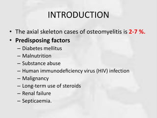
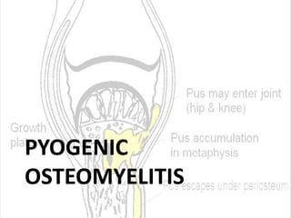
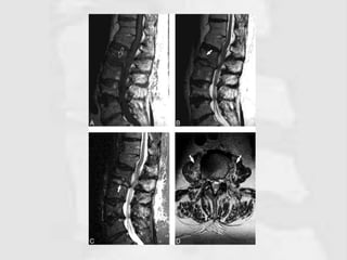
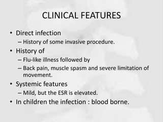
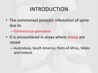
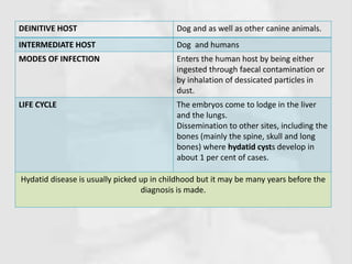
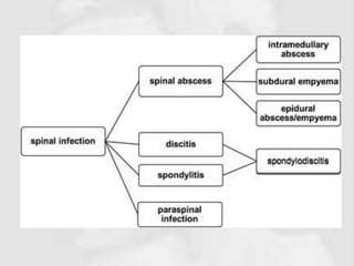
OTHER SPINE
INFECTIONS
NUR FARRA NAJWA
BINTI ABDUL AZIM
082015100035
LEARNING OBJECTIVES
By the end of seminar, students should be able to
explain
• Definition
• Etiology
• Risk factor
• Clinical feature
• Management
1. Pyogenic
osteomyelitis
2. Discitis
3. Fungal spine infection
4. Parasitic infection
INTRODUCTION
• The axial skeleton cases of osteomyelitis is 2-7 %.
• Predisposing factors
– Diabetes mellitus
– Malnutrition
– Substance abuse
– Human immunodeficiency virus (HIV) infection
– Malignancy
– Long-term use of steroids
– Renal failure
– Septicaemia.
PYOGENIC
OSTEOMYELITIS
INTRODUCTION
• Acute pyogenic infection of the spine is
uncommon
• Predisposing factor
– The elderly
– Chronically ill and immuno-deficient patients
PATHOLOGY
• Staphylococcus aureus (50–60%)
• Immunosuppressed patients
– Gram negative organisms
• E. coli and pseudomonas
• Sources of infection are
– Haematogenous spread from a distant focus of
infection
– Inoculation during invasive procedures
Infection begins in the
vertebral endplates
1) Secondary
spread to the
disc and
adjacent
vertebra
2) Spread
along the
anterior
longitudinal
ligament to
an adjacent
vertebra
3) Outwards
into the
paravertebral
soft tissues
Cont.
• Spinal canal is rarely involved
– Once infected, form an epidural abscess
(surgical emergency!)
• Even rapid surgical decompression,
– some degree of permanent paralysis present
From the thoracic spine along the
psoas to the groin
From the lumbar region to the
buttock, the sacroiliac joint or the
hip.
3) Outwards into the paravertebral soft tissues
SPINAL DECOMPRESSION SURGERY
• A general term that refers to various
procedures intended to relieve symptoms
caused by pressure, or compression, on the
spinal cord and/or nerve roots, ex,
– a corpectomy,
– a diskectomy,
– a laminotomy,
– a foraminotomy,
– or osteophyte removal.
CLINICAL FEATURES
• History
– Invasive spinal procedure
– A distant infection during the preceding few
weeks
• A careful history and general examination to
exclude a focus of infection
– Skin, ENT, chest, pelvis.
Cont.Localized
pain (the
cardinal
symptom)
Intense
Unremitting
Associated with
muscle spasm and
restricted
movement
Point tenderness
over the affected
vertebra.
Intercostal
neuralgia is a
frequent
symptom with
thoracic spine
involvement.
Systemic sign
pyrexia and
tachycardia.
IN CHILDREN
The diagnosis is
difficult
Awkward gait with a
stiff spine
If lumbar spine is
involved they can
present with
abdominal symptoms
and signs
IMAGING
• X-rays
– No change for several weeks;
– Delayed diagnosis, the examination should be repeated.
• Early signs are
– Loss of disc height
– Irregularity of the disc space
– Erosion of the vertebral end-plate
– Reactive new bone formation
– Soft-tissue swelling may be visible
• The early loss of disc height distinguishes vertebral
osteomyelitis from metastatic disease, where the disc
can remain intact despite advanced bony destruction.
https://www.orthobullets.com/spine/2025/adult-pyogenic-vertebral-osteomyelitis
https://www.nejm.org/doi/full/10.1056/NEJMcp0910753
Cont.
• Radionuclide scanning will show increased
activity at the site but this is non-specific.
• MRI may show characteristic changes in the
– Vertebral end-plates, intervertebral disc and
paravertebral tissues;
– This investigation is highly sensitive but not specific.
– Similar features may be seen in discitis.
• Needle biopsy may help with diagnosis, but often
no organism is found.
Cont.
• Other investigations
– The white cell count
– C-reactive protein (CRP) level
– Erythrocyte sedimentation rate (ESR)
– Anti-staphylococcal antibodies
– Agglutination tests for salmonella and brucella
(endemic regions).
– Blood culture is essential in patients who are
febrile though it is often negative in the early
stages of infection.
TREATMENT
• If the blood culture is negative a closed needle biopsy is performed
• Treatment is started
– Basis of a clinical diagnosis of infection
– Includes bed rest, pain relief and intravenous antibiotic administration
• The duration of antibiotic treatment depends on the
– Clinical, haematological and radiological findings.
• Intravenous antibiotics
– For 4–6 weeks;
– If there is a good response
– Oral antibiotics are then used for a further 6– 8 weeks and the patient
is mobilized in a spinal brace.
• As methicillin-resistant staphylococcus aureus (MRSA)
– Vancomycin or linezolid may be required.
• During this period nutritional support and management of co-
morbidities
OPERATIVE TREATMENT
• Seldom needed
• The indications for an open biopsy and decompression are
– Failure to obtain a positive yield from a closed needle biopsy
and a poor response to conservative treatment
– The presence of neurological signs
– The need to drain a soft-tissue abscess
• An anterior approach is preferred
– Necrotic and infected material is removed
– If necessary, the cord is decompressed
– The anterior column defect is reconstructed with rib or iliac
grafts
• For a primary epidural abscess, laminectomy is indicated.
https://www.aans.org/Patients/Neurosurgical-Conditions-and-Treatments/Spinal-Infections
Cont.
• In the elderly and in immuno-compromised
patients
– posterolateral extraplueral /retroperitoneal
decompression and instrumentation is effective.
• Unstable spine
– posterior fixation may be necessary
• Postoperatively
– spine is supported in a brace until healing occurs.
• Spontaneous fusion of infected vertebrae is a
common radiological feature of healed
staphylococcal osteomyelitis.
DISCITIS
INTRODUCTION
• Infection limited to the intervertebral disc is
rare
• Usually due to direct inoculation
– Following discography
– Chemo-nucleolysis
– Discectomy
• The vertebral end-plates are rapidly attacked
and the infection then spreads into the
vertebral body
https://en.wikipedia.org/wiki/Discitis
CLINICAL FEATURES
• Direct infection
– History of some invasive procedure.
• History of
– Flu-like illness followed by
– Back pain, muscle spasm and severe limitation of
movement.
• Systemic features
– Mild, but the ESR is elevated.
• In children the infection : blood borne.
INVESTIGATIONS
• X-rays, radioscintigraphy and MRI show the
same features as in pyogenic spondylitis.
RADIOGRAPHIC FEATURES
PLAIN RADIOGRAPH CT
• Insensitive to the early changes of
diskitis/osteomyelitis,
• Normal appearances being maintained
for up to 2-4 weeks.
• CT findings are similar to plain film
• More sensitive to earlier changes.
• Thereafter disc space narrowing and
irregularity or ill definition of the
vertebral endplates can be seen.
• Additionally, surrounding soft tissue
swelling, intervertebral disc
enhancement with contrast, collections
• In untreated cases, bony sclerosis may
begin to appear in 10-12 weeks.
TREATMENT
PREVENTION IS ALWAYS BETTER THAN CURE
• After an injection into the disc
– Broad-spectrum antibiotic should be administered
intravenously.
• Non-iatrogenic discitis
– Self-limiting
• Acute stage
– Bed rest and analgesics are essential
• If symptoms do not resolve rapidly
– Needle biopsy is advisable
• Only if there are signs of abscess formation or cord or
nerve root pressure
– Surgical evacuation or decompression indicated.
FUNGAL INFECTION
INTRODUCTION
• These are opportunistic infections occurring in an
immuno-compromised host however, may also
affect a normal host
• Air-borne fungi that initially affect the lungs
– Ex: Aspergillosis and Cryptococcus
– Spine is involved by haematogenous spread.
• In children with chronic granulomatous disease,
– Thoracic spine involvement is due to contiguous
spread from the lungs.
CLINICAL PRESENTATION, CLINICAL
FINDINGS AND RADIOGRAPHIC FEATURES
• May mimic those of TB.
• The chest x-ray may show
– A fungal ball or pneumonia.
• The diagnosis by
– Sputum examination and bronchoscopy.
• The immuno-diffusion test is specific for
Aspergillosis and the latex agglutination test for
Cryptococcus.
• A biopsy is performed to confirm the diagnosis.
http://www.ijoonline.com/article.asp?issn=0019-
5413;year=2012;volume=46;issue=2;spage=246;epage=250;aulast=Sethi
https://www.researchgate.net/figure/CT-scan-and-chest-X-ray-showing-
pulmonary-invasive-aspergillosis-associated-with-spinal_fig12_227992990
TREATMENT
• Neurological deficit is an indication for
operative decompression.
• Specific treatment includes
– 5-flucytosine and Amphotericin B
– Synthetic oral antifungals.
• Concurrent treatment of the underlying
immuno-comprised state is essential
PARASITIC INFESTATION
INTRODUCTION
• The commonest parasitic infestation of spine
due to
– Echinococcus granulosis
• It is encountered in areas where sheep are
raised
– Australasia, South America, Parts of Africa, Wales
and Iceland.
DEINITIVE HOST Dog and as well as other canine animals.
INTERMEDIATE HOST Dog and humans
MODES OF INFECTION Enters the human host by being either
ingested through faecal contamination or
by inhalation of dessicated particles in
dust.
LIFE CYCLE The embryos come to lodge in the liver
and the lungs.
Dissemination to other sites, including the
bones (mainly the spine, skull and long
bones) where hydatid cysts develop in
about 1 per cent of cases.
Hydatid disease is usually picked up in childhood but it may be many years before the
diagnosis is made.
CLINICAL PRESENTATION AND
CLINICAL FEATURES
• Similar to those of other forms of spondylitis.
• X-rays may
– Reveal a translucent area with a sclerotic margin in
the affected vertebral body
• In untreated cases
– Lead to bone destruction
• Significant morbidity and mortality due to
– Neurological deficit
– The difficulty in eradicating the disease
– The tendency to recurrence make for
http://www.jotr.in/article.asp?issn=0975-
7341;year=2017;volume=9;issue=2;spage=134;epage=138;aulast=Jain
Figure 1: Anteroposterior (a)
and lateral (b) lower dorsal
spine radiographic views of the
patient showing paraspinal
fusiform swelling from D6 to
D11, along with the right
hemicollapse of D9 and
reduced D9–D10 intervertebral
disc space
Figure 2: Sagittal T2 (a) and T1 (b) with coronal T2 (c) and transverse (d and e) magnetic
resonance images dorsolumbar spine of the patient showing two oval cysts measuring
10 cm × 9 cm × 5 cm contained multiple round grape bunch-like daughter cysts, in the
right pre- and para-vertebral region along with erosion of the right half of D8 and D9.
The cysts are hypointense in T1 and hyperintense in T2 sequences
http://www.jotr.in/article.asp?issn=0975-
7341;year=2017;volume=9;issue=2;spage=134;epage=138;aulast=Jain
Figure 3: Coronal (a) and transverse (b and c) computer tomographic views of patient
showing the normal liver and chest with long two well-circumscribed hypoattenuating
cystic lesions in the posterior mediastinum with multiple daughter cysts with erosion of
D9 vertebra http://www.jotr.in/article.asp?issn=0975-
7341;year=2017;volume=9;issue=2;spage=134;epage=138;aulast=Jain
Cont.
• Systemic treatment
– Albendazole
– Which is active against the larvae and the cysts
– Three cycles of 25 days each is the usual
recommendation.
• Operative treatment to achieve spinal
decompression may be called for
– Spillage of cyst contents must be avoided.
SUMMARY
Definition
Etiology
Risk factor
Clinical feature
Management
 Pyogenic
osteomyelitis
 Discitis
 Fungal spine infection
 Parasitic infection
REFERENCES
• https://www.aans.org/Pa
tients/Neurosurgical-
Conditions-and-
Treatments/Spinal-
Infections
• https://radiopaedia.org/
articles/spondylodiscitis?
lang=us
Other spine infections

Other spine infections

  • 1.
    OTHER SPINE INFECTIONS NUR FARRANAJWA BINTI ABDUL AZIM 082015100035
  • 2.
    LEARNING OBJECTIVES By theend of seminar, students should be able to explain • Definition • Etiology • Risk factor • Clinical feature • Management 1. Pyogenic osteomyelitis 2. Discitis 3. Fungal spine infection 4. Parasitic infection
  • 3.
    INTRODUCTION • The axialskeleton cases of osteomyelitis is 2-7 %. • Predisposing factors – Diabetes mellitus – Malnutrition – Substance abuse – Human immunodeficiency virus (HIV) infection – Malignancy – Long-term use of steroids – Renal failure – Septicaemia.
  • 4.
  • 5.
    INTRODUCTION • Acute pyogenicinfection of the spine is uncommon • Predisposing factor – The elderly – Chronically ill and immuno-deficient patients
  • 6.
    PATHOLOGY • Staphylococcus aureus(50–60%) • Immunosuppressed patients – Gram negative organisms • E. coli and pseudomonas • Sources of infection are – Haematogenous spread from a distant focus of infection – Inoculation during invasive procedures
  • 7.
    Infection begins inthe vertebral endplates 1) Secondary spread to the disc and adjacent vertebra 2) Spread along the anterior longitudinal ligament to an adjacent vertebra 3) Outwards into the paravertebral soft tissues
  • 9.
    Cont. • Spinal canalis rarely involved – Once infected, form an epidural abscess (surgical emergency!) • Even rapid surgical decompression, – some degree of permanent paralysis present From the thoracic spine along the psoas to the groin From the lumbar region to the buttock, the sacroiliac joint or the hip. 3) Outwards into the paravertebral soft tissues
  • 11.
    SPINAL DECOMPRESSION SURGERY •A general term that refers to various procedures intended to relieve symptoms caused by pressure, or compression, on the spinal cord and/or nerve roots, ex, – a corpectomy, – a diskectomy, – a laminotomy, – a foraminotomy, – or osteophyte removal.
  • 12.
    CLINICAL FEATURES • History –Invasive spinal procedure – A distant infection during the preceding few weeks • A careful history and general examination to exclude a focus of infection – Skin, ENT, chest, pelvis.
  • 13.
    Cont.Localized pain (the cardinal symptom) Intense Unremitting Associated with musclespasm and restricted movement Point tenderness over the affected vertebra. Intercostal neuralgia is a frequent symptom with thoracic spine involvement. Systemic sign pyrexia and tachycardia.
  • 14.
    IN CHILDREN The diagnosisis difficult Awkward gait with a stiff spine If lumbar spine is involved they can present with abdominal symptoms and signs
  • 15.
    IMAGING • X-rays – Nochange for several weeks; – Delayed diagnosis, the examination should be repeated. • Early signs are – Loss of disc height – Irregularity of the disc space – Erosion of the vertebral end-plate – Reactive new bone formation – Soft-tissue swelling may be visible • The early loss of disc height distinguishes vertebral osteomyelitis from metastatic disease, where the disc can remain intact despite advanced bony destruction.
  • 16.
  • 17.
  • 18.
    Cont. • Radionuclide scanningwill show increased activity at the site but this is non-specific. • MRI may show characteristic changes in the – Vertebral end-plates, intervertebral disc and paravertebral tissues; – This investigation is highly sensitive but not specific. – Similar features may be seen in discitis. • Needle biopsy may help with diagnosis, but often no organism is found.
  • 19.
    Cont. • Other investigations –The white cell count – C-reactive protein (CRP) level – Erythrocyte sedimentation rate (ESR) – Anti-staphylococcal antibodies – Agglutination tests for salmonella and brucella (endemic regions). – Blood culture is essential in patients who are febrile though it is often negative in the early stages of infection.
  • 20.
    TREATMENT • If theblood culture is negative a closed needle biopsy is performed • Treatment is started – Basis of a clinical diagnosis of infection – Includes bed rest, pain relief and intravenous antibiotic administration • The duration of antibiotic treatment depends on the – Clinical, haematological and radiological findings. • Intravenous antibiotics – For 4–6 weeks; – If there is a good response – Oral antibiotics are then used for a further 6– 8 weeks and the patient is mobilized in a spinal brace. • As methicillin-resistant staphylococcus aureus (MRSA) – Vancomycin or linezolid may be required. • During this period nutritional support and management of co- morbidities
  • 21.
    OPERATIVE TREATMENT • Seldomneeded • The indications for an open biopsy and decompression are – Failure to obtain a positive yield from a closed needle biopsy and a poor response to conservative treatment – The presence of neurological signs – The need to drain a soft-tissue abscess • An anterior approach is preferred – Necrotic and infected material is removed – If necessary, the cord is decompressed – The anterior column defect is reconstructed with rib or iliac grafts • For a primary epidural abscess, laminectomy is indicated.
  • 22.
  • 23.
    Cont. • In theelderly and in immuno-compromised patients – posterolateral extraplueral /retroperitoneal decompression and instrumentation is effective. • Unstable spine – posterior fixation may be necessary • Postoperatively – spine is supported in a brace until healing occurs. • Spontaneous fusion of infected vertebrae is a common radiological feature of healed staphylococcal osteomyelitis.
  • 24.
  • 25.
    INTRODUCTION • Infection limitedto the intervertebral disc is rare • Usually due to direct inoculation – Following discography – Chemo-nucleolysis – Discectomy • The vertebral end-plates are rapidly attacked and the infection then spreads into the vertebral body
  • 27.
  • 28.
    CLINICAL FEATURES • Directinfection – History of some invasive procedure. • History of – Flu-like illness followed by – Back pain, muscle spasm and severe limitation of movement. • Systemic features – Mild, but the ESR is elevated. • In children the infection : blood borne.
  • 29.
    INVESTIGATIONS • X-rays, radioscintigraphyand MRI show the same features as in pyogenic spondylitis.
  • 30.
    RADIOGRAPHIC FEATURES PLAIN RADIOGRAPHCT • Insensitive to the early changes of diskitis/osteomyelitis, • Normal appearances being maintained for up to 2-4 weeks. • CT findings are similar to plain film • More sensitive to earlier changes. • Thereafter disc space narrowing and irregularity or ill definition of the vertebral endplates can be seen. • Additionally, surrounding soft tissue swelling, intervertebral disc enhancement with contrast, collections • In untreated cases, bony sclerosis may begin to appear in 10-12 weeks.
  • 31.
    TREATMENT PREVENTION IS ALWAYSBETTER THAN CURE • After an injection into the disc – Broad-spectrum antibiotic should be administered intravenously. • Non-iatrogenic discitis – Self-limiting • Acute stage – Bed rest and analgesics are essential • If symptoms do not resolve rapidly – Needle biopsy is advisable • Only if there are signs of abscess formation or cord or nerve root pressure – Surgical evacuation or decompression indicated.
  • 32.
  • 33.
    INTRODUCTION • These areopportunistic infections occurring in an immuno-compromised host however, may also affect a normal host • Air-borne fungi that initially affect the lungs – Ex: Aspergillosis and Cryptococcus – Spine is involved by haematogenous spread. • In children with chronic granulomatous disease, – Thoracic spine involvement is due to contiguous spread from the lungs.
  • 34.
    CLINICAL PRESENTATION, CLINICAL FINDINGSAND RADIOGRAPHIC FEATURES • May mimic those of TB. • The chest x-ray may show – A fungal ball or pneumonia. • The diagnosis by – Sputum examination and bronchoscopy. • The immuno-diffusion test is specific for Aspergillosis and the latex agglutination test for Cryptococcus. • A biopsy is performed to confirm the diagnosis.
  • 35.
  • 36.
  • 37.
    TREATMENT • Neurological deficitis an indication for operative decompression. • Specific treatment includes – 5-flucytosine and Amphotericin B – Synthetic oral antifungals. • Concurrent treatment of the underlying immuno-comprised state is essential
  • 38.
  • 39.
    INTRODUCTION • The commonestparasitic infestation of spine due to – Echinococcus granulosis • It is encountered in areas where sheep are raised – Australasia, South America, Parts of Africa, Wales and Iceland.
  • 40.
    DEINITIVE HOST Dogand as well as other canine animals. INTERMEDIATE HOST Dog and humans MODES OF INFECTION Enters the human host by being either ingested through faecal contamination or by inhalation of dessicated particles in dust. LIFE CYCLE The embryos come to lodge in the liver and the lungs. Dissemination to other sites, including the bones (mainly the spine, skull and long bones) where hydatid cysts develop in about 1 per cent of cases. Hydatid disease is usually picked up in childhood but it may be many years before the diagnosis is made.
  • 41.
    CLINICAL PRESENTATION AND CLINICALFEATURES • Similar to those of other forms of spondylitis. • X-rays may – Reveal a translucent area with a sclerotic margin in the affected vertebral body • In untreated cases – Lead to bone destruction • Significant morbidity and mortality due to – Neurological deficit – The difficulty in eradicating the disease – The tendency to recurrence make for
  • 42.
    http://www.jotr.in/article.asp?issn=0975- 7341;year=2017;volume=9;issue=2;spage=134;epage=138;aulast=Jain Figure 1: Anteroposterior(a) and lateral (b) lower dorsal spine radiographic views of the patient showing paraspinal fusiform swelling from D6 to D11, along with the right hemicollapse of D9 and reduced D9–D10 intervertebral disc space
  • 43.
    Figure 2: SagittalT2 (a) and T1 (b) with coronal T2 (c) and transverse (d and e) magnetic resonance images dorsolumbar spine of the patient showing two oval cysts measuring 10 cm × 9 cm × 5 cm contained multiple round grape bunch-like daughter cysts, in the right pre- and para-vertebral region along with erosion of the right half of D8 and D9. The cysts are hypointense in T1 and hyperintense in T2 sequences http://www.jotr.in/article.asp?issn=0975- 7341;year=2017;volume=9;issue=2;spage=134;epage=138;aulast=Jain
  • 44.
    Figure 3: Coronal(a) and transverse (b and c) computer tomographic views of patient showing the normal liver and chest with long two well-circumscribed hypoattenuating cystic lesions in the posterior mediastinum with multiple daughter cysts with erosion of D9 vertebra http://www.jotr.in/article.asp?issn=0975- 7341;year=2017;volume=9;issue=2;spage=134;epage=138;aulast=Jain
  • 46.
    Cont. • Systemic treatment –Albendazole – Which is active against the larvae and the cysts – Three cycles of 25 days each is the usual recommendation. • Operative treatment to achieve spinal decompression may be called for – Spillage of cyst contents must be avoided.
  • 47.
    SUMMARY Definition Etiology Risk factor Clinical feature Management Pyogenic osteomyelitis  Discitis  Fungal spine infection  Parasitic infection
  • 49.