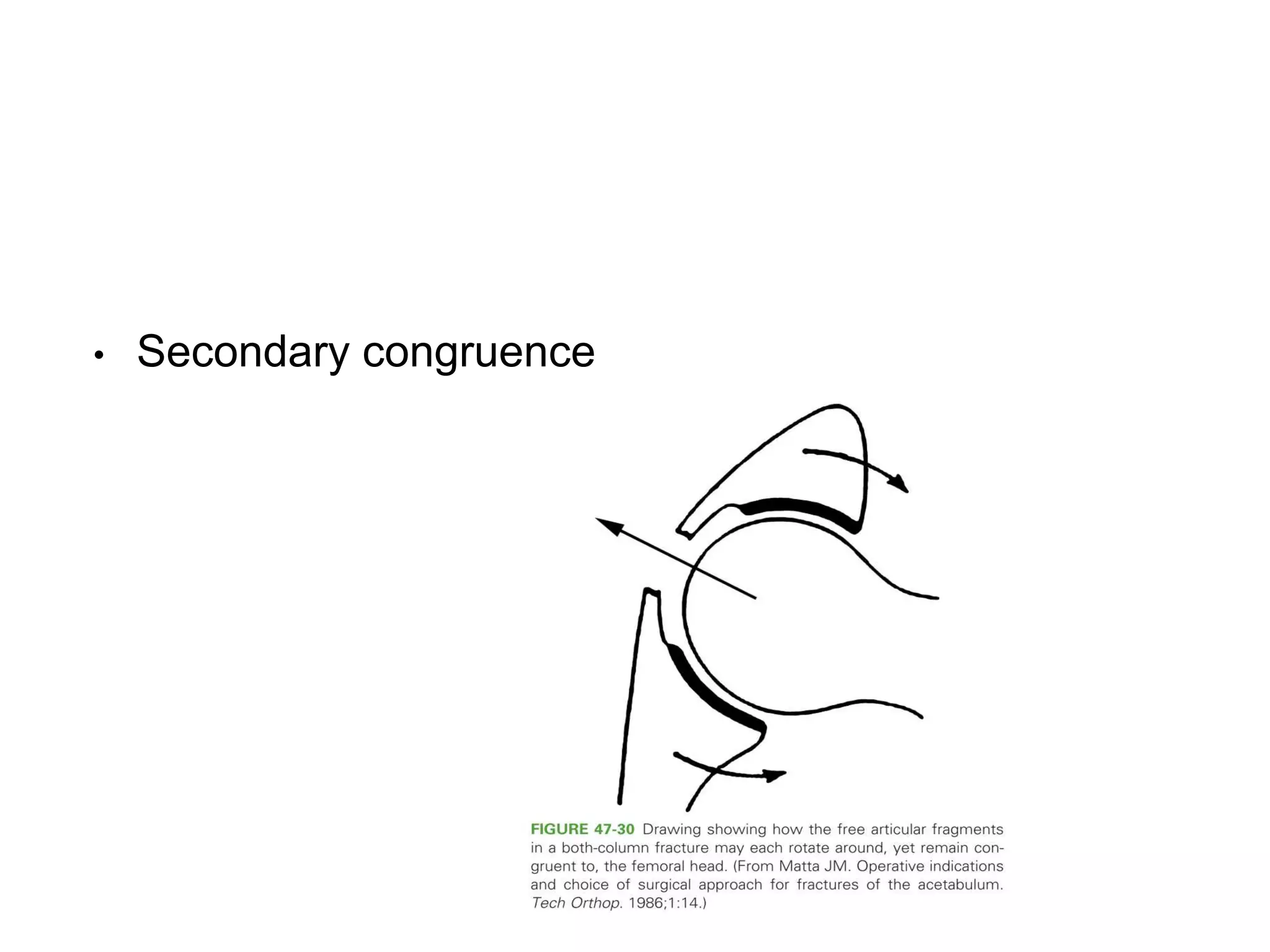
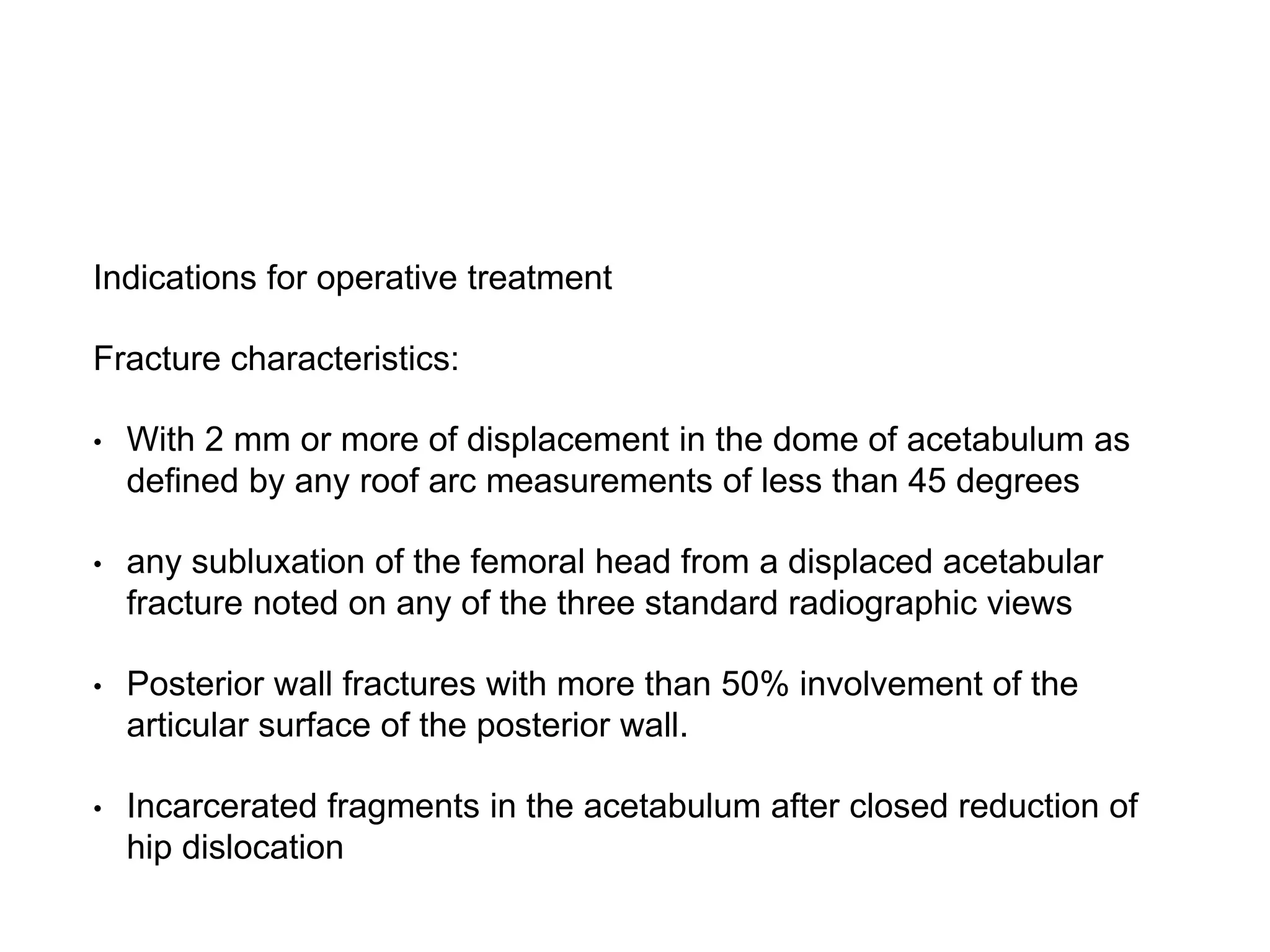
Acetabulum Fracture
Dr Sijan Bhattachan
3rd year resident
Orthopaedics & Trauma Surgery, NAMS
Introduction
• Treatment of acetabular fractures is a complex area
of orthopaedics that is being continually refined.
• Caused by high energy trauma and associated
injuries are frequent.
• Management of entire patient should follow
accepted ATLS protocol.
Anatomy
• Acetabulum; Incomplete hemispherical socket with
an inverted horseshoe shaped articular surface
surrounding the nonarticular cotyloid fossa.
• Articular socket supported by two columns of bone,
described by Letournel and Judet as an inverted Y.
Columns
Dome
Quadrilateral Plate
• Sciatic nerve & Superior gluteal vessels
Corona Mortis
Mechanism of injury
• Impact of femoral head with acetabular articular
surface
Radiographic evaluation
• AP view
• Judet views (45 degrees oblique views)
-Iliac oblique view
-Obturator oblique view
Roof Arc
• Matta et al developed a system for roughly
quantifying the acetabular dome after fracture, which
they called the ‘Roof arc” measurement.
• Determines if the remaining intact acetabulum is sufficient to
maintain a stable and congruous relationship with femoral head.
• If any of the roof arc measurements in a displaced fracture are
less than 45 degrees, operative treatment should be considered
• CT scan is invaluable in the treatment of acetabular
fractures.
Classification
• Letournel & Judet;
Posterior Wall Fracture
• 25% of all acetabular fractures
Posterior Column Fracture
Both column Fracture
• 23% of all acetabular fractures
• Acetabulum completely disconnected from axial skeleton.
• Central dislocation of femoral head
• Spur Sign; External cortex of most caudal portion of
intact ilium.
• Secondary congruence
Treatment Protocol
• Radiographs allow proper fracture classification
• Fracture location and displacement determine need
for surgery
• Fracture Pattern determines Approach.
Non Operative ; Indications
• Nondisplaced and minimally displaced fractures (<2 mm)
• Fractures with significant displacement but in which the region of the joint
involved is judged to be unimportant prognostically (roof arc).
• Secondary congruence in displaced both column fractures
• Medical contraindications to surgery
• Local soft tissue problems, such as infection, wounds and soft tissue lesions
• Elderly patients with osteoporotic bone in whom open reduction may not be
feasible
Non Operative Treatment Techniques;
• Bed Rest with joint mobilisation.
• When there is adequate fracture healing , usually by
6-12 weeks , gradually progress to full weight
bearing..
• Prolonged traction treatment for those patients with
operative indications related to fracture displacement
but having contraindications to surgical intervention.
Indications for operative treatment
Fracture characteristics:
• With 2 mm or more of displacement in the dome of acetabulum as
defined by any roof arc measurements of less than 45 degrees
• any subluxation of the femoral head from a displaced acetabular
fracture noted on any of the three standard radiographic views
• Posterior wall fractures with more than 50% involvement of the
articular surface of the posterior wall.
• Incarcerated fragments in the acetabulum after closed reduction of
hip dislocation
• Urgent surgical interventions
-Irreducible hip dislocation
-Open fracture
-Vascular compromise
-Worsening neurologic deficit
• No delay beyond 15 days for elementary fractures and 10 days
for associated types
Surgical Approach
• Anterior
-Ilioinguinal
-Modified Stoppa
• Posterior (Kocher-Langenbeck approach)
• Extensile approaches
-Extended iliofemoral
-Triradiate approach
-T approach
• Combined Anterior & Posterior Approach
Selection of Surgical approach
• Fracture type
• Elapsed time from injury to operative intervention
• Magnitude and location of maximal fracture
displacement
Fracture Reduction & Fixation;
• First reduce and stabilise the
displaced columns , if present and
then reduce any wall fracture.
• After definitive fixation of the
reduced fragments, the entire
construct is stabilised with
buttress plates.
Percutaneous Treatment
• Mini open exposure through lateral window of ilioinguinal
incision.
Indications;
• To prevent potential further fracture displacement.
• Displaced fractures in elderly.
• Simple fractures with minimal displacement
• As an adjunct to standard ORIF techniques
• Severe injuries that prevent formal ORIF
Kocher-Langenbeck
Approach
• Indications;
Dissection
Complications;
• Infection
• Sciatic nerve palsy
• Heterotopic ossification
Special considerations
• Transecting the piriformis & obturator internus tendons
1.5 cm from GT.
• Quadratus femoris & obturator externus intact.
• Sciatic nerve directly visualised & protected; Recognize
anatomical variations.
• Sciatic nerve & Piriformis;
Ilioinguinal Approach
• Developed by Letournel after extensive cadaveric anatomical
study.
• Indications;
Access
Dissection
• Iliopectineal fascia separates Lacuna musculorum and
Lacuna vasorum.
• 3 windows;
Complications;
• Infection
• Femoral nerve palsy
• Injury to Lateral femoral cutaneous nerve
• Vascular injury
Modified Stoppa Approach
• Exposes internal surface of the anterior column and
the quadrilateral surface.
• It can be used for many fractures previously treated
through ilioinguinal approach.
Dissection
• Use of Stoppa Approach with the Lateral window of
the ilioinguinal approach has been promoted as a
way of avoiding the dissection of the middle window
of the ilioinguinal approach and thus exposure of
femoral vessels and nerve.
Complications
• Overall mortality rates (0 - 2.5%)
• Post traumatic arthritis & osteonecrosis of femoral head
• Infections
• Sciatic nerve palsy (10-15% ;2-6%)
• Heterotopic ossification
• Thromboembolic complications
• Intra articular hardware
THR
• In older patients with extremely poor prognoses.
• Indications include intraarticular comminution,
full thickness abrasive loss of articular cartilage,
impaction of femoral head, impaction of dome,
associated femoral neck fracture and
preexistent arthritis.
• Fractures can be fixed with percutaneous
screws, plates or cables and fixation augmented
with multiple screw fixation of the ingrowth cup.
• 45 yr/M ; Left Acetabulum Fracture
• Modified Stoppa with lateral window of Ilioinguinal
Approach
References
• Campbell’s operative orthopaedics, 13th edition.
• Rockwood & Green’s Fractures in Adults, 8th
edition.
• OTA lecture series III (Acetabulum Fracture).

Acetabulum Fracture

  • 1.
    Acetabulum Fracture Dr SijanBhattachan 3rd year resident Orthopaedics & Trauma Surgery, NAMS
  • 2.
    Introduction • Treatment ofacetabular fractures is a complex area of orthopaedics that is being continually refined. • Caused by high energy trauma and associated injuries are frequent. • Management of entire patient should follow accepted ATLS protocol.
  • 3.
    Anatomy • Acetabulum; Incompletehemispherical socket with an inverted horseshoe shaped articular surface surrounding the nonarticular cotyloid fossa. • Articular socket supported by two columns of bone, described by Letournel and Judet as an inverted Y.
  • 4.
  • 5.
  • 6.
  • 7.
    • Sciatic nerve& Superior gluteal vessels
  • 8.
  • 9.
    Mechanism of injury •Impact of femoral head with acetabular articular surface
  • 10.
    Radiographic evaluation • APview • Judet views (45 degrees oblique views) -Iliac oblique view -Obturator oblique view
  • 13.
    Roof Arc • Mattaet al developed a system for roughly quantifying the acetabular dome after fracture, which they called the ‘Roof arc” measurement.
  • 14.
    • Determines ifthe remaining intact acetabulum is sufficient to maintain a stable and congruous relationship with femoral head. • If any of the roof arc measurements in a displaced fracture are less than 45 degrees, operative treatment should be considered
  • 15.
    • CT scanis invaluable in the treatment of acetabular fractures.
  • 16.
  • 17.
    Posterior Wall Fracture •25% of all acetabular fractures
  • 19.
  • 21.
    Both column Fracture •23% of all acetabular fractures • Acetabulum completely disconnected from axial skeleton. • Central dislocation of femoral head
  • 22.
    • Spur Sign;External cortex of most caudal portion of intact ilium.
  • 23.
  • 24.
    Treatment Protocol • Radiographsallow proper fracture classification • Fracture location and displacement determine need for surgery • Fracture Pattern determines Approach.
  • 25.
    Non Operative ;Indications • Nondisplaced and minimally displaced fractures (<2 mm) • Fractures with significant displacement but in which the region of the joint involved is judged to be unimportant prognostically (roof arc). • Secondary congruence in displaced both column fractures • Medical contraindications to surgery • Local soft tissue problems, such as infection, wounds and soft tissue lesions • Elderly patients with osteoporotic bone in whom open reduction may not be feasible
  • 26.
    Non Operative TreatmentTechniques; • Bed Rest with joint mobilisation. • When there is adequate fracture healing , usually by 6-12 weeks , gradually progress to full weight bearing.. • Prolonged traction treatment for those patients with operative indications related to fracture displacement but having contraindications to surgical intervention.
  • 27.
    Indications for operativetreatment Fracture characteristics: • With 2 mm or more of displacement in the dome of acetabulum as defined by any roof arc measurements of less than 45 degrees • any subluxation of the femoral head from a displaced acetabular fracture noted on any of the three standard radiographic views • Posterior wall fractures with more than 50% involvement of the articular surface of the posterior wall. • Incarcerated fragments in the acetabulum after closed reduction of hip dislocation
  • 28.
    • Urgent surgicalinterventions -Irreducible hip dislocation -Open fracture -Vascular compromise -Worsening neurologic deficit • No delay beyond 15 days for elementary fractures and 10 days for associated types
  • 29.
    Surgical Approach • Anterior -Ilioinguinal -ModifiedStoppa • Posterior (Kocher-Langenbeck approach) • Extensile approaches -Extended iliofemoral -Triradiate approach -T approach • Combined Anterior & Posterior Approach
  • 30.
    Selection of Surgicalapproach • Fracture type • Elapsed time from injury to operative intervention • Magnitude and location of maximal fracture displacement
  • 31.
    Fracture Reduction &Fixation; • First reduce and stabilise the displaced columns , if present and then reduce any wall fracture. • After definitive fixation of the reduced fragments, the entire construct is stabilised with buttress plates.
  • 32.
    Percutaneous Treatment • Miniopen exposure through lateral window of ilioinguinal incision. Indications; • To prevent potential further fracture displacement. • Displaced fractures in elderly. • Simple fractures with minimal displacement • As an adjunct to standard ORIF techniques • Severe injuries that prevent formal ORIF
  • 35.
  • 37.
  • 40.
    Complications; • Infection • Sciaticnerve palsy • Heterotopic ossification
  • 41.
    Special considerations • Transectingthe piriformis & obturator internus tendons 1.5 cm from GT. • Quadratus femoris & obturator externus intact. • Sciatic nerve directly visualised & protected; Recognize anatomical variations.
  • 42.
    • Sciatic nerve& Piriformis;
  • 43.
    Ilioinguinal Approach • Developedby Letournel after extensive cadaveric anatomical study. • Indications;
  • 44.
  • 45.
  • 47.
    • Iliopectineal fasciaseparates Lacuna musculorum and Lacuna vasorum.
  • 48.
  • 49.
    Complications; • Infection • Femoralnerve palsy • Injury to Lateral femoral cutaneous nerve • Vascular injury
  • 50.
    Modified Stoppa Approach •Exposes internal surface of the anterior column and the quadrilateral surface. • It can be used for many fractures previously treated through ilioinguinal approach.
  • 51.
  • 56.
    • Use ofStoppa Approach with the Lateral window of the ilioinguinal approach has been promoted as a way of avoiding the dissection of the middle window of the ilioinguinal approach and thus exposure of femoral vessels and nerve.
  • 57.
    Complications • Overall mortalityrates (0 - 2.5%) • Post traumatic arthritis & osteonecrosis of femoral head • Infections • Sciatic nerve palsy (10-15% ;2-6%) • Heterotopic ossification • Thromboembolic complications • Intra articular hardware
  • 58.
    THR • In olderpatients with extremely poor prognoses. • Indications include intraarticular comminution, full thickness abrasive loss of articular cartilage, impaction of femoral head, impaction of dome, associated femoral neck fracture and preexistent arthritis. • Fractures can be fixed with percutaneous screws, plates or cables and fixation augmented with multiple screw fixation of the ingrowth cup.
  • 59.
    • 45 yr/M; Left Acetabulum Fracture
  • 60.
    • Modified Stoppawith lateral window of Ilioinguinal Approach
  • 61.
    References • Campbell’s operativeorthopaedics, 13th edition. • Rockwood & Green’s Fractures in Adults, 8th edition. • OTA lecture series III (Acetabulum Fracture).