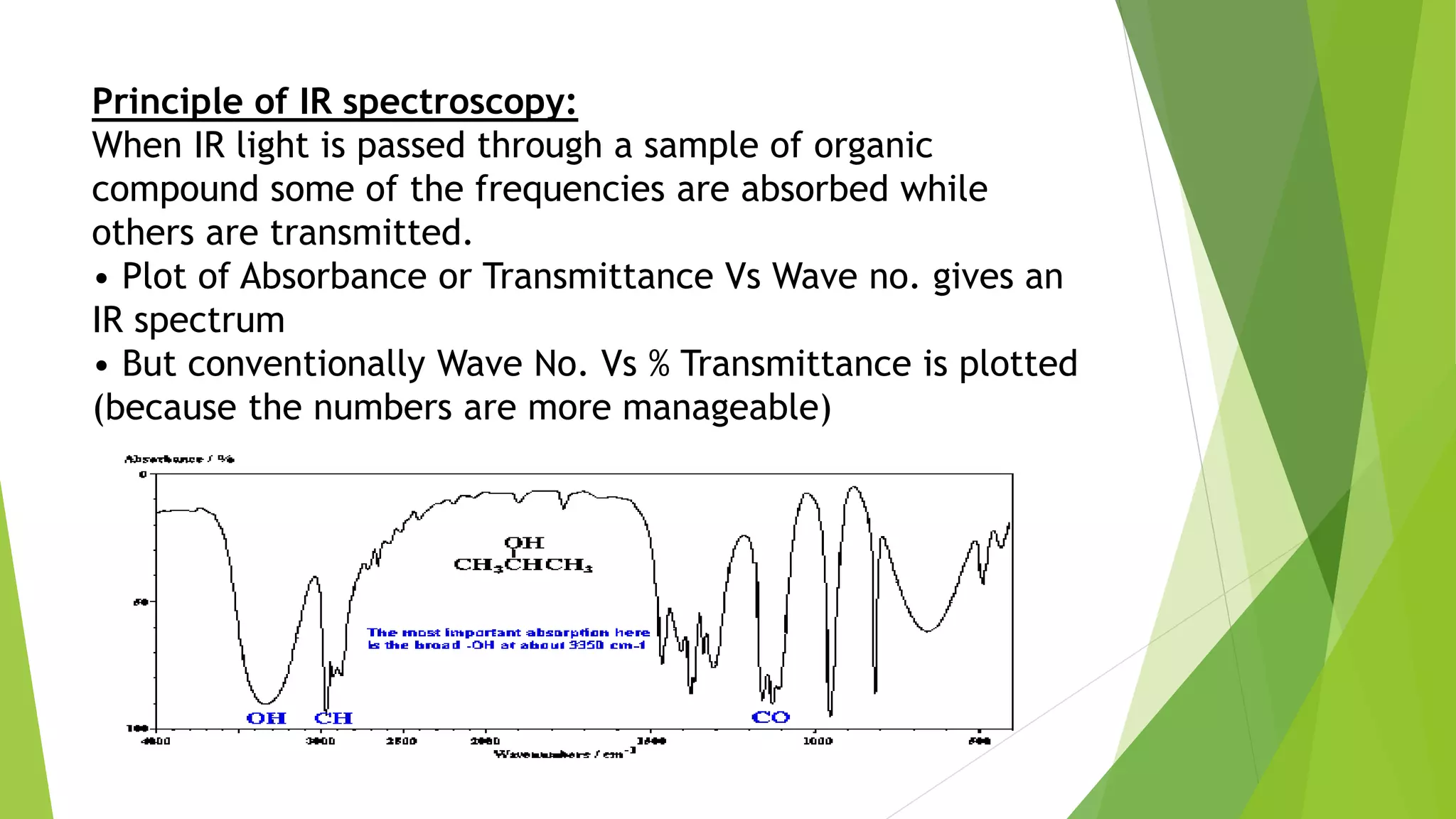
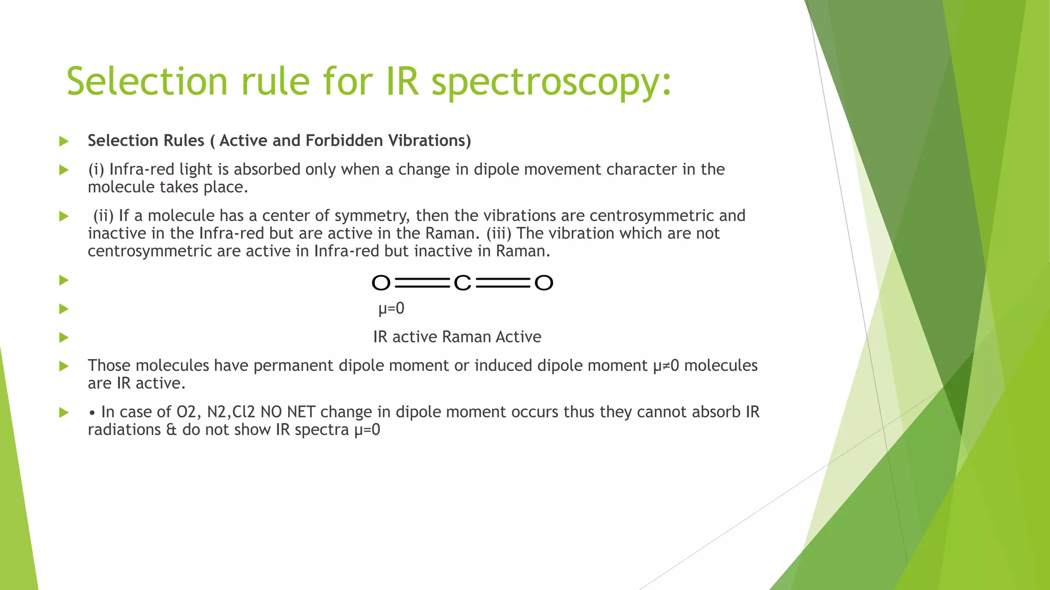
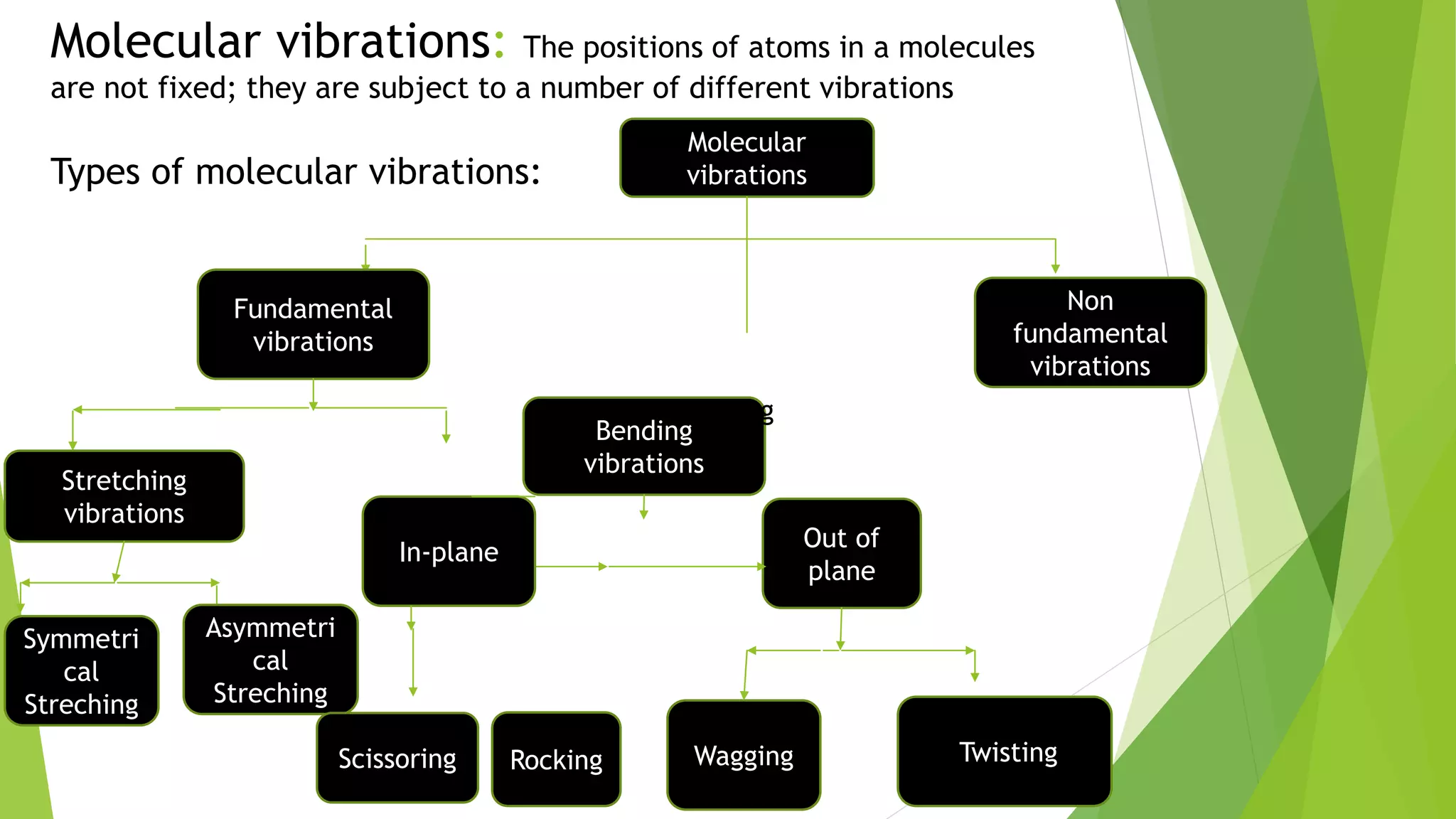
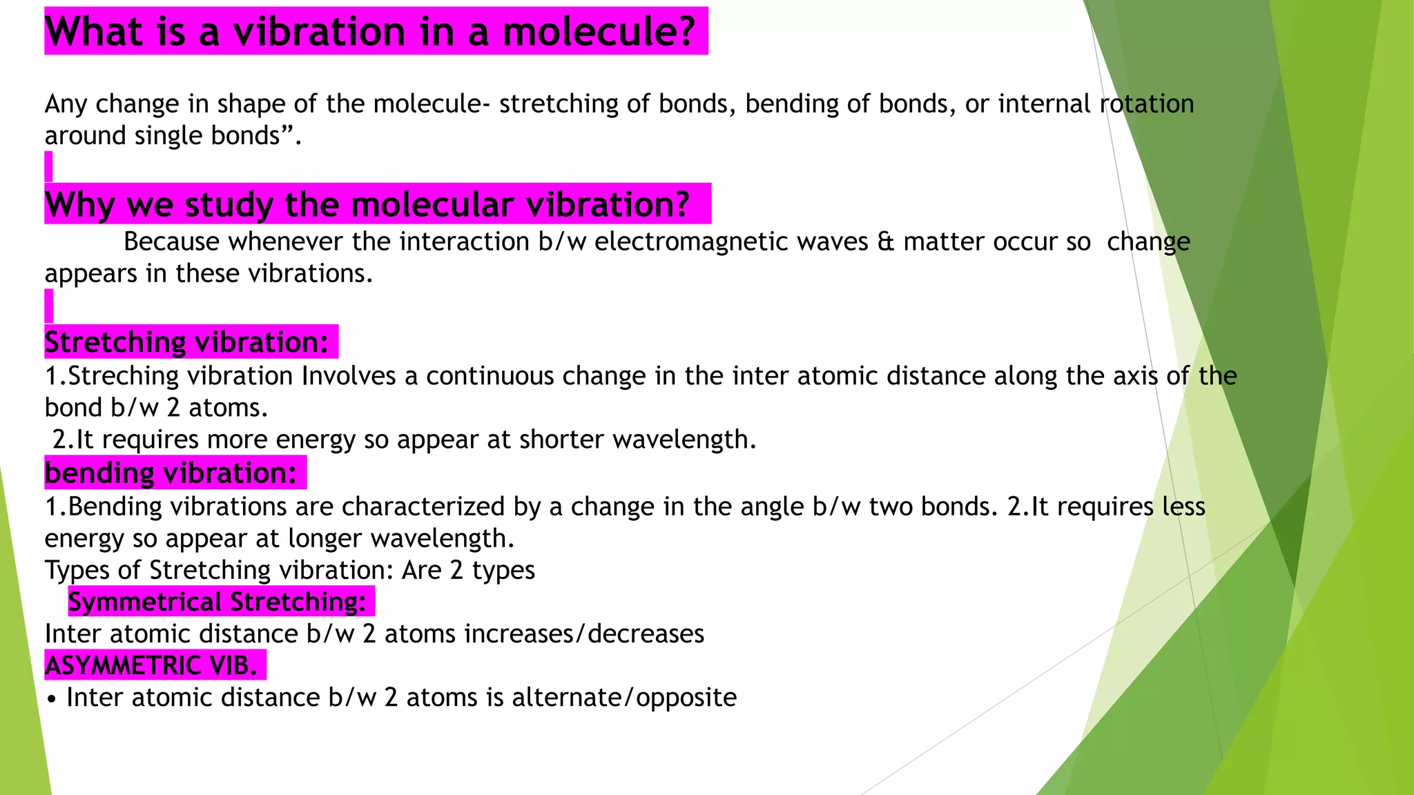
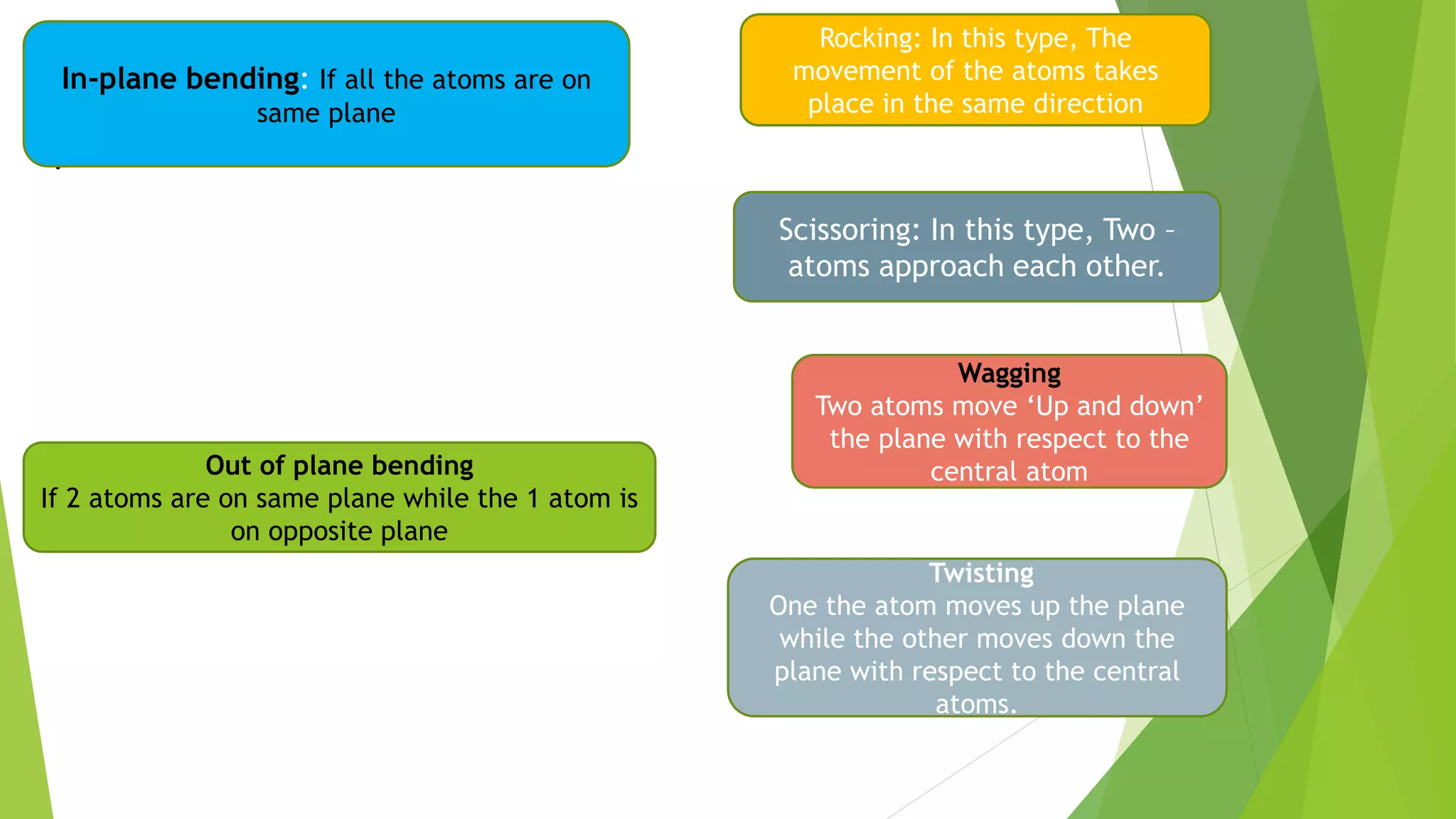
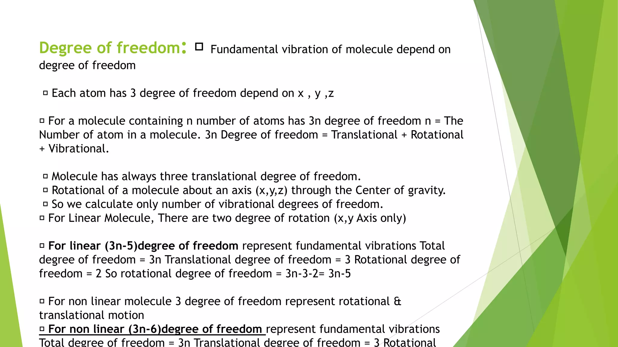
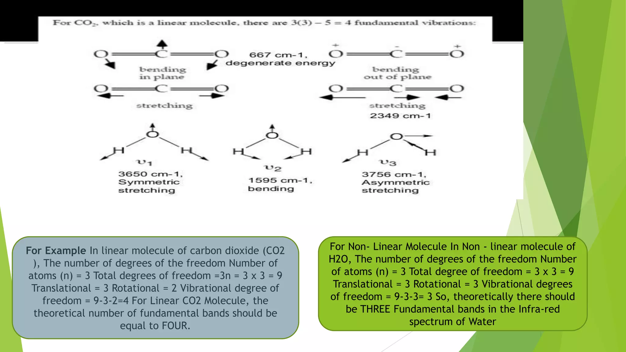
Spectroscopic techniques
UNIT-V
P.HARIPRASAD
Introduction to Spectroscopy
 Spectroscopy means measurement of radiation
 Spectro = Radiation Scopy = measurement
 ‘’Spectroscopy is branch of science which deals with the study of
interaction of Electromagnetic radiation with matter’’.
 Electro magnetic radiation can behave as both wave nature and
particle like nature.
 Spectroscopy is ‘’ non Destructive technique’’
 It requires small amount of sample(10mg)
 It is very economical in long time run.
 It is one the best method for the detection of structure of the
molecule and different functional groups which are present in the
sample.
 Electromagnetic Radiation
 • Electromagnetic radiation consist of discrete packages
of energy which are called as photons.
 • A photon consists of an oscillating electric field (E) & an
oscillating magnetic field (B) which are perpendicular to
each other.
Wave number: the number of waves per unit distance of radiant energy of a
given wavelength : the reciprocal of the wavelength.
Principles of Spectroscopy
based on the measurement of spectrum of a sample containing atoms /
molecules.
• Spectrum is a graph of intensity of absorbed or emitted radiation by
sample verses frequency (ν) or wavelength (λ).
• Spectrometer is an instrument design to measure the spectrum of a
compound. Principles of Spectroscopy
1. Absorption Spectroscopy: • An analytical technique which
concerns with the measurement of absorption of electromagnetic
radiation. • e.g. UV (185 - 400 nm) / Visible (400 - 800 nm) Spectroscopy,
IR Spectroscopy (0.76 - 15 μm)
2. Emission Spectroscopy: • An analytical technique in which
emission (of a particle or radiation) is dispersed according to some
property of the emission & the amount of dispersion is measured.
• e.g. Mass Spectrometry
Interaction of EMR with matter
1. Electronic Energy Levels: • At room temperature the molecules are in the
lowest energy levels E0.
• When the molecules absorb UV-visible light from EMR, one of the outermost
bond / lone pair electron is promoted to higher energy state such as E1, E2, …En,
etc is called as electronic transition and the difference is as: ∆E = h ν = En - E0
where (n = 1, 2, 3, … etc) ∆E = 35 to 71 kcal/mole
2. Vibrational Energy Levels: • These are less energy level than electronic
energy levels.
• The spacing between energy levels are relatively small i.e. 0.01 to 10
kcal/mole.
• e.g. when IR radiation is absorbed, molecules are excited from one vibrational
level to another or it vibrates with higher amplitude
3. Rotational Energy Levels: • These energy levels are quantized & discrete.
• The spacing between energy levels are even smaller than vibrational energy
levels.
∆E rotational < ∆E vibrational < ∆E electronic
UV-VISIBLE Spectroscopy(electronic
Spectroscopy)
 Principle • The UV radiation region extends from 10 nm to 400 nm
and the visible radiation region extends from 400 nm to 800 nm. Near
UV Region: 200 nm to 400 nm Far UV Region: below 200 nm •
 Far UV spectroscopy is studied under vacuum condition.
 • The common solvent used for preparing sample to be analyzed is
either ethyl alcohol or hexane.
Selection rules for UV-Visible spectroscopy:
1.Allowed transition: the electronic transitions which are takes place with higher probability are
allowed transitions. It gives more intense spectrum.
2.Forbidden transition: the electronic transition which are takes place with low probability are called
Forbidden transition.
3.Spin selection rule: The transitions which are involves there is no change in Spin multiplicity.(2S+1)
The total spin cannot change, ΔS=0
Singlet to singlet and triplet to triplet are allowed transitions (OR)
Singlet to triplet are forbidden transitions
4.Laporte Selection rule: those transitions which involves where there is no change in parity of d-
orbital symmetry is known as laporte selection rule.
ΔL=0, ± 1
Electronic transitions:
1.σ → σ* transition:
σ electron from orbital is excited to corresponding anti-bonding orbital σ*. • The energy required is large
for this transition. • e.g. Methane (CH4 ) has C-H bond only and can undergo σ → σ* transition and shows
absorbance maxima at 125 nm
2. π → π* transitions: π electron in a bonding orbital is excited to corresponding anti-bonding orbital π*.
• Compounds containing multiple bonds like alkenes, alkynes, carbonyl, nitriles, aromatic compounds, etc
undergo
π → π* transitions.
• e.g. Alkenes generally absorb in the region 170 to 205 nm
3. n → σ* transition.
Saturated compounds containing atoms with lone pair of electrons like O, N, S and
halogens are capable of n → σ* transition. • These transitions usually requires less energy
than σ → σ* transitions. • The number of organic functional groups with n → σ* peaks in
UV region is small (150 – 250 nm)
4. n → π* transitions
An electron from non-bonding orbital is promoted to anti-bonding π* orbital. • Compounds
containing double bond involving hetero atoms (C=O, C≡N, N=O) undergo such transitions. •
n → π* transitions require minimum energy and show absorption at longer wavelength
around 300 nm.
Chromophore:
The functional groups containing multiple bonds capable of absorbing radiations above
200nm to 800nm these functional groups are called chromophore.
due to n → π* & π → π* transitions.
e.g. NO2 , N=O, C=O, C=N, C≡N, C=C, C=S, etc
When double bonds are conjugated in a compound λmax is shifted to longer wavelength.
Crotonaldehyde has λmax = 290 nm
H2C=CH2 Ethylene has λmax = 171 nm
e.g. 1,5 - hexadiene has λmax = 178 nm
2,4 - hexadiene has λmax = 227 nm
Auxo chrome: The Atom or groups which can not directly absorb the UV-Visible
range of radiation but these groups are attached to the chromophore can increase the
wavelength max.
Λmax=254nm Λmax=270nm Λmax=280nm Λmax=284nm
Acetone which has
λmax = 279 nm λmax =291nm
Applications
1• Qualitative & Quantitative Analysis: – It is used for characterizing aromatic
compounds and conjugated olefins. – It can be used to find out molar
concentration of the solute under study.
2.Detection of impurities: – It is one of the important method to detect
impurities in organic solvents.
3• Detection of isomers are possible. • Determination of molecular weight using
Beer’s law.
4.Characterization of aromatic compounds and conjugated dienes or other
olefins.
5. Determination of unknown concentration.
IR Spectroscopy:
Infrared spectroscopy (IR spectroscopy) is the spectroscopy that deals
with the infrared region of the electromagnetic spectrum, that is light
with a longer wavelength and lower frequency than visible light
Infrared Spectroscopy is the analysis of infrared light interacting with
a molecule.
It is based on absorption spectroscopy
INFRARED REGIONS RANGE:
Near infrared region 0.8-2.5 µ(12,500-4000 cm-1)
Main infrared region 2.5-15 µ(4000-667cm-1)
Far infrared region 15-200 m µ(667-100 cm-1)
Principle of IR spectroscopy:
When IR light is passed through a sample of organic
compound some of the frequencies are absorbed while
others are transmitted.
• Plot of Absorbance or Transmittance Vs Wave no. gives an
IR spectrum
• But conventionally Wave No. Vs % Transmittance is plotted
(because the numbers are more manageable)
Selection rule for IR spectroscopy:
 Selection Rules ( Active and Forbidden Vibrations)
 (i) Infra-red light is absorbed only when a change in dipole movement character in the
molecule takes place.
 (ii) If a molecule has a center of symmetry, then the vibrations are centrosymmetric and
inactive in the Infra-red but are active in the Raman. (iii) The vibration which are not
centrosymmetric are active in Infra-red but inactive in Raman.

 µ=0
 IR active Raman Active
 Those molecules have permanent dipole moment or induced dipole moment µ≠0 molecules
are IR active.
 • In case of O2, N2,Cl2 NO NET change in dipole moment occurs thus they cannot absorb IR
radiations & do not show IR spectra µ=0
Molecular vibrations: The positions of atoms in a molecules
are not fixed; they are subject to a number of different vibrations
Types of molecular vibrations:
Molecular
vibrations
Fundamental
vibrations
Non
fundamental
vibrations
Stretching
vibrations
Bending
vibrations
Asymmetri
cal
Streching
Symmetri
cal
Streching
Out of
plane
In-plane
TwistingWaggingScissoring Rocking
Rocking
What is a vibration in a molecule?
Any change in shape of the molecule- stretching of bonds, bending of bonds, or internal rotation
around single bonds”.
Why we study the molecular vibration?
Because whenever the interaction b/w electromagnetic waves & matter occur so change
appears in these vibrations.
Stretching vibration:
1.Streching vibration Involves a continuous change in the inter atomic distance along the axis of the
bond b/w 2 atoms.
2.It requires more energy so appear at shorter wavelength.
bending vibration:
1.Bending vibrations are characterized by a change in the angle b/w two bonds. 2.It requires less
energy so appear at longer wavelength.
Types of Stretching vibration: Are 2 types
Symmetrical Stretching:
Inter atomic distance b/w 2 atoms increases/decreases
ASYMMETRIC VIB.
• Inter atomic distance b/w 2 atoms is alternate/opposite
In-plane of bending
vibrations
Out of plane Bending
vibrations
•.
.
Twisting
One the atom moves up the plane
while the other moves down the
plane with respect to the central
atoms.
Wagging
Two atoms move ‘Up and down’
the plane with respect to the
central atomOut of plane bending
If 2 atoms are on same plane while the 1 atom is
on opposite plane
In-plane bending: If all the atoms are on
same plane
Scissoring: In this type, Two –
atoms approach each other.
Rocking: In this type, The
movement of the atoms takes
place in the same direction
Degree of freedom: Fundamental vibration of molecule depend on
degree of freedom
Each atom has 3 degree of freedom depend on x , y ,z
For a molecule containing n number of atoms has 3n degree of freedom n = The
Number of atom in a molecule. 3n Degree of freedom = Translational + Rotational
+ Vibrational.
Molecule has always three translational degree of freedom.
Rotational of a molecule about an axis (x,y,z) through the Center of gravity.
So we calculate only number of vibrational degrees of freedom.
For Linear Molecule, There are two degree of rotation (x,y Axis only)
For linear (3n-5)degree of freedom represent fundamental vibrations Total
degree of freedom = 3n Translational degree of freedom = 3 Rotational degree of
freedom = 2 So rotational degree of freedom = 3n-3-2= 3n-5
For non linear molecule 3 degree of freedom represent rotational &
translational motion
For non linear (3n-6)degree of freedom represent fundamental vibrations
Total degree of freedom = 3n Translational degree of freedom = 3 Rotational
For Example In linear molecule of carbon dioxide (CO2
), The number of degrees of the freedom Number of
atoms (n) = 3 Total degrees of freedom =3n = 3 x 3 = 9
Translational = 3 Rotational = 2 Vibrational degree of
freedom = 9-3-2=4 For Linear CO2 Molecule, the
theoretical number of fundamental bands should be
equal to FOUR.
For Non- Linear Molecule In Non - linear molecule of
H2O, The number of degrees of the freedom Number
of atoms (n) = 3 Total degree of freedom = 3 x 3 = 9
Translational = 3 Rotational = 3 Vibrational degrees
of freedom = 9-3-3= 3 So, theoretically there should
be THREE Fundamental bands in the Infra-red
spectrum of Water
Applications of IR spectroscopy:
1. Identifications of an Organic Compounds: Most Organic Compounds is
conformed in Finger print region (1400-667cm-1)
2. Structure Determination: This technique helps to establish the structure of an
unknown compounds.
3. Qualitative analysis of functional groups: The Presence or absence of
absorption bands help in predicting the presence of certain functional group in
the compounds. Presence of Oxygen may be –OH, C=O, COOR, -COOH etc. But an
absorption band between 3600-3200 cm-1
4. Distinction between two types of hydrogen bonding: To find the Inter Or Intra
molecular H- Bonding.
5. Quantitative analysis: It help to make a quantitative estimation of an organic
mixture. For Example Xylene commercial is mixture of Ortho, Meta, Para
Compound. The separate of the mixture can not be easily done. But percentage
composition of the mixture can be determine.
6. Conformational Analysis: Chair or Boat Form
7. Geometrical Isomerism: Cis or Trans , Syn or Anti
NMR Spectroscopy
The study of absorption of radiofrequency radiation by nuclei in a magnetic field is
called Nuclear Magnetic Resonance.
• Nuclear magnetic resonance spectroscopy is basically another form of absorption
spectrometry. It involve change of the spin state of a nucleus, when the nucleus
absorb electromagnetic radiation in a strong magnetic field.
• The source of energy in NMR is radio waves which have long wavelengths, and
thus low energy and frequency.
• When low-energy radio waves interact with a molecule, they can change the
nuclear spins of some elements having spin state 1/2, including 1H and 13C
•
.
•
All nuclei carry a charge. In same nuclei charge spins on the
nuclear axis and this circular, charge generates a magnetic dipole
along the axis. The angular momentumof the spinning charge
can be described in terms of quantum number I, “ I = 0, ½, 1,
3/2 5/2…….. and so on. If spin I = 0 no spin and hence no 1H
NMRphenomenon.
 As shown in figure. Twoenergy levels one is lower energy states i.e, aligned or parallel I
= +1/2 whose population is N other is higher energy state I = -1/2, antiparallel whose
population is N then N > N in accordancewith the boltzmandistribution.
 therefore
 Where ,
 h  Planck’s constant. ∆E =h    Bo
   Frequency of oscillator
 B o  Applied external magnetic field strength.
   Gyromagnetic ratio ( being ratio between the nuclear magnetic
moment)
Spin active nuclei have permanent magnetic moments and quantized nuclear spin
states. The number of spin states for a given nucleus is given by the expression (2I +1) where
I is the overall nuclear spin.
In a magnetic field, there are now two energy states for a proton: a lower energy
state with the nucleus aligned in the same direction as B0, and a higher energy
state in which the nucleus aligned against B0
Alignment withthe field(Lower energy stateor parallel to the field I =+1/2)
Alignmentagainst the field (Higherenergystateor antiparallel to the field. I =-1/2) NMR
Active Nuclei: nuclear spin quantum number (I) atomic mass and atomic number
Number of spin states = 2I + 1 (number of possible energy levels
Even mass nuclei that have even number of neutron have I = 0 (NMR inactive) Even
mass nuclei that have odd number of neutrons have an integer spin quantum number (I
= 1, 2, 3, etc) Odd mass nuclei have half-integer spin quantum number (I = 1/2, 3/2, 5/2,
etc)
I= 1/2: 1H, 13C, 19F, 31P
I= 1: 2H, 14N I= 3/2: 15N
I= 0: 12C, 16O
Chemical shift: The difference in the absorption of position of a particular
proton from the absorption position of a reference proton is called Chemicalshift.
Chemical shifts position are normally expressed in
(delta) units, which are defined as proportional differences, in parts per million(ppm),
from an appropriate reference standard (TMS in case of protonNMR).
Chemical shift = frequency of sample – frequency of reference. ×10^6/Applied external
magnetic field(B0). Ppm
*Tetramethyl silane is used as refrence due to its stable state , low Deshielding and non
reactive or its inert nature
*In TMS 12 chemical equivalent protons are present
Splitting of Signal (spin –spin coupling)
Splitting of signal is caused by the neighboring protons . The relation is given by 2nI+1
Where, N= number of neighboring proton
I = spin angle of quantum number For H atom l= ½ Then, 2×n×½+1 = n+1.
Eg. In CH₃–CH₂–Br for first CH₃ its neighboring proton is 2 so 2+1=3 Which give triplet
peak for NMR
Example ethyl alcohol NMR spectrum
NMR spectrum of
ethyl bromide
CH3-CH2-OH
Magnetic Imaging Resonance :
MRI is specialist application of multi dimensional Fourier transformation NMR
1. MRI is a medical imaging technique used in radiology to form pictures of the anatomy
and the physiological processes of the body in both health and disease.
2. MRI scanners use strong magnetic fields, magnetic fields gradient and radio waves to
generate images of the organs in the body
3.
4. Hydrogen nucleus( single proton) present in water molecules and fats.tissues
5. The hydrogen nuclei partially aligned by a strong magnetic field in the scanner
6. The nuclei can be related using radio waves and they subsequent oscillate in the
magnetic filed.
7. Simultaneously they emit a radio signal this is detected using antennas( coils ) gives
very detailed images can be made of soft tissues
Applications of NMR Spectroscopy
NMR is used in biology to study the biofluids, cells, perfused organs and bio
macromolecules such as nucleic acids (DNA. RNA) carbohydrates, proteins and
peptides, And also labeling studies is biochemistry.
NMR is used physics and physical chemistry to study high pressure diffusion,
Liquid crystals, liquid crystal solutions, membranes, rigid solids.
 NMR is used in pharmaceutical science to study
pharmaceutical and drug metabolism.
Thank you

Spectroscpoic techniques

  • 1.
  • 2.
    Introduction to Spectroscopy Spectroscopy means measurement of radiation  Spectro = Radiation Scopy = measurement  ‘’Spectroscopy is branch of science which deals with the study of interaction of Electromagnetic radiation with matter’’.  Electro magnetic radiation can behave as both wave nature and particle like nature.  Spectroscopy is ‘’ non Destructive technique’’  It requires small amount of sample(10mg)  It is very economical in long time run.  It is one the best method for the detection of structure of the molecule and different functional groups which are present in the sample.
  • 3.
     Electromagnetic Radiation • Electromagnetic radiation consist of discrete packages of energy which are called as photons.  • A photon consists of an oscillating electric field (E) & an oscillating magnetic field (B) which are perpendicular to each other.
  • 5.
    Wave number: thenumber of waves per unit distance of radiant energy of a given wavelength : the reciprocal of the wavelength.
  • 6.
    Principles of Spectroscopy basedon the measurement of spectrum of a sample containing atoms / molecules. • Spectrum is a graph of intensity of absorbed or emitted radiation by sample verses frequency (ν) or wavelength (λ). • Spectrometer is an instrument design to measure the spectrum of a compound. Principles of Spectroscopy 1. Absorption Spectroscopy: • An analytical technique which concerns with the measurement of absorption of electromagnetic radiation. • e.g. UV (185 - 400 nm) / Visible (400 - 800 nm) Spectroscopy, IR Spectroscopy (0.76 - 15 μm) 2. Emission Spectroscopy: • An analytical technique in which emission (of a particle or radiation) is dispersed according to some property of the emission & the amount of dispersion is measured. • e.g. Mass Spectrometry
  • 7.
    Interaction of EMRwith matter 1. Electronic Energy Levels: • At room temperature the molecules are in the lowest energy levels E0. • When the molecules absorb UV-visible light from EMR, one of the outermost bond / lone pair electron is promoted to higher energy state such as E1, E2, …En, etc is called as electronic transition and the difference is as: ∆E = h ν = En - E0 where (n = 1, 2, 3, … etc) ∆E = 35 to 71 kcal/mole 2. Vibrational Energy Levels: • These are less energy level than electronic energy levels. • The spacing between energy levels are relatively small i.e. 0.01 to 10 kcal/mole. • e.g. when IR radiation is absorbed, molecules are excited from one vibrational level to another or it vibrates with higher amplitude 3. Rotational Energy Levels: • These energy levels are quantized & discrete. • The spacing between energy levels are even smaller than vibrational energy levels. ∆E rotational < ∆E vibrational < ∆E electronic
  • 8.
    UV-VISIBLE Spectroscopy(electronic Spectroscopy)  Principle• The UV radiation region extends from 10 nm to 400 nm and the visible radiation region extends from 400 nm to 800 nm. Near UV Region: 200 nm to 400 nm Far UV Region: below 200 nm •  Far UV spectroscopy is studied under vacuum condition.  • The common solvent used for preparing sample to be analyzed is either ethyl alcohol or hexane.
  • 9.
    Selection rules forUV-Visible spectroscopy: 1.Allowed transition: the electronic transitions which are takes place with higher probability are allowed transitions. It gives more intense spectrum. 2.Forbidden transition: the electronic transition which are takes place with low probability are called Forbidden transition. 3.Spin selection rule: The transitions which are involves there is no change in Spin multiplicity.(2S+1) The total spin cannot change, ΔS=0 Singlet to singlet and triplet to triplet are allowed transitions (OR) Singlet to triplet are forbidden transitions 4.Laporte Selection rule: those transitions which involves where there is no change in parity of d- orbital symmetry is known as laporte selection rule. ΔL=0, ± 1 Electronic transitions: 1.σ → σ* transition: σ electron from orbital is excited to corresponding anti-bonding orbital σ*. • The energy required is large for this transition. • e.g. Methane (CH4 ) has C-H bond only and can undergo σ → σ* transition and shows absorbance maxima at 125 nm 2. π → π* transitions: π electron in a bonding orbital is excited to corresponding anti-bonding orbital π*. • Compounds containing multiple bonds like alkenes, alkynes, carbonyl, nitriles, aromatic compounds, etc undergo π → π* transitions. • e.g. Alkenes generally absorb in the region 170 to 205 nm
  • 10.
    3. n →σ* transition. Saturated compounds containing atoms with lone pair of electrons like O, N, S and halogens are capable of n → σ* transition. • These transitions usually requires less energy than σ → σ* transitions. • The number of organic functional groups with n → σ* peaks in UV region is small (150 – 250 nm) 4. n → π* transitions An electron from non-bonding orbital is promoted to anti-bonding π* orbital. • Compounds containing double bond involving hetero atoms (C=O, C≡N, N=O) undergo such transitions. • n → π* transitions require minimum energy and show absorption at longer wavelength around 300 nm.
  • 11.
    Chromophore: The functional groupscontaining multiple bonds capable of absorbing radiations above 200nm to 800nm these functional groups are called chromophore. due to n → π* & π → π* transitions. e.g. NO2 , N=O, C=O, C=N, C≡N, C=C, C=S, etc When double bonds are conjugated in a compound λmax is shifted to longer wavelength. Crotonaldehyde has λmax = 290 nm H2C=CH2 Ethylene has λmax = 171 nm e.g. 1,5 - hexadiene has λmax = 178 nm 2,4 - hexadiene has λmax = 227 nm Auxo chrome: The Atom or groups which can not directly absorb the UV-Visible range of radiation but these groups are attached to the chromophore can increase the wavelength max. Λmax=254nm Λmax=270nm Λmax=280nm Λmax=284nm Acetone which has λmax = 279 nm λmax =291nm
  • 12.
    Applications 1• Qualitative &Quantitative Analysis: – It is used for characterizing aromatic compounds and conjugated olefins. – It can be used to find out molar concentration of the solute under study. 2.Detection of impurities: – It is one of the important method to detect impurities in organic solvents. 3• Detection of isomers are possible. • Determination of molecular weight using Beer’s law. 4.Characterization of aromatic compounds and conjugated dienes or other olefins. 5. Determination of unknown concentration.
  • 13.
    IR Spectroscopy: Infrared spectroscopy(IR spectroscopy) is the spectroscopy that deals with the infrared region of the electromagnetic spectrum, that is light with a longer wavelength and lower frequency than visible light Infrared Spectroscopy is the analysis of infrared light interacting with a molecule. It is based on absorption spectroscopy INFRARED REGIONS RANGE: Near infrared region 0.8-2.5 µ(12,500-4000 cm-1) Main infrared region 2.5-15 µ(4000-667cm-1) Far infrared region 15-200 m µ(667-100 cm-1)
  • 14.
    Principle of IRspectroscopy: When IR light is passed through a sample of organic compound some of the frequencies are absorbed while others are transmitted. • Plot of Absorbance or Transmittance Vs Wave no. gives an IR spectrum • But conventionally Wave No. Vs % Transmittance is plotted (because the numbers are more manageable)
  • 15.
    Selection rule forIR spectroscopy:  Selection Rules ( Active and Forbidden Vibrations)  (i) Infra-red light is absorbed only when a change in dipole movement character in the molecule takes place.  (ii) If a molecule has a center of symmetry, then the vibrations are centrosymmetric and inactive in the Infra-red but are active in the Raman. (iii) The vibration which are not centrosymmetric are active in Infra-red but inactive in Raman.   µ=0  IR active Raman Active  Those molecules have permanent dipole moment or induced dipole moment µ≠0 molecules are IR active.  • In case of O2, N2,Cl2 NO NET change in dipole moment occurs thus they cannot absorb IR radiations & do not show IR spectra µ=0
  • 16.
    Molecular vibrations: Thepositions of atoms in a molecules are not fixed; they are subject to a number of different vibrations Types of molecular vibrations: Molecular vibrations Fundamental vibrations Non fundamental vibrations Stretching vibrations Bending vibrations Asymmetri cal Streching Symmetri cal Streching Out of plane In-plane TwistingWaggingScissoring Rocking Rocking
  • 17.
    What is avibration in a molecule? Any change in shape of the molecule- stretching of bonds, bending of bonds, or internal rotation around single bonds”. Why we study the molecular vibration? Because whenever the interaction b/w electromagnetic waves & matter occur so change appears in these vibrations. Stretching vibration: 1.Streching vibration Involves a continuous change in the inter atomic distance along the axis of the bond b/w 2 atoms. 2.It requires more energy so appear at shorter wavelength. bending vibration: 1.Bending vibrations are characterized by a change in the angle b/w two bonds. 2.It requires less energy so appear at longer wavelength. Types of Stretching vibration: Are 2 types Symmetrical Stretching: Inter atomic distance b/w 2 atoms increases/decreases ASYMMETRIC VIB. • Inter atomic distance b/w 2 atoms is alternate/opposite
  • 18.
    In-plane of bending vibrations Outof plane Bending vibrations
  • 19.
    •. . Twisting One the atommoves up the plane while the other moves down the plane with respect to the central atoms. Wagging Two atoms move ‘Up and down’ the plane with respect to the central atomOut of plane bending If 2 atoms are on same plane while the 1 atom is on opposite plane In-plane bending: If all the atoms are on same plane Scissoring: In this type, Two – atoms approach each other. Rocking: In this type, The movement of the atoms takes place in the same direction
  • 20.
    Degree of freedom:Fundamental vibration of molecule depend on degree of freedom Each atom has 3 degree of freedom depend on x , y ,z For a molecule containing n number of atoms has 3n degree of freedom n = The Number of atom in a molecule. 3n Degree of freedom = Translational + Rotational + Vibrational. Molecule has always three translational degree of freedom. Rotational of a molecule about an axis (x,y,z) through the Center of gravity. So we calculate only number of vibrational degrees of freedom. For Linear Molecule, There are two degree of rotation (x,y Axis only) For linear (3n-5)degree of freedom represent fundamental vibrations Total degree of freedom = 3n Translational degree of freedom = 3 Rotational degree of freedom = 2 So rotational degree of freedom = 3n-3-2= 3n-5 For non linear molecule 3 degree of freedom represent rotational & translational motion For non linear (3n-6)degree of freedom represent fundamental vibrations Total degree of freedom = 3n Translational degree of freedom = 3 Rotational
  • 21.
    For Example Inlinear molecule of carbon dioxide (CO2 ), The number of degrees of the freedom Number of atoms (n) = 3 Total degrees of freedom =3n = 3 x 3 = 9 Translational = 3 Rotational = 2 Vibrational degree of freedom = 9-3-2=4 For Linear CO2 Molecule, the theoretical number of fundamental bands should be equal to FOUR. For Non- Linear Molecule In Non - linear molecule of H2O, The number of degrees of the freedom Number of atoms (n) = 3 Total degree of freedom = 3 x 3 = 9 Translational = 3 Rotational = 3 Vibrational degrees of freedom = 9-3-3= 3 So, theoretically there should be THREE Fundamental bands in the Infra-red spectrum of Water
  • 22.
    Applications of IRspectroscopy: 1. Identifications of an Organic Compounds: Most Organic Compounds is conformed in Finger print region (1400-667cm-1) 2. Structure Determination: This technique helps to establish the structure of an unknown compounds. 3. Qualitative analysis of functional groups: The Presence or absence of absorption bands help in predicting the presence of certain functional group in the compounds. Presence of Oxygen may be –OH, C=O, COOR, -COOH etc. But an absorption band between 3600-3200 cm-1 4. Distinction between two types of hydrogen bonding: To find the Inter Or Intra molecular H- Bonding. 5. Quantitative analysis: It help to make a quantitative estimation of an organic mixture. For Example Xylene commercial is mixture of Ortho, Meta, Para Compound. The separate of the mixture can not be easily done. But percentage composition of the mixture can be determine. 6. Conformational Analysis: Chair or Boat Form 7. Geometrical Isomerism: Cis or Trans , Syn or Anti
  • 23.
    NMR Spectroscopy The studyof absorption of radiofrequency radiation by nuclei in a magnetic field is called Nuclear Magnetic Resonance. • Nuclear magnetic resonance spectroscopy is basically another form of absorption spectrometry. It involve change of the spin state of a nucleus, when the nucleus absorb electromagnetic radiation in a strong magnetic field. • The source of energy in NMR is radio waves which have long wavelengths, and thus low energy and frequency. • When low-energy radio waves interact with a molecule, they can change the nuclear spins of some elements having spin state 1/2, including 1H and 13C
  • 24.
  • 25.
    All nuclei carrya charge. In same nuclei charge spins on the nuclear axis and this circular, charge generates a magnetic dipole along the axis. The angular momentumof the spinning charge can be described in terms of quantum number I, “ I = 0, ½, 1, 3/2 5/2…….. and so on. If spin I = 0 no spin and hence no 1H NMRphenomenon.
  • 26.
     As shownin figure. Twoenergy levels one is lower energy states i.e, aligned or parallel I = +1/2 whose population is N other is higher energy state I = -1/2, antiparallel whose population is N then N > N in accordancewith the boltzmandistribution.  therefore  Where ,  h  Planck’s constant. ∆E =h    Bo    Frequency of oscillator  B o  Applied external magnetic field strength.    Gyromagnetic ratio ( being ratio between the nuclear magnetic moment)
  • 27.
    Spin active nucleihave permanent magnetic moments and quantized nuclear spin states. The number of spin states for a given nucleus is given by the expression (2I +1) where I is the overall nuclear spin. In a magnetic field, there are now two energy states for a proton: a lower energy state with the nucleus aligned in the same direction as B0, and a higher energy state in which the nucleus aligned against B0 Alignment withthe field(Lower energy stateor parallel to the field I =+1/2) Alignmentagainst the field (Higherenergystateor antiparallel to the field. I =-1/2) NMR Active Nuclei: nuclear spin quantum number (I) atomic mass and atomic number Number of spin states = 2I + 1 (number of possible energy levels Even mass nuclei that have even number of neutron have I = 0 (NMR inactive) Even mass nuclei that have odd number of neutrons have an integer spin quantum number (I = 1, 2, 3, etc) Odd mass nuclei have half-integer spin quantum number (I = 1/2, 3/2, 5/2, etc) I= 1/2: 1H, 13C, 19F, 31P I= 1: 2H, 14N I= 3/2: 15N I= 0: 12C, 16O
  • 28.
    Chemical shift: Thedifference in the absorption of position of a particular proton from the absorption position of a reference proton is called Chemicalshift. Chemical shifts position are normally expressed in (delta) units, which are defined as proportional differences, in parts per million(ppm), from an appropriate reference standard (TMS in case of protonNMR). Chemical shift = frequency of sample – frequency of reference. ×10^6/Applied external magnetic field(B0). Ppm *Tetramethyl silane is used as refrence due to its stable state , low Deshielding and non reactive or its inert nature *In TMS 12 chemical equivalent protons are present Splitting of Signal (spin –spin coupling) Splitting of signal is caused by the neighboring protons . The relation is given by 2nI+1 Where, N= number of neighboring proton I = spin angle of quantum number For H atom l= ½ Then, 2×n×½+1 = n+1. Eg. In CH₃–CH₂–Br for first CH₃ its neighboring proton is 2 so 2+1=3 Which give triplet peak for NMR
  • 29.
    Example ethyl alcoholNMR spectrum NMR spectrum of ethyl bromide CH3-CH2-OH
  • 30.
    Magnetic Imaging Resonance: MRI is specialist application of multi dimensional Fourier transformation NMR 1. MRI is a medical imaging technique used in radiology to form pictures of the anatomy and the physiological processes of the body in both health and disease. 2. MRI scanners use strong magnetic fields, magnetic fields gradient and radio waves to generate images of the organs in the body 3. 4. Hydrogen nucleus( single proton) present in water molecules and fats.tissues 5. The hydrogen nuclei partially aligned by a strong magnetic field in the scanner 6. The nuclei can be related using radio waves and they subsequent oscillate in the magnetic filed. 7. Simultaneously they emit a radio signal this is detected using antennas( coils ) gives very detailed images can be made of soft tissues
  • 31.
    Applications of NMRSpectroscopy NMR is used in biology to study the biofluids, cells, perfused organs and bio macromolecules such as nucleic acids (DNA. RNA) carbohydrates, proteins and peptides, And also labeling studies is biochemistry. NMR is used physics and physical chemistry to study high pressure diffusion, Liquid crystals, liquid crystal solutions, membranes, rigid solids.  NMR is used in pharmaceutical science to study pharmaceutical and drug metabolism.
  • 32.