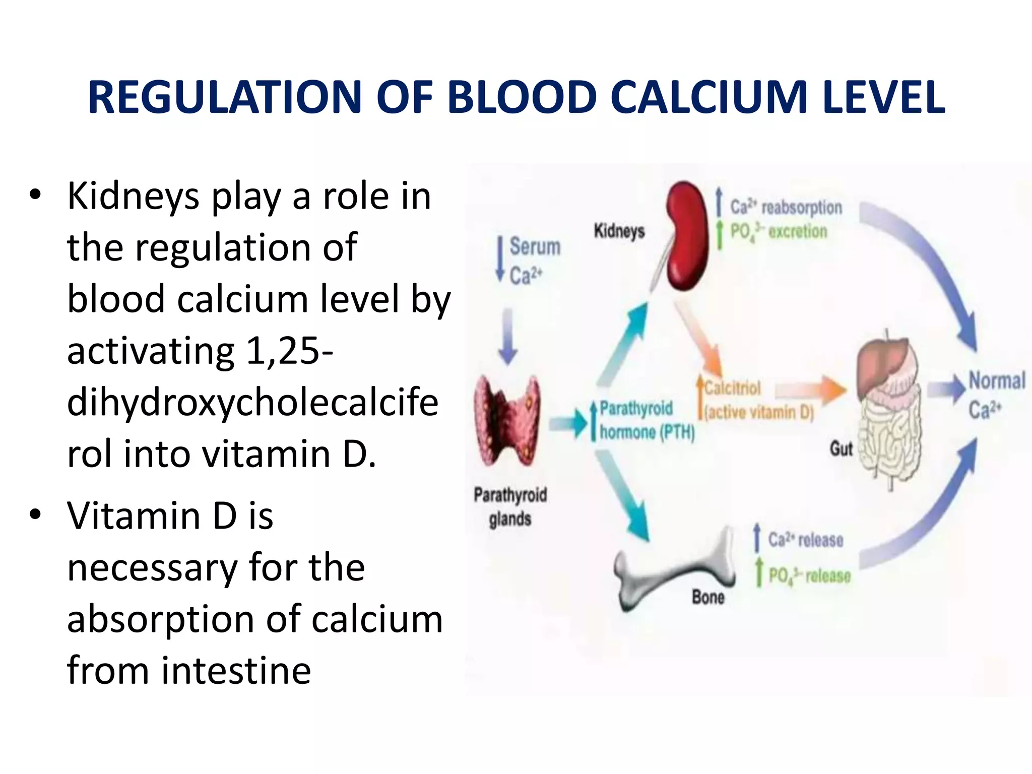
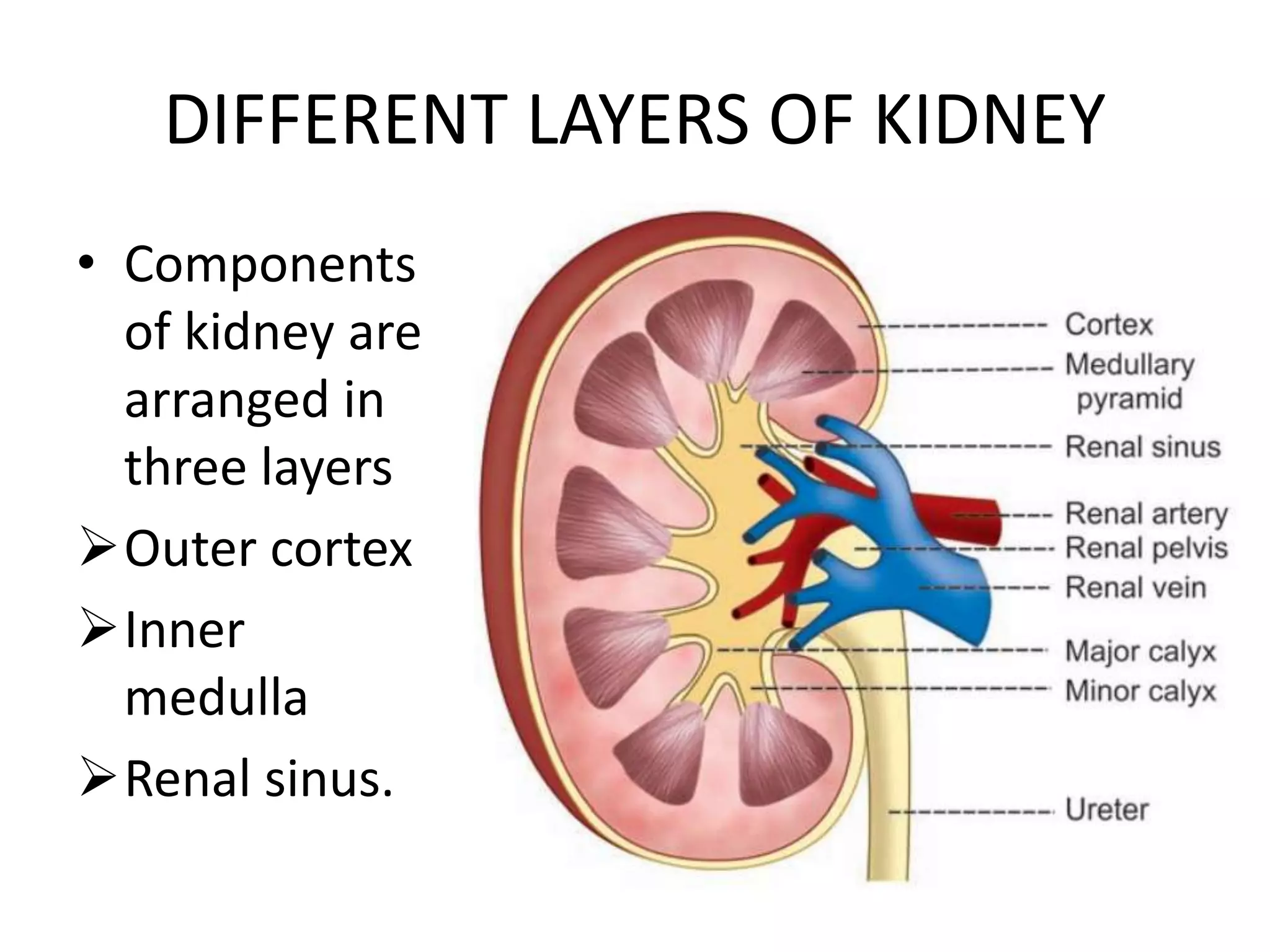
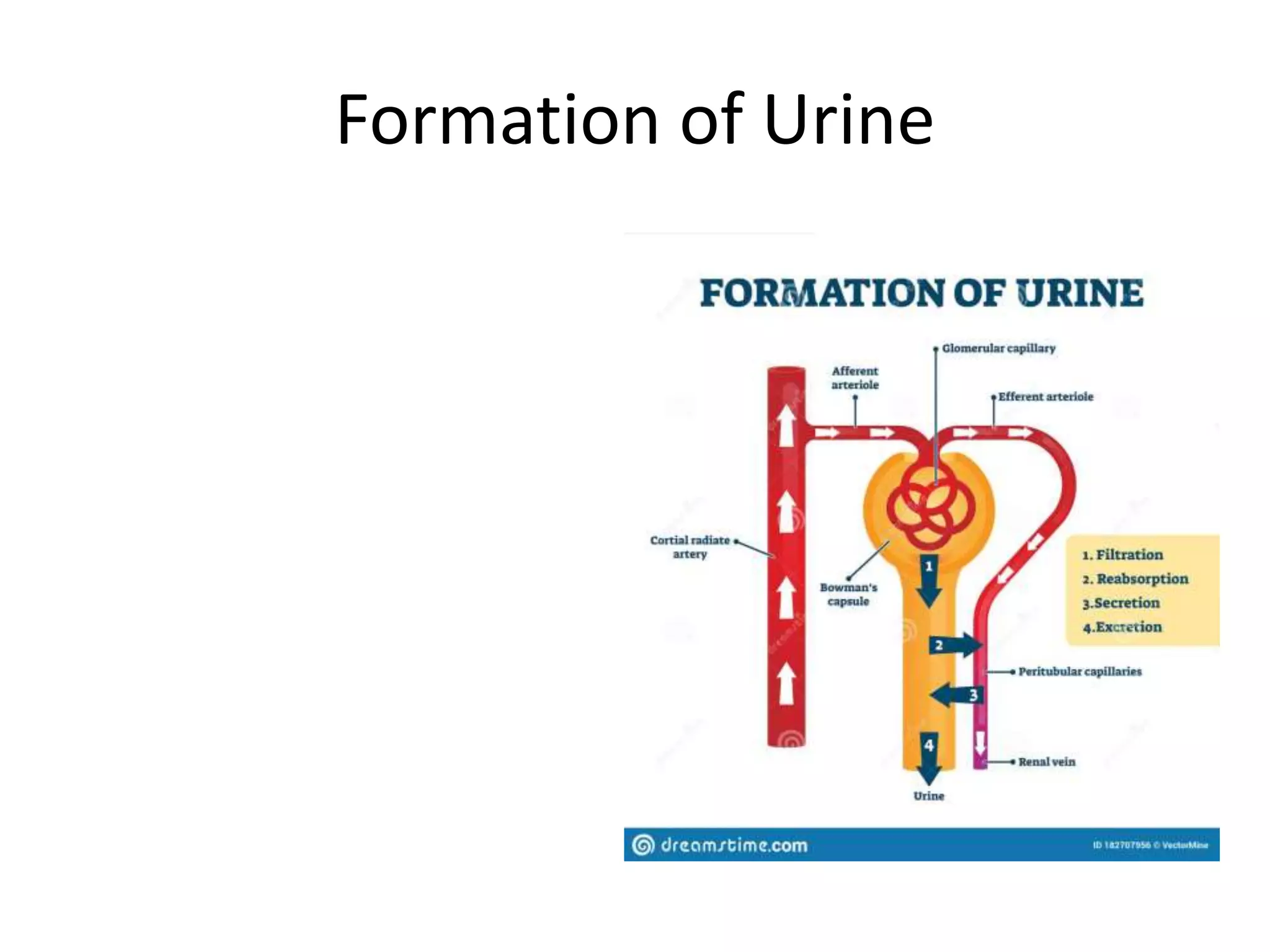
URINARY SYSTEM
SURESH BABU EMANDI M.Pharm
Vikas Institute of Pharmaceutical Sciences
Near Air Port, Rajahmundry,
Andhrapradesh-533102.
EXCREATION
• Excretion is the process by which the unwanted
substances and metabolic wastes are eliminated
from the body.
• A large amount of waste materials and carbon
dioxide are produced in the tissues during
metabolic process.
• In addition, residue of undigested food, heavy
metals, drugs, toxic substances and pathogenic
organisms like bacteria are also present in the
body.
1.Digestive system excretes food residues in the
form of feces. Some bacteria and toxic
substances also are excreted through feces
2. Lungs remove carbon dioxide and water vapor
3. Skin excretes water, salts and some wastes.
It also removes heat from the body
4. Liver excretes many substances like bile
pigments, heavy metals, drugs, toxins, bacteria,
etc. through bile.
FUNCTIONS OF KIDNEY
• Kidneys perform several vital functions
besides formation of urine.
• By excreting urine, kidneys play the principal
role in homeostasis.
• Hemopoietic function.
• Endocrine function.
• Regulation of blood pressure.
• Regulation of blood calcium level.
ROLE IN HOMEOSTASIS
• Primary function
of kidneys is
homeostasis.
• It is accomplished
by the formation
of urine.
• During the
formation of
urine, kidneys
regulate various
activities in the
body
i. Excretion of Waste Products
• Kidneys excrete the unwanted waste products,
which are formed during metabolic activities.
• Kidneys also excrete harmful foreign chemical
substances such as toxins, drugs, heavy metals
pesticides, etc.
iii. Maintenance of Electrolyte Balance
• Maintenance of
electrolyte balance,
especially sodium is
in relation to water
balance.
• Kidneys retain
sodium if the
osmolarity of body
water decreases and
eliminate sodium
when osmolarity
increases.
ii. Maintenance of Water Balance
• Kidneys maintain the
water balance in the
body by conserving
water when it is
decreased and excreting
water when it is excess in
the body.
• This is an important
process for homeostasis
iv. Maintenance of Acid–Base Balance
• The pH of the blood and
body fluids should be
maintained within
narrow range for healthy
living. It is achieved by
the function of kidneys.
• Body is under constant
threat to develop
acidosis, because of
production of lot of
acids during metabolic
activities.
HEMOPOIETIC FUNCTION
• Kidneys stimulate the
production of erythrocytes
by secreting erythropoietin.
• Erythropoietin is the
important stimulating factor
for erythropoiesis.
• Kidney also secretes another
factor called thrombopoietin,
which stimulates the
production of thrombocytes
ENDOCRINE FUNCTION
• Kidneys secrete many hormonal substances in
addition to erythropoietin and thrombopoietin.
• Hormones secreted by kidneys
i. Erythropoietin
ii. Thrombopoietin
iii. Renin
iv. 1,25-dihydroxycholecalciferol (calcitriol)
v. Prostaglandins
REGULATION OF BLOOD PRESSURE
• Kidneys play an
important role in
the long-term
regulation of
arterial blood
pressure by two
ways:
• i. By regulating the
volume of
extracellular fluid
• ii. Through renin-
angiotensin
mechanism.
REGULATION OF BLOOD CALCIUM LEVEL
• Kidneys play a role in
the regulation of
blood calcium level by
activating 1,25-
dihydroxycholecalcife
rol into vitamin D.
• Vitamin D is
necessary for the
absorption of calcium
from intestine
ANATOMY OF KIDNEY
• Kidney is a compound tubular gland covered
by a connective tissue capsule.
• There is a depression on the medial border of
kidney called hilum, through which renal
artery, renal veins, nerves and ureter pass. „
.
DIFFERENT LAYERS OF KIDNEY
• Components
of kidney are
arranged in
three layers
Outer cortex
Inner
medulla
Renal sinus.
DIFFERENT LAYERS OF KIDNEY
• Components of kidney are arranged in three
1. Outer cortex
2. Inner medulla
3. Renal sinus
• 1. Outer Cortex
Cortex is dark and granular in appearance. It
contains renal corpuscles and convoluted tubules.
• At intervals, cortical tissue penetrates medulla in
the form of columns, which are called renal
columns or columns of Bertini.
2. Inner Medulla
• Medulla contains tubular and vascular
structures arranged in parallel radial lines.
Medullary mass is divided into 8 to 18 medullary
or Malpighian pyramids.
• Broad base of each pyramid is in contact with
cortex and the apex projects into minor calyx.
3. Renal Sinus
• Renal sinus consists of the following
structures:
i. Upper expanded part of ureter called renal
pelvis
ii. Subdivisions of pelvis: 2 or 3 major calyces
and about 8 minor calyces
iii. Branches of nerves, arteries and tributaries of
veins
iv. Loose connective tissues and fat.
Formation of Urine
TUBULAR STRUCTURES OF KIDNEY
• Kidney is made up of closely arranged tubular
structures called uriniferous tubules.
• Blood vessels and interstitial connective tissues
are interposed between these tubules.
Uriniferous tubules include:
1. Terminal or secretary tubules called nephrons,
which are concerned with formation of urine.
2. Collecting ducts or tubules, which are concerned
with transport of urine from nephrons to pelvis of
ureter. Collecting ducts unite to form ducts of
Bellini, which open into minor calyces through
papilla.
Nephron
Nephron is defined as the structural and functional unit of kidney. Each kidney consists of 1
to 1.3 millions of nephrons.
Each nephron is formed by two parts .
1. A blind end called renal corpuscle or Malpighian corpuscle .
2. A tubular portion called renal tubule.
RENAL CORPUSCLE
• Renal corpuscle or
Malpighian corpuscle is a
spheroidal and slightly
flattened structure with a
diameter of about 200 µ.
• Function of the renal
corpuscle is the filtration
of blood which forms the
first phase of urine
formation
SITUATION OF RENAL CORPUSCLE AND TYPES OF NEPHRON
• Renal corpuscle is situated in the cortex of the kidney
either near the periphery or near the medulla.
Classification of Nephrons Based on the situation of
renal corpuscle, the nephrons are classified into two
types:
• 1. Cortical nephrons or superficial nephrons: Nephrons
having the corpuscles in outer cortex of the kidney
near the periphery . In human kidneys, 85% nephrons
are cortical nephrons.
• 2. Juxtamedullary nephrons: Nephrons having the
corpuscles in inner cortex near medulla or
corticomedullary junction.
STRUCTURE OF RENAL CORPUSCLE
Renal corpuscle is formed by two
portions:
1. Glomerulus
2. Bowman capsule.
Glomerulus
Glomerulus is a tuft of capillaries
enclosed by Bowman capsule. It
consists of glomerular capillaries
interposed between afferent arteriole
on one end and efferent arteriole on
the other end. Thus, the vascular
system in the glomerulus is purely
arterial.
Glomerular capillaries arise from the
afferent arteriole. After entering the
Bowman capsule, the affarteriole
divides into 4 or 5 large capillaries.
Bowman Capsule
• Bowman capsule is a capsular structure, which encloses the
glomerulus.
• It is formed by two layers:
• i. Inner visceral layer
• ii. Outer parietal layer. Visceral layer covers the glomerular
capillaries. It is continued as the parietal layer at the visceral
pole.
• Parietal layer is continued with the wall of the tubular
portion of nephron.
• The cleft like space between the visceral and parietal layers
is continued as the lumen of the tubular portion.
• Functional anatomy of Bowman capsule resembles a funnel
with filter paper. Diameter of Bowman capsule is 200 µ.
Tubular portion of nephron
• Tubular portion of nephron is the continuation
of Bowman capsule.
• It is made up of three parts:
• 1. Proximal convoluted tubule
• 2. Loop of Henle
• 3. Distal convoluted tubule.
PROXIMAL CONVOLUTED TUBULE
• Proximal convoluted tubule is the coiled
portion arising from Bowman capsule. It is
situated in the cortex. It is continued as
descending limb of loop of Henle. Length of
proximal convoluted tubule is 14 mm and the
diameter is 55 µ. Proximal convoluted tubule
is continued as loop of Henle
LOOP OF HENLE
• Loop of Henle consists of:
• i. Descending limb
• ii. Hairpin bend
• iii. Ascending limb.
• i. Descending Limb
• Descending limb of loop of Henle is made up of two
segments:
• a. Thick descending segment
• b. Thin descending segment.
• Thick descending segment Thick descending segment is
the direct continuation of the proximal convoluted tubule.
• It descends down into medulla. It has a length of 6 mm
and a diameter of 55 µ.
• It is formed by brush bordered cuboidal epithelial cells.
• Thin descending segment Thick descending segment is
continued as thin descending segment.
• It is formed by flattened epithelial cells without brush
border and it is continued as hairpin bend of the loop.
• ii. Hairpin Bend
• Hairpin bend formed by flattened epithelial cells
without brush border and it is continued as the
ascending limb of loop of Henle.
• iii. Ascending Limb
Ascending limb or segment of Henle loop has two
parts:
• a. Thin ascending segment
• b. Thick ascending segmen
DISTAL CONVOLUTED TUBULE
• Distal convoluted tubule is the continuation of
thick ascending segment and occupies the
cortex of kidney.
• It is continued as collecting duct. The length of
the distal convoluted tubule is 14.5 to 15 mm.
It has a diameter of 22 to 50 µ.
COLLECTING DUCT
• Distal convoluted tubule continues as the
initial or arched collecting duct, which is in
cortex. The lower part of the collecting duct
lies in medulla.
• Seven to ten initial collecting ducts unite to
form the straight collecting duct, which passes
through medulla.
• Thin ascending segment Thin ascending segment is
the continuation of hairpin bend.
• It is also lined by flattened epithelial cells without
brush border.
• Total length of thin descending segment, hairpin
bend and thin ascending segment of Henle loop is
10 mm to 15 mm and the diameter is 15 µ.
• Thin ascending segment is continued as thick
ascending segment.
• Thick ascending segment Thick ascending segment
is about 9 mm long with a diameter of 30 µ.
• Thick ascending segment is lined by cuboidal
epithelial cells without brush border.
Size and cells of different parts of nephron and collecting duct

URINARY SYSTEM

  • 1.
    URINARY SYSTEM SURESH BABUEMANDI M.Pharm Vikas Institute of Pharmaceutical Sciences Near Air Port, Rajahmundry, Andhrapradesh-533102.
  • 2.
    EXCREATION • Excretion isthe process by which the unwanted substances and metabolic wastes are eliminated from the body. • A large amount of waste materials and carbon dioxide are produced in the tissues during metabolic process. • In addition, residue of undigested food, heavy metals, drugs, toxic substances and pathogenic organisms like bacteria are also present in the body.
  • 3.
    1.Digestive system excretesfood residues in the form of feces. Some bacteria and toxic substances also are excreted through feces 2. Lungs remove carbon dioxide and water vapor 3. Skin excretes water, salts and some wastes. It also removes heat from the body 4. Liver excretes many substances like bile pigments, heavy metals, drugs, toxins, bacteria, etc. through bile.
  • 5.
    FUNCTIONS OF KIDNEY •Kidneys perform several vital functions besides formation of urine. • By excreting urine, kidneys play the principal role in homeostasis. • Hemopoietic function. • Endocrine function. • Regulation of blood pressure. • Regulation of blood calcium level.
  • 6.
    ROLE IN HOMEOSTASIS •Primary function of kidneys is homeostasis. • It is accomplished by the formation of urine. • During the formation of urine, kidneys regulate various activities in the body
  • 7.
    i. Excretion ofWaste Products • Kidneys excrete the unwanted waste products, which are formed during metabolic activities. • Kidneys also excrete harmful foreign chemical substances such as toxins, drugs, heavy metals pesticides, etc.
  • 8.
    iii. Maintenance ofElectrolyte Balance • Maintenance of electrolyte balance, especially sodium is in relation to water balance. • Kidneys retain sodium if the osmolarity of body water decreases and eliminate sodium when osmolarity increases.
  • 9.
    ii. Maintenance ofWater Balance • Kidneys maintain the water balance in the body by conserving water when it is decreased and excreting water when it is excess in the body. • This is an important process for homeostasis
  • 10.
    iv. Maintenance ofAcid–Base Balance • The pH of the blood and body fluids should be maintained within narrow range for healthy living. It is achieved by the function of kidneys. • Body is under constant threat to develop acidosis, because of production of lot of acids during metabolic activities.
  • 11.
    HEMOPOIETIC FUNCTION • Kidneysstimulate the production of erythrocytes by secreting erythropoietin. • Erythropoietin is the important stimulating factor for erythropoiesis. • Kidney also secretes another factor called thrombopoietin, which stimulates the production of thrombocytes
  • 12.
    ENDOCRINE FUNCTION • Kidneyssecrete many hormonal substances in addition to erythropoietin and thrombopoietin. • Hormones secreted by kidneys i. Erythropoietin ii. Thrombopoietin iii. Renin iv. 1,25-dihydroxycholecalciferol (calcitriol) v. Prostaglandins
  • 14.
    REGULATION OF BLOODPRESSURE • Kidneys play an important role in the long-term regulation of arterial blood pressure by two ways: • i. By regulating the volume of extracellular fluid • ii. Through renin- angiotensin mechanism.
  • 15.
    REGULATION OF BLOODCALCIUM LEVEL • Kidneys play a role in the regulation of blood calcium level by activating 1,25- dihydroxycholecalcife rol into vitamin D. • Vitamin D is necessary for the absorption of calcium from intestine
  • 16.
    ANATOMY OF KIDNEY •Kidney is a compound tubular gland covered by a connective tissue capsule. • There is a depression on the medial border of kidney called hilum, through which renal artery, renal veins, nerves and ureter pass. „ .
  • 17.
    DIFFERENT LAYERS OFKIDNEY • Components of kidney are arranged in three layers Outer cortex Inner medulla Renal sinus.
  • 18.
    DIFFERENT LAYERS OFKIDNEY • Components of kidney are arranged in three 1. Outer cortex 2. Inner medulla 3. Renal sinus
  • 19.
    • 1. OuterCortex Cortex is dark and granular in appearance. It contains renal corpuscles and convoluted tubules. • At intervals, cortical tissue penetrates medulla in the form of columns, which are called renal columns or columns of Bertini.
  • 20.
    2. Inner Medulla •Medulla contains tubular and vascular structures arranged in parallel radial lines. Medullary mass is divided into 8 to 18 medullary or Malpighian pyramids. • Broad base of each pyramid is in contact with cortex and the apex projects into minor calyx.
  • 21.
    3. Renal Sinus •Renal sinus consists of the following structures: i. Upper expanded part of ureter called renal pelvis ii. Subdivisions of pelvis: 2 or 3 major calyces and about 8 minor calyces iii. Branches of nerves, arteries and tributaries of veins iv. Loose connective tissues and fat.
  • 23.
  • 24.
    TUBULAR STRUCTURES OFKIDNEY • Kidney is made up of closely arranged tubular structures called uriniferous tubules. • Blood vessels and interstitial connective tissues are interposed between these tubules. Uriniferous tubules include: 1. Terminal or secretary tubules called nephrons, which are concerned with formation of urine. 2. Collecting ducts or tubules, which are concerned with transport of urine from nephrons to pelvis of ureter. Collecting ducts unite to form ducts of Bellini, which open into minor calyces through papilla.
  • 26.
    Nephron Nephron is definedas the structural and functional unit of kidney. Each kidney consists of 1 to 1.3 millions of nephrons. Each nephron is formed by two parts . 1. A blind end called renal corpuscle or Malpighian corpuscle . 2. A tubular portion called renal tubule.
  • 27.
    RENAL CORPUSCLE • Renalcorpuscle or Malpighian corpuscle is a spheroidal and slightly flattened structure with a diameter of about 200 µ. • Function of the renal corpuscle is the filtration of blood which forms the first phase of urine formation
  • 28.
    SITUATION OF RENALCORPUSCLE AND TYPES OF NEPHRON • Renal corpuscle is situated in the cortex of the kidney either near the periphery or near the medulla. Classification of Nephrons Based on the situation of renal corpuscle, the nephrons are classified into two types: • 1. Cortical nephrons or superficial nephrons: Nephrons having the corpuscles in outer cortex of the kidney near the periphery . In human kidneys, 85% nephrons are cortical nephrons. • 2. Juxtamedullary nephrons: Nephrons having the corpuscles in inner cortex near medulla or corticomedullary junction.
  • 29.
    STRUCTURE OF RENALCORPUSCLE Renal corpuscle is formed by two portions: 1. Glomerulus 2. Bowman capsule. Glomerulus Glomerulus is a tuft of capillaries enclosed by Bowman capsule. It consists of glomerular capillaries interposed between afferent arteriole on one end and efferent arteriole on the other end. Thus, the vascular system in the glomerulus is purely arterial. Glomerular capillaries arise from the afferent arteriole. After entering the Bowman capsule, the affarteriole divides into 4 or 5 large capillaries.
  • 30.
    Bowman Capsule • Bowmancapsule is a capsular structure, which encloses the glomerulus. • It is formed by two layers: • i. Inner visceral layer • ii. Outer parietal layer. Visceral layer covers the glomerular capillaries. It is continued as the parietal layer at the visceral pole. • Parietal layer is continued with the wall of the tubular portion of nephron. • The cleft like space between the visceral and parietal layers is continued as the lumen of the tubular portion. • Functional anatomy of Bowman capsule resembles a funnel with filter paper. Diameter of Bowman capsule is 200 µ.
  • 32.
    Tubular portion ofnephron • Tubular portion of nephron is the continuation of Bowman capsule. • It is made up of three parts: • 1. Proximal convoluted tubule • 2. Loop of Henle • 3. Distal convoluted tubule.
  • 33.
    PROXIMAL CONVOLUTED TUBULE •Proximal convoluted tubule is the coiled portion arising from Bowman capsule. It is situated in the cortex. It is continued as descending limb of loop of Henle. Length of proximal convoluted tubule is 14 mm and the diameter is 55 µ. Proximal convoluted tubule is continued as loop of Henle
  • 34.
    LOOP OF HENLE •Loop of Henle consists of: • i. Descending limb • ii. Hairpin bend • iii. Ascending limb.
  • 35.
    • i. DescendingLimb • Descending limb of loop of Henle is made up of two segments: • a. Thick descending segment • b. Thin descending segment. • Thick descending segment Thick descending segment is the direct continuation of the proximal convoluted tubule. • It descends down into medulla. It has a length of 6 mm and a diameter of 55 µ. • It is formed by brush bordered cuboidal epithelial cells. • Thin descending segment Thick descending segment is continued as thin descending segment. • It is formed by flattened epithelial cells without brush border and it is continued as hairpin bend of the loop.
  • 36.
    • ii. HairpinBend • Hairpin bend formed by flattened epithelial cells without brush border and it is continued as the ascending limb of loop of Henle. • iii. Ascending Limb Ascending limb or segment of Henle loop has two parts: • a. Thin ascending segment • b. Thick ascending segmen
  • 37.
    DISTAL CONVOLUTED TUBULE •Distal convoluted tubule is the continuation of thick ascending segment and occupies the cortex of kidney. • It is continued as collecting duct. The length of the distal convoluted tubule is 14.5 to 15 mm. It has a diameter of 22 to 50 µ.
  • 38.
    COLLECTING DUCT • Distalconvoluted tubule continues as the initial or arched collecting duct, which is in cortex. The lower part of the collecting duct lies in medulla. • Seven to ten initial collecting ducts unite to form the straight collecting duct, which passes through medulla.
  • 39.
    • Thin ascendingsegment Thin ascending segment is the continuation of hairpin bend. • It is also lined by flattened epithelial cells without brush border. • Total length of thin descending segment, hairpin bend and thin ascending segment of Henle loop is 10 mm to 15 mm and the diameter is 15 µ. • Thin ascending segment is continued as thick ascending segment. • Thick ascending segment Thick ascending segment is about 9 mm long with a diameter of 30 µ. • Thick ascending segment is lined by cuboidal epithelial cells without brush border.
  • 40.
    Size and cellsof different parts of nephron and collecting duct