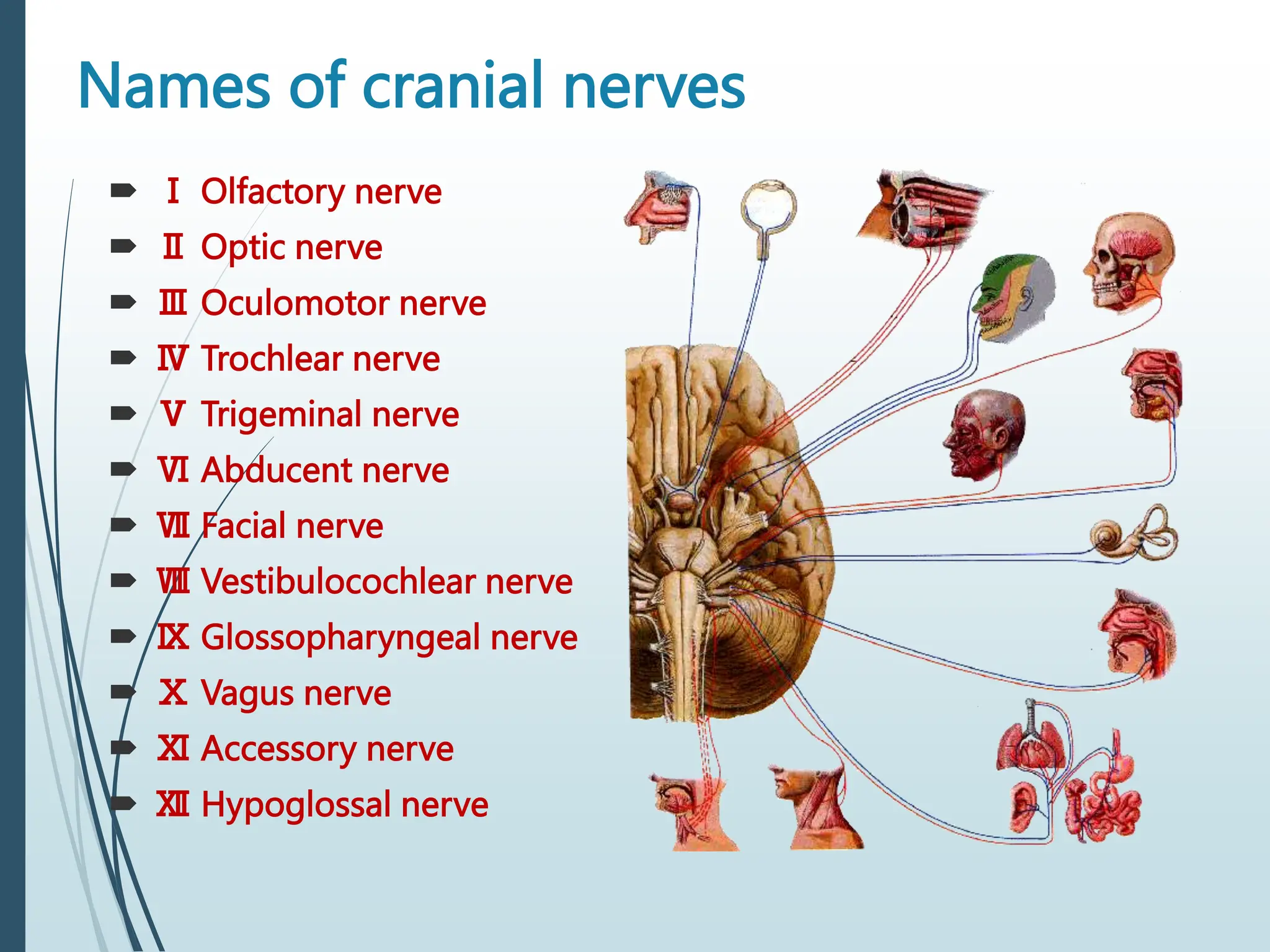
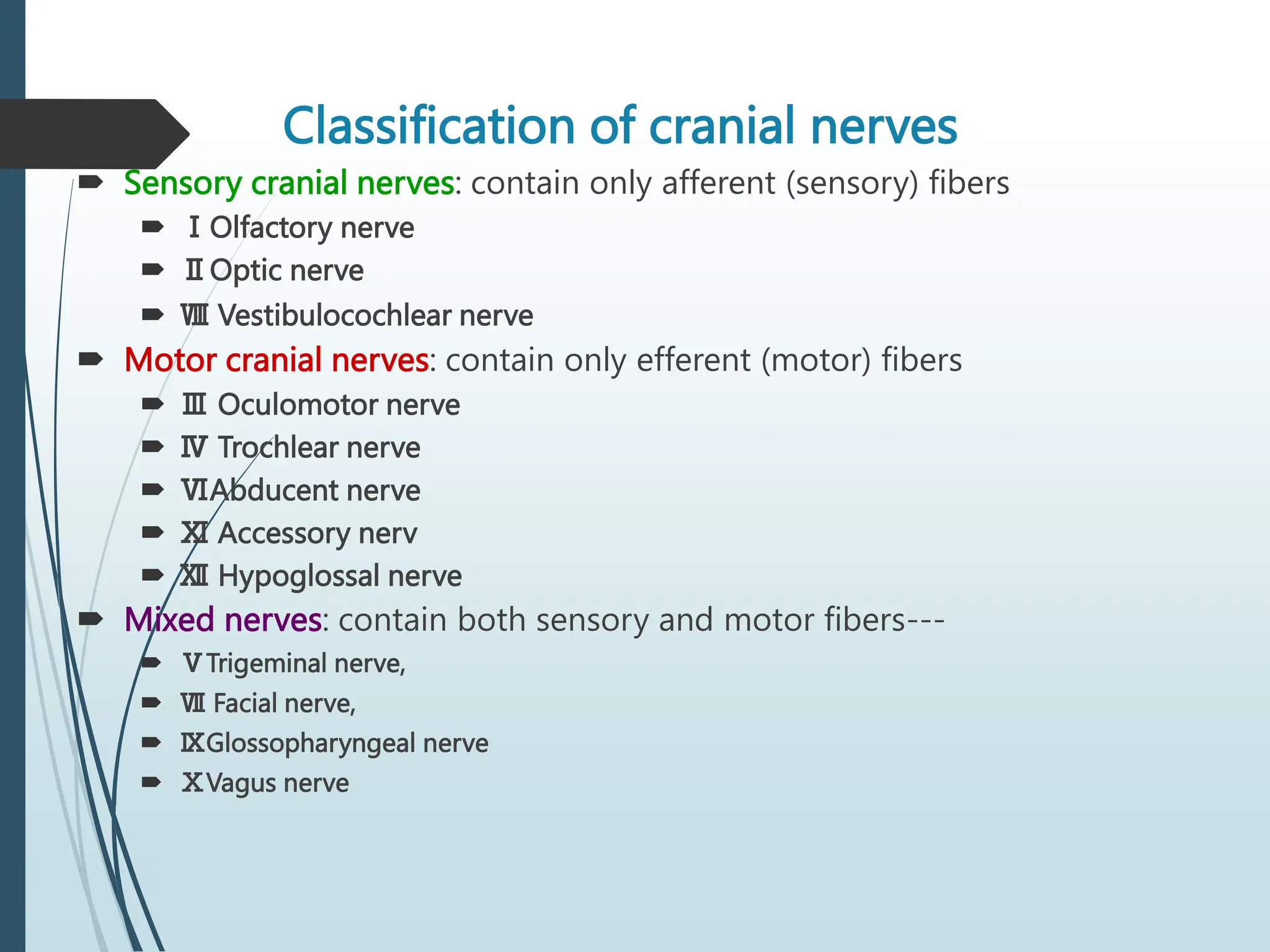
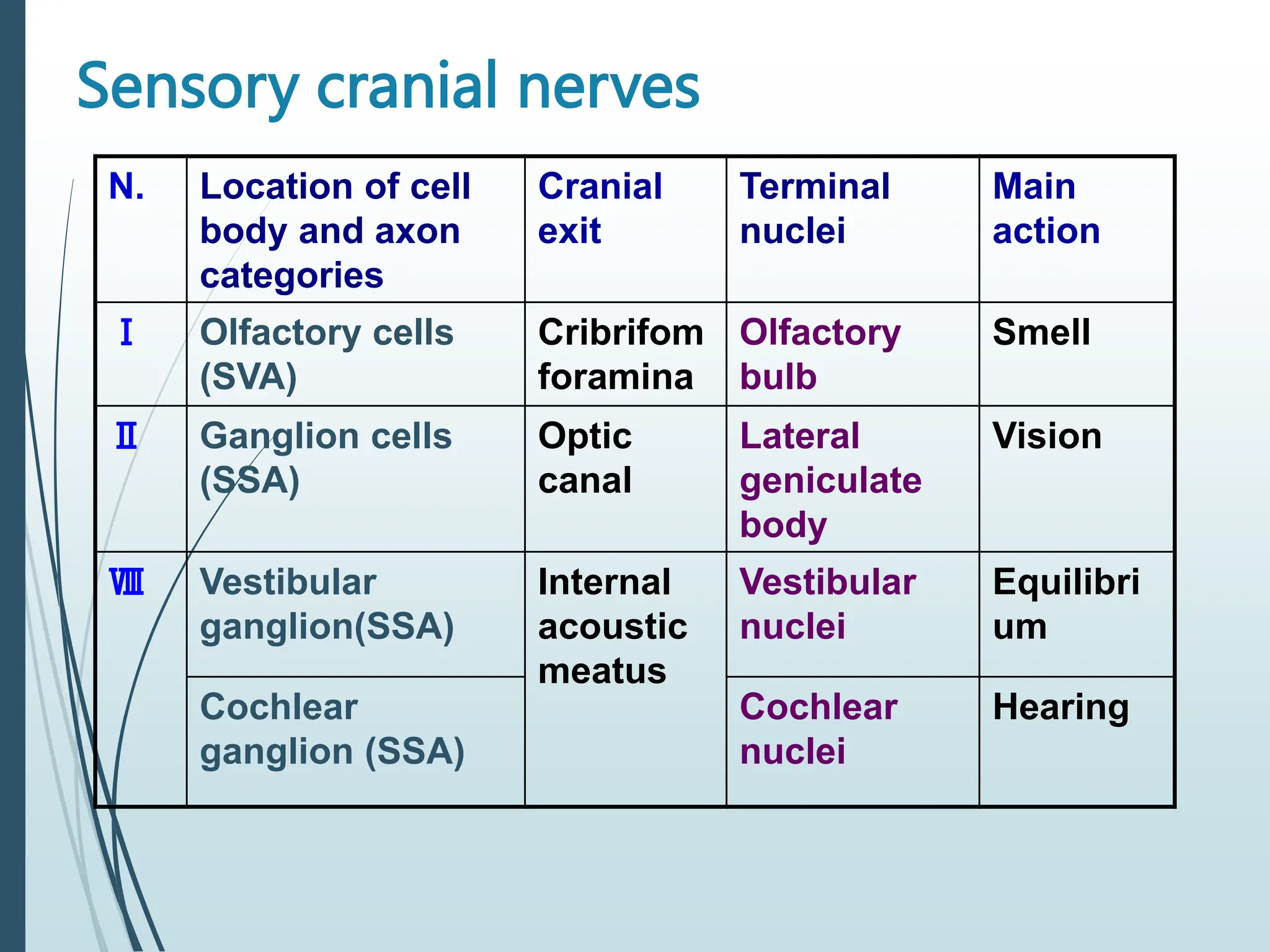
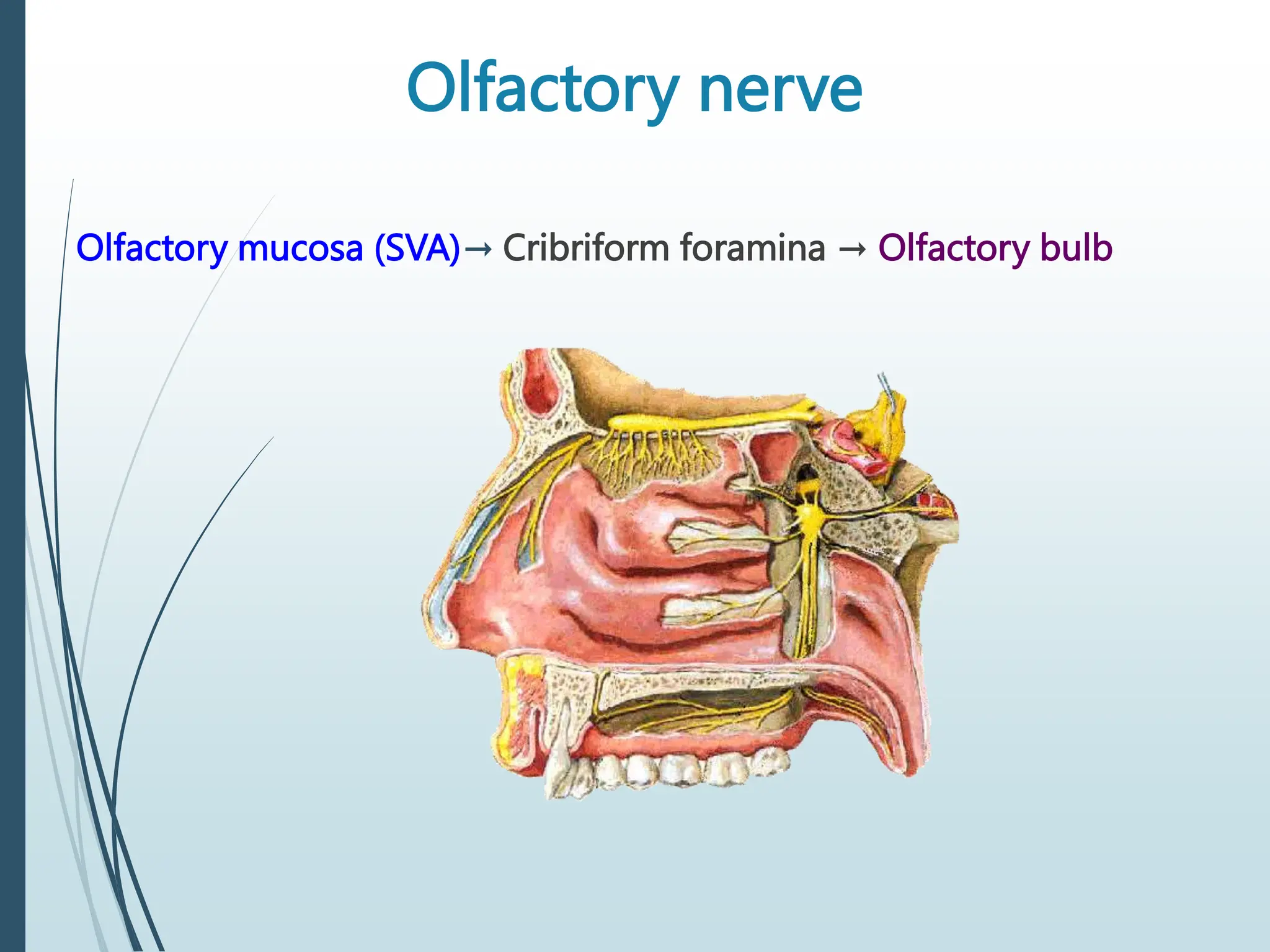
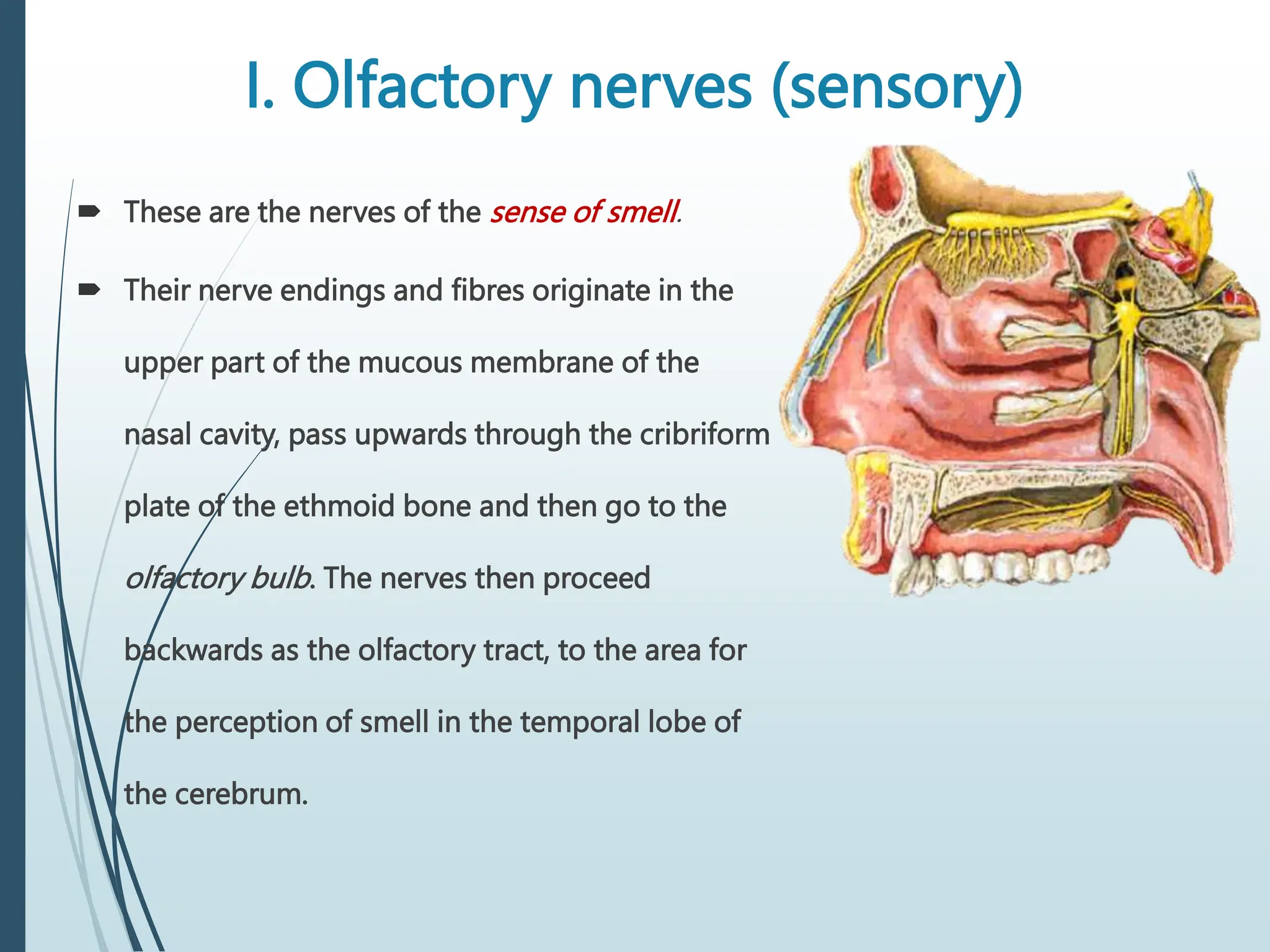
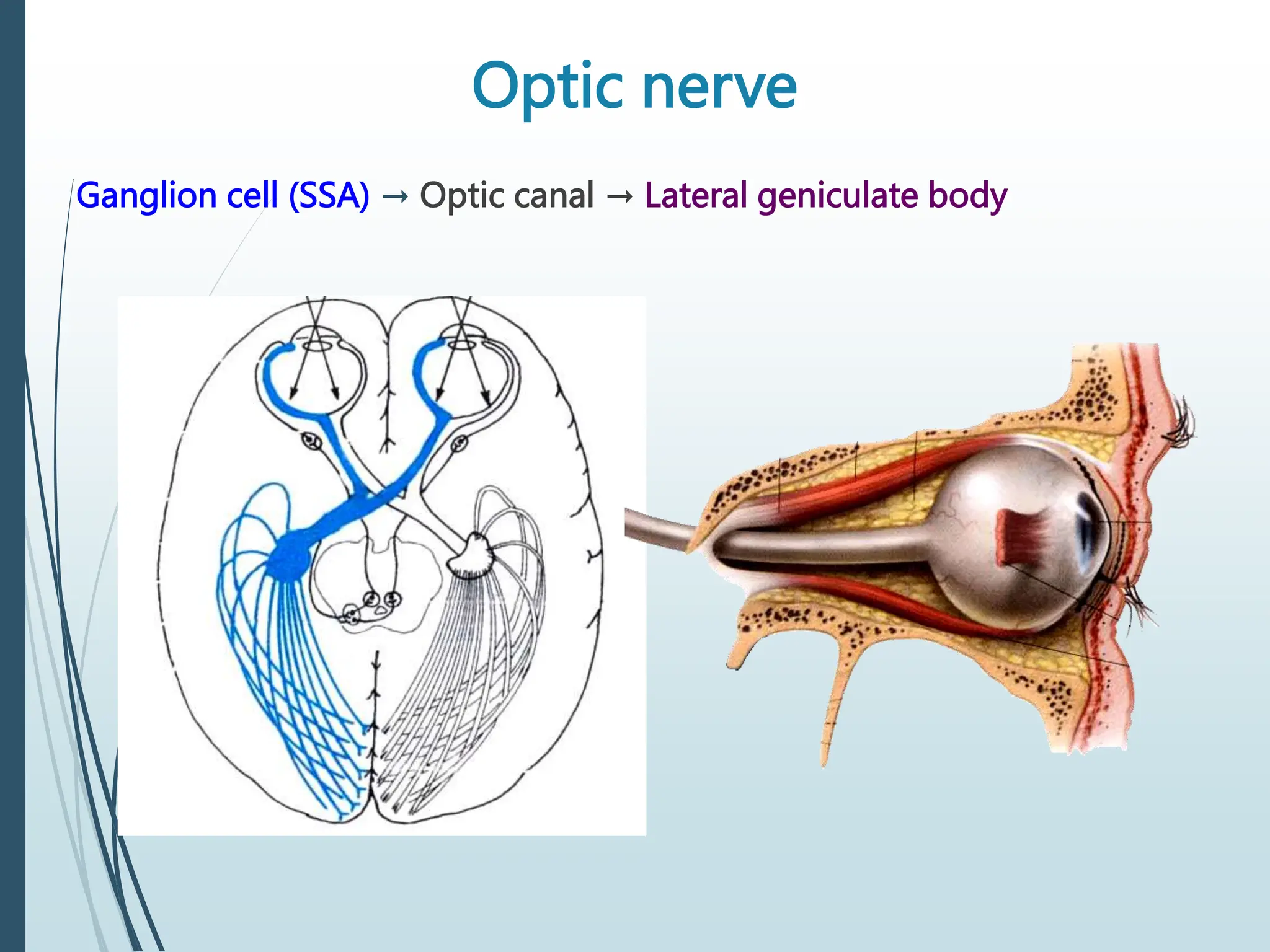
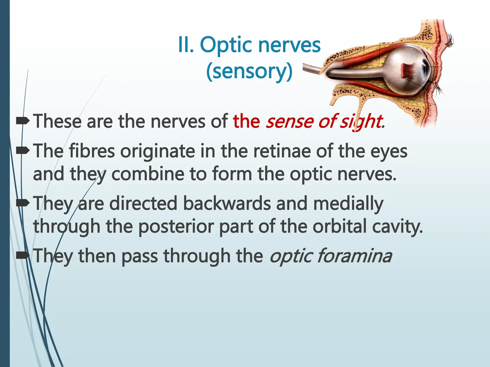
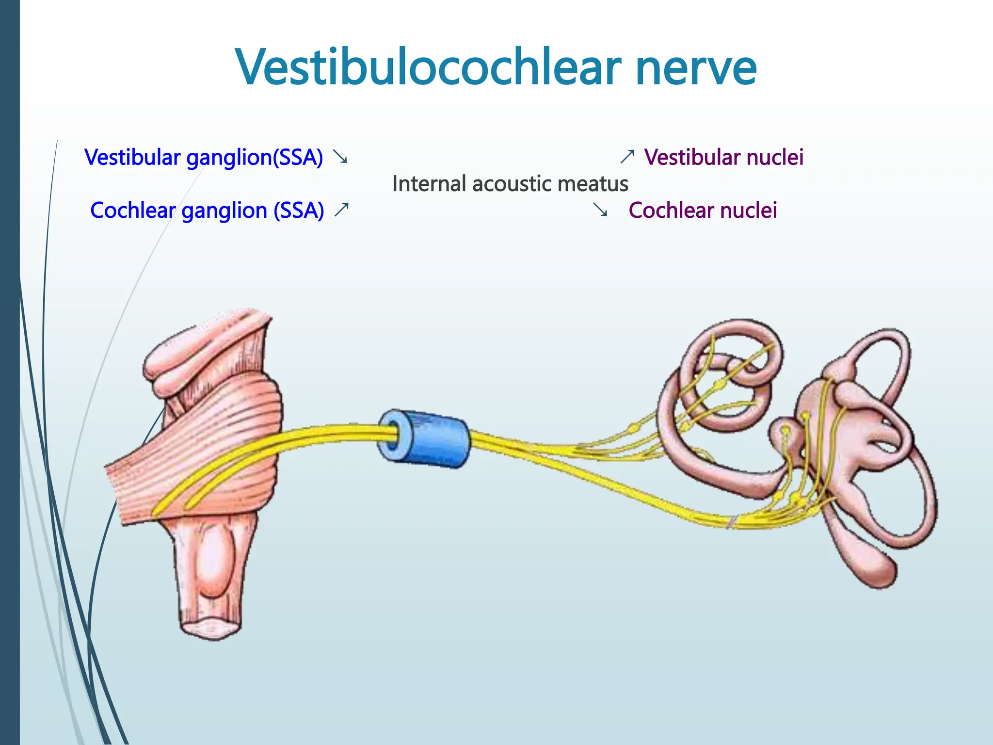
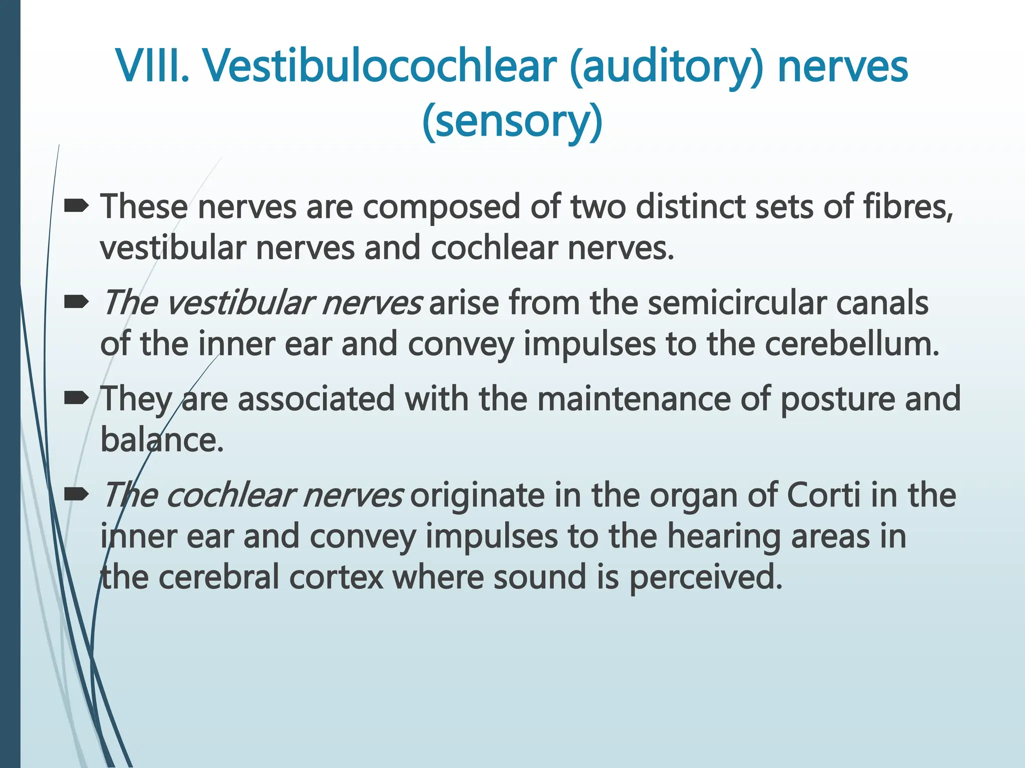
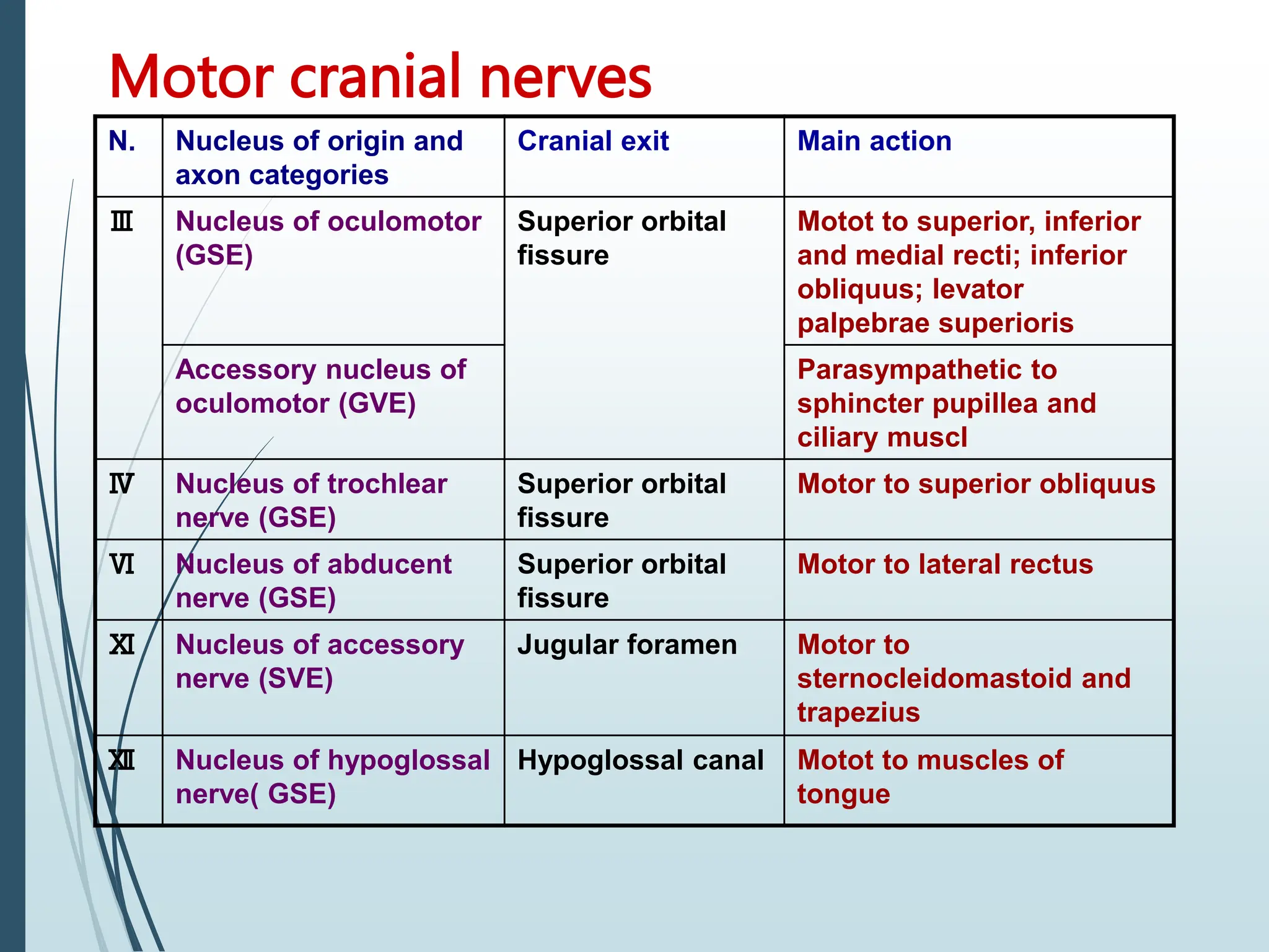
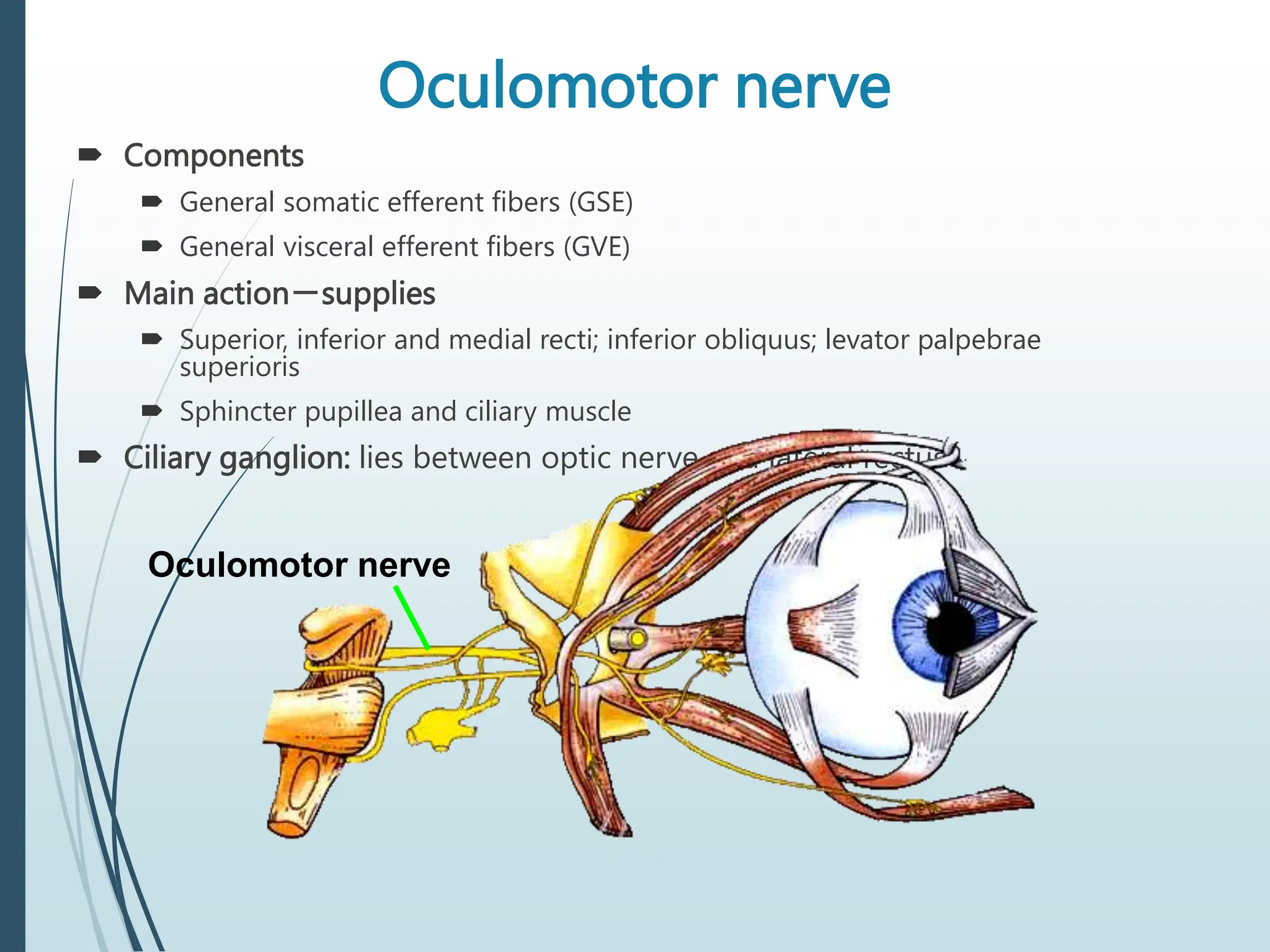
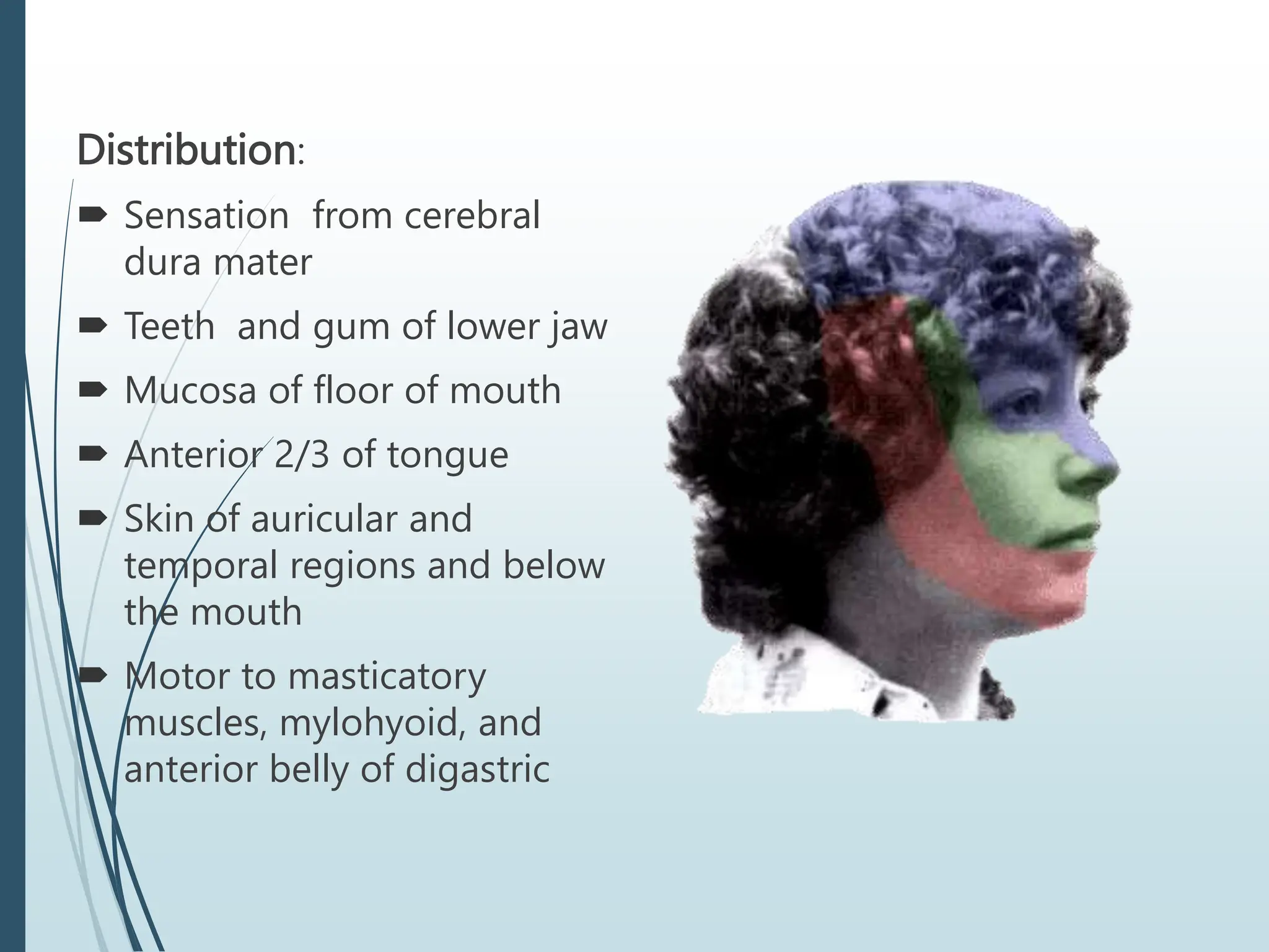
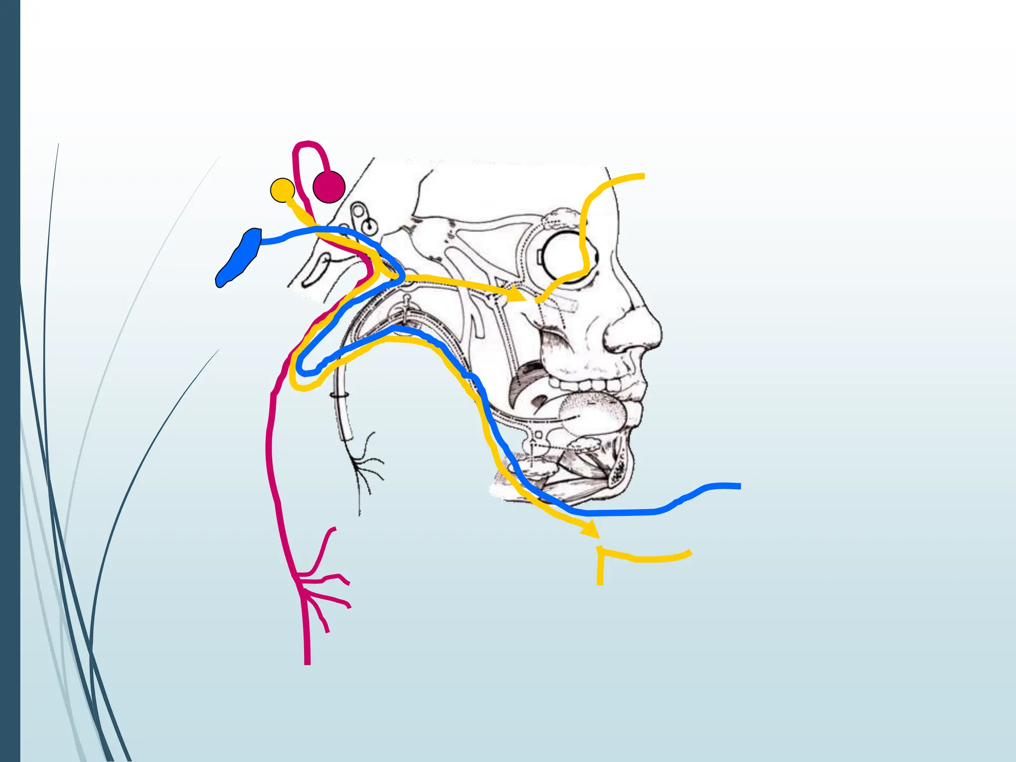
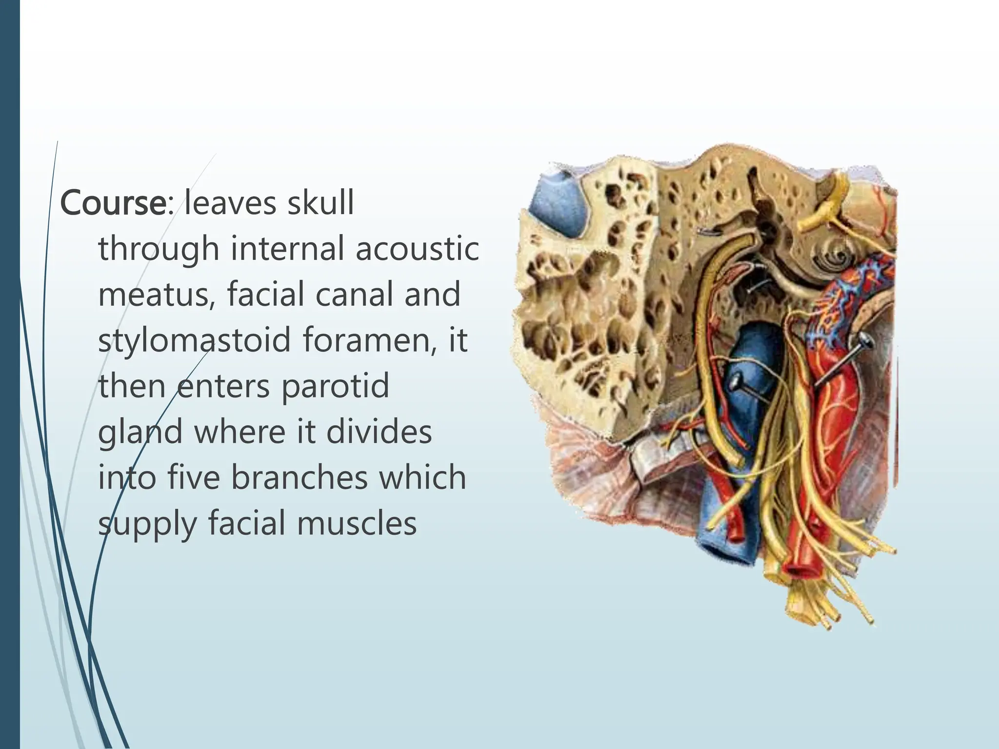
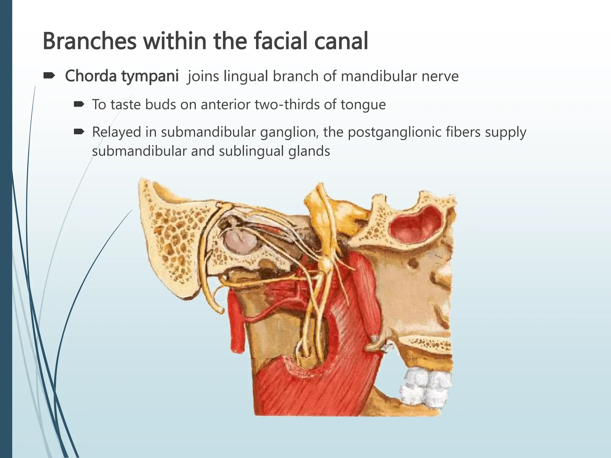
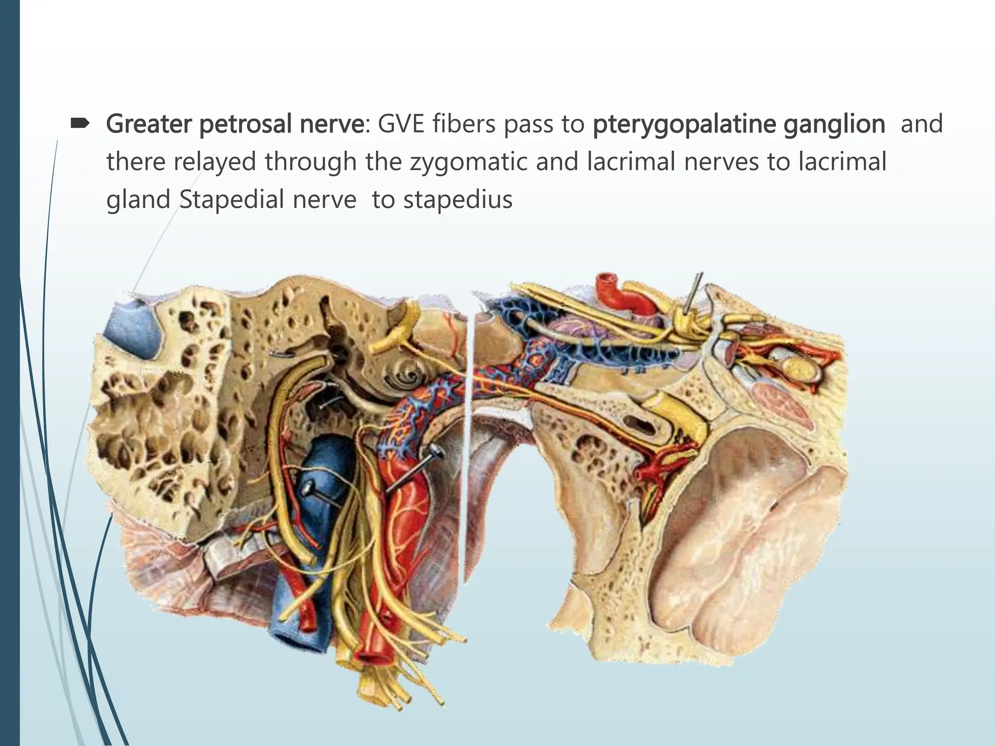
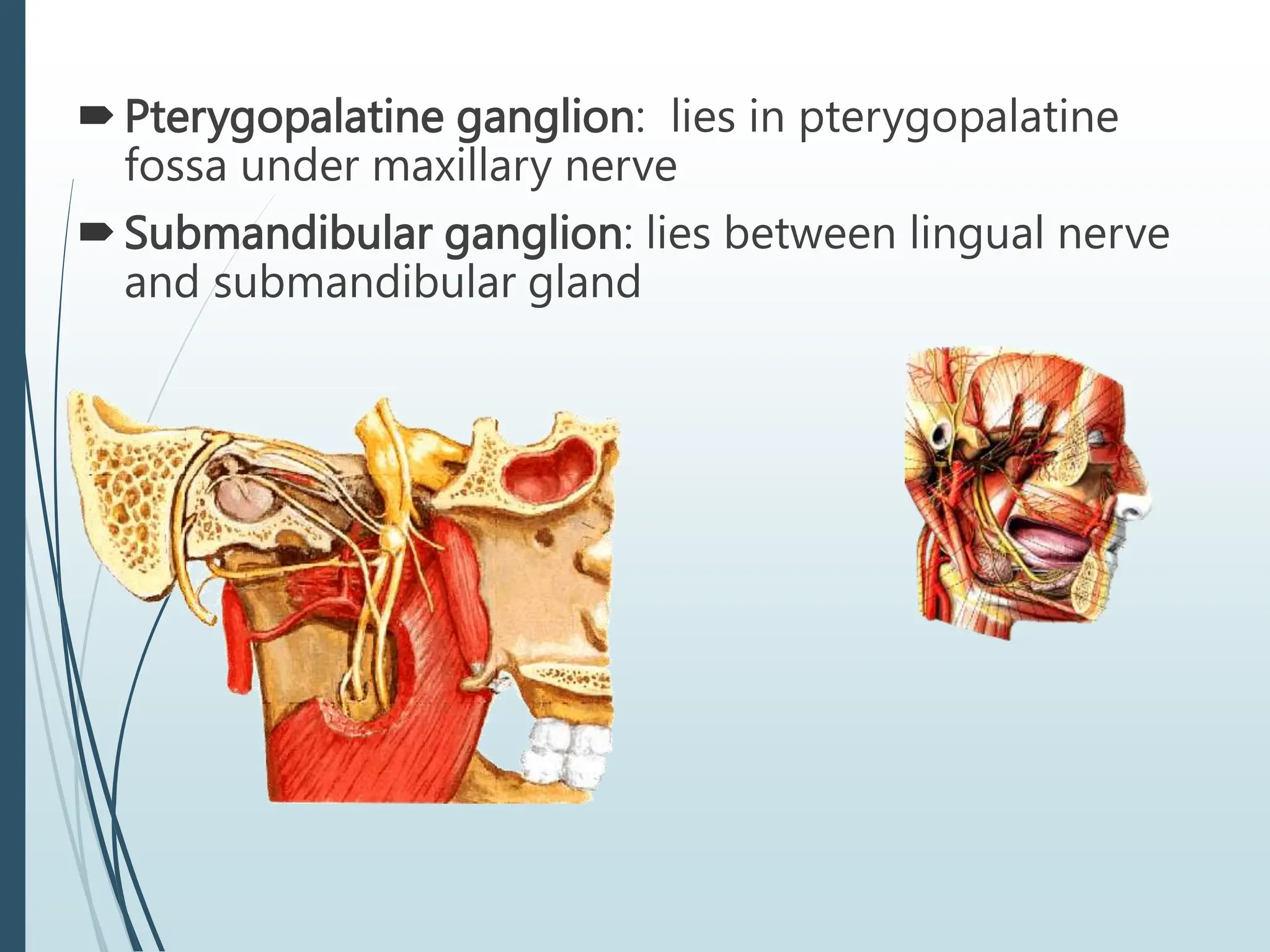
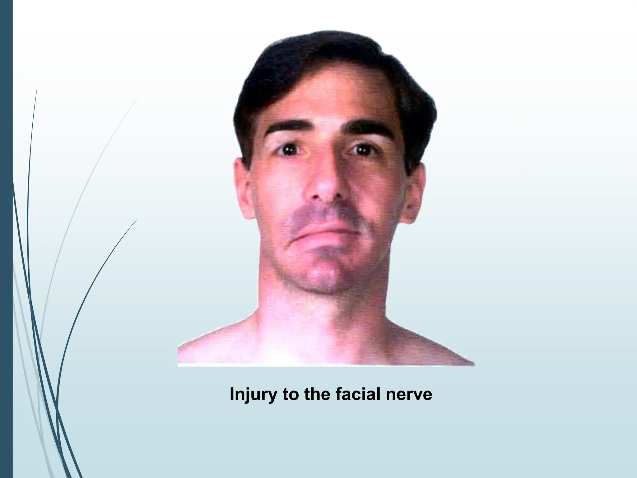
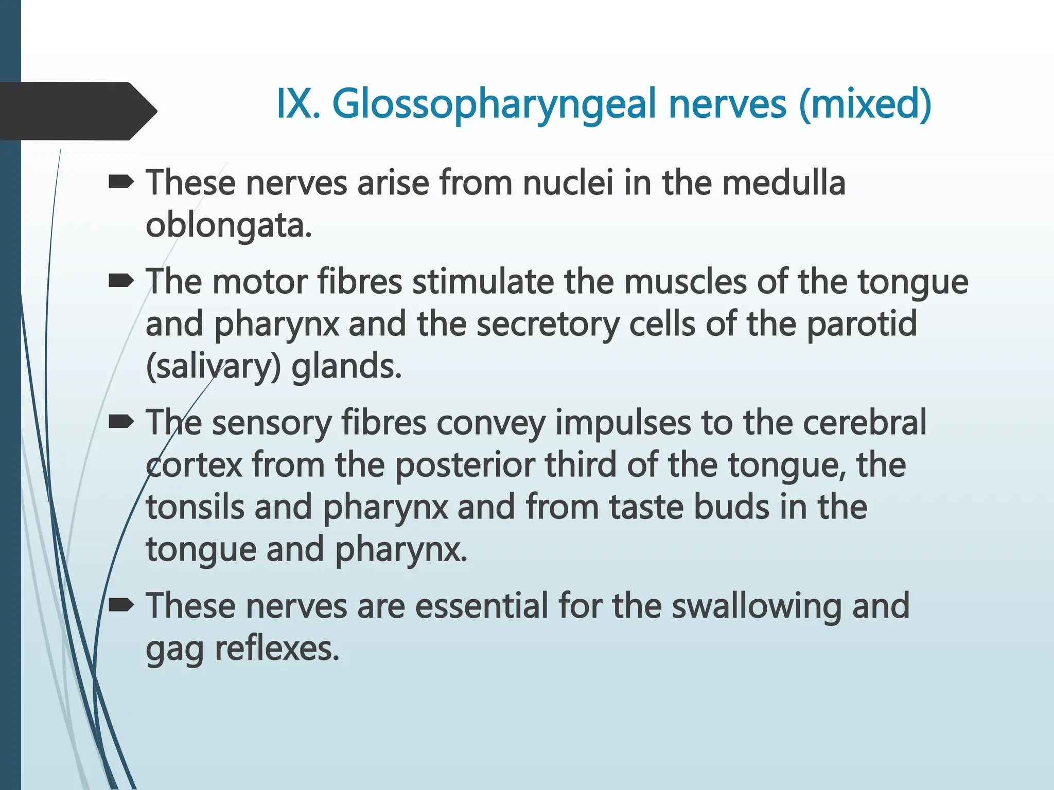
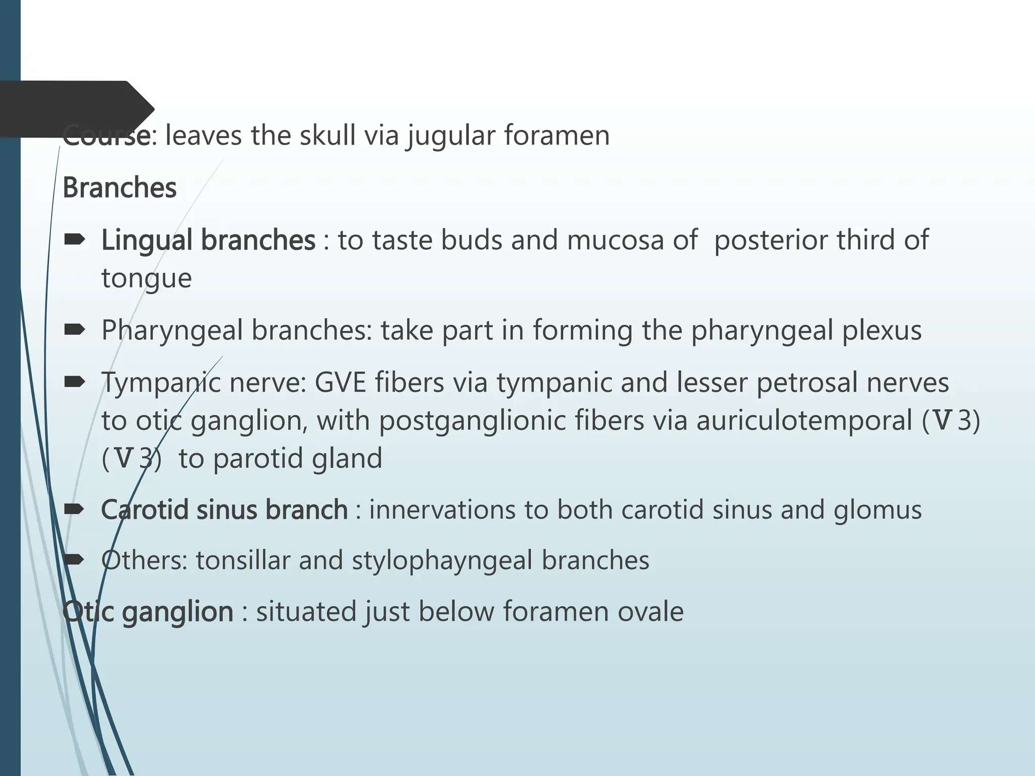
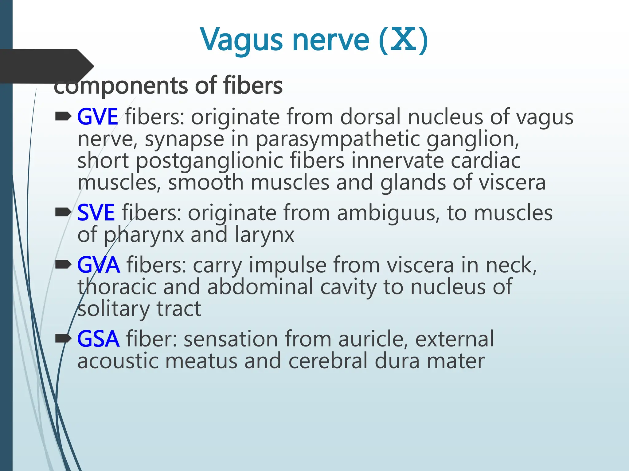
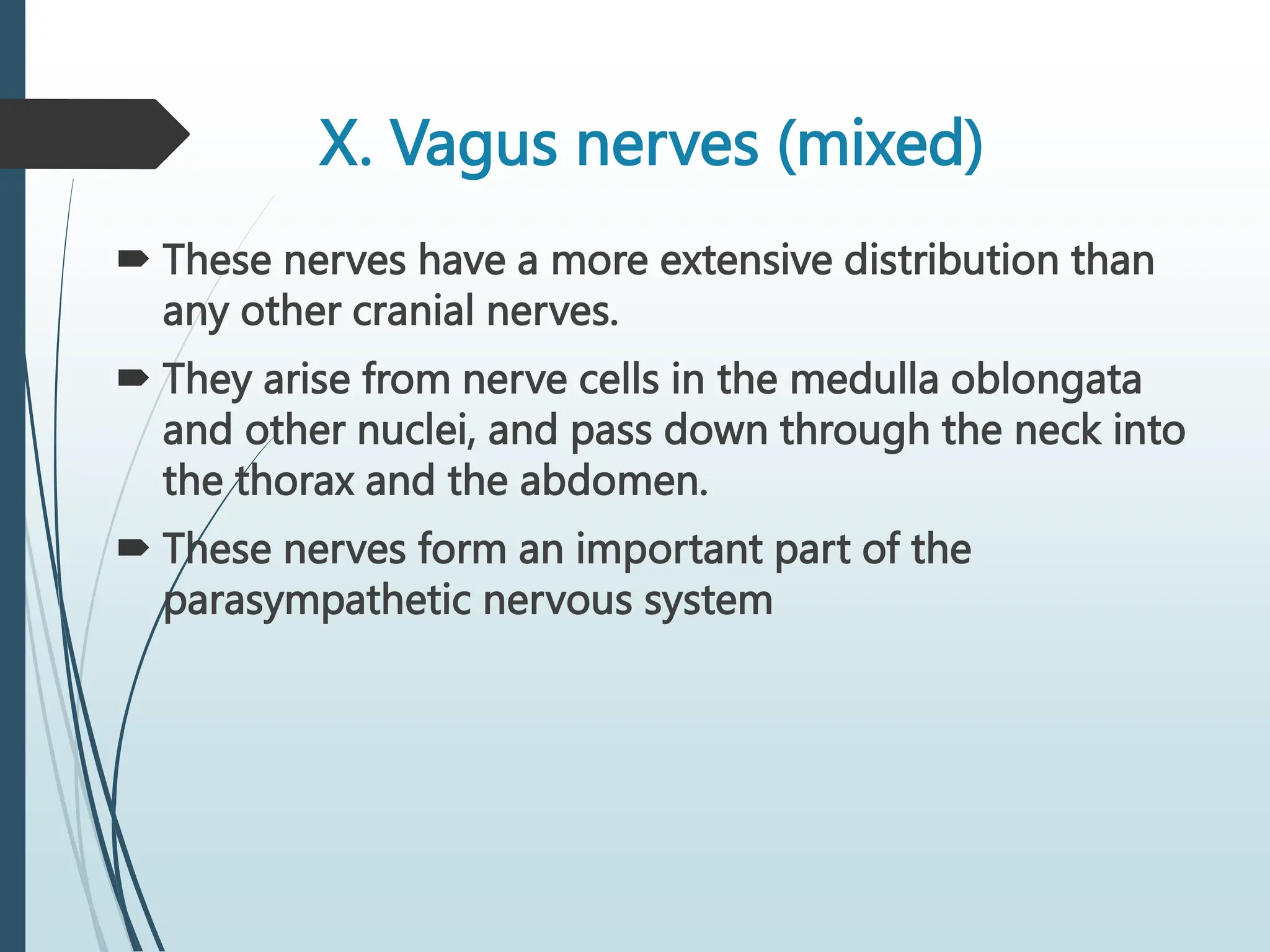
1. Cranial Nerves
SURESH BABU EMANDI M.Pharm
Department of Pharmacognosy,
Vikas Institute of Pharmaceutical Sciences
Near Air Port, Rajahmundry,
Andhrapradesh-533102.
6. I. Olfactory nerves (sensory)
These are the nerves of the sense of smell.
Their nerve endings and fibres originate in the
upper part of the mucous membrane of the
nasal cavity, pass upwards through the cribriform
plate of the ethmoid bone and then go to the
olfactory bulb. The nerves then proceed
backwards as the olfactory tract, to the area for
the perception of smell in the temporal lobe of
the cerebrum.
8. II. Optic nerves
(sensory)
These are the nerves of the sense of sight.
The fibres originate in the retinae of the eyes
and they combine to form the optic nerves.
They are directed backwards and medially
through the posterior part of the orbital cavity.
They then pass through the optic foramina
10. VIII. Vestibulocochlear (auditory) nerves
(sensory)
These nerves are composed of two distinct sets of fibres,
vestibular nerves and cochlear nerves.
The vestibular nerves arise from the semicircular canals
of the inner ear and convey impulses to the cerebellum.
They are associated with the maintenance of posture and
balance.
The cochlear nerves originate in the organ of Corti in the
inner ear and convey impulses to the hearing areas in
the cerebral cortex where sound is perceived.
11. Motor cranial nerves
N. Nucleus of origin and
axon categories
Cranial exit Main action
Ⅲ Nucleus of oculomotor
(GSE)
Superior orbital
fissure
Motot to superior, inferior
and medial recti; inferior
obliquus; levator
palpebrae superioris
Accessory nucleus of
oculomotor (GVE)
Parasympathetic to
sphincter pupillea and
ciliary muscl
Ⅳ Nucleus of trochlear
nerve (GSE)
Superior orbital
fissure
Motor to superior obliquus
Ⅵ Nucleus of abducent
nerve (GSE)
Superior orbital
fissure
Motor to lateral rectus
Ⅺ Nucleus of accessory
nerve (SVE)
Jugular foramen Motor to
sternocleidomastoid and
trapezius
Ⅻ Nucleus of hypoglossal
nerve( GSE)
Hypoglossal canal Motot to muscles of
tongue
12.
13. Oculomotor nerve
Components
General somatic efferent fibers (GSE)
General visceral efferent fibers (GVE)
Main action-supplies
Superior, inferior and medial recti; inferior obliquus; levator palpebrae
superioris
Sphincter pupillea and ciliary muscle
Ciliary ganglion: lies between optic nerve and lateral rectus
Oculomotor nerve
14. III. Oculomotor nerves (motor)
These nerves arise from nerve cells near the cerebral
aqueduct.
They supply:
four extraocular muscles, which move the eyeball, i.e. the
superior, medial and inferior recti and the inferior oblique
muscle
intraocular muscles:
— ciliary muscles which alter the shape of the lens,
changing its refractive power
— circular muscles of the iris which constrict the pupil
the levator palpebrae muscle which raises the upper eyelid.
16. IV. Trochlear nerves (motor)
These nerves arise from nerve cells near the
cerebral aqueduct.
They supply the superior oblique muscles of the
eyes.
17. VI. Abducent nerves (motor)
These nerves arise from a group of nerve cells
lying under the floor of the fourth ventricle.
They supply the lateral rectus muscles of the
eyeballs
19. XII. Hypoglossal nerves (motor)
These nerves arise from cells in the medulla
oblongata.
They supply the muscles of the tongue and
muscles.
surrounding the hyoid bone and contribute
to swallowing and speech.
22. Trigeminal nerve
Components of fibers
SVE fibers: originate from motor nucleus of
trigeminal nerve, and supply masticatory
muscles
GSA fibers: transmit facial sensation to sensory
nuclei of trigeminal nerve, the GSA fibers have
their cell bodies in trigeminal ganglion, which
lies on the apex of petrous part of temporal
bone
23. V. Trigeminal nerves (mixed)
These nerves contain motor and sensory fibres and
are among the largest of the cranial nerves.
They are the chief sensory nerves for the face and
head (including the oral and nasal cavities and
teeth), receiving impulses of pain, temperature and
touch.
The motor fibres stimulate the muscles of
mastication.There are three main branches of the
trigeminal nerves. The dermatomes supplied by the
sensory fibres
24. .
The ophthalmic nerves are sensory only and supply the lacrimal glands,
conjunctiva of the eyes, forehead, eyelids, anterior aspect of the scalp
and mucous membrane of the nose.
The maxillary nerves are sensory only and supply the cheeks, upper
gums, upper teeth and lower eyelids.
The mandibular nerves contain both sensory and motor fibres. These
are the largest of the three divisions and they supply the teeth and
gums of the lower jaw, pinnae of the ears, lower lip and tongue. The
motor fibres supply the muscles of mastication
25.
26. Branches
Ophthalmic nerve
(Ⅴ1, sensory) leave
the skull through the
superior orbital fissure, to
enter orbital cavity
Branches
Frontal nerve:
Supratrochlear nerve
Supraorbital nerve
Lacrimal nerve
Nasociliary nerve
27. Distribution:
Sensation from cerebral
dura mater
Visual organ
Mucosa of nose
Skin above the eye and
back of nose
30. Mandibular nerve (Ⅴ3, mixed)
Leave the skull through the
foramen ovale to enter the
infratemporal fossa
Branches
Auriculo-temporal nerve
Buccal nerve
Lingual nerve
Inferior alveolar nerve
Nerve of masticatory muscles
31.
32. Distribution:
Sensation from cerebral
dura mater
Teeth and gum of lower jaw
Mucosa of floor of mouth
Anterior 2/3 of tongue
Skin of auricular and
temporal regions and below
the mouth
Motor to masticatory
muscles, mylohyoid, and
anterior belly of digastric
33. Facial nerve (Ⅶ)
Components of fibers
SVE fibers originate from nucleus of facial nerve, and supply
facial muscles
GVE fibers derived from superior salivatory nucleus and relayed
in pterygopalatine ganglion and submandibular ganglion. The
postganglionic fibers supply lacrimal, submandibular and
sublingual glands
SVA fiber from taste buds of anterior two-thirds of tongue which
cell bodies are in the geniculate ganglion of the facial nerve and
end by synapsing with cells of nucleus of solitary tract
GSA fibers from skin of external ear
34.
35. VII. Facial nerves (mixed)
These nerves are composed of both motor and
sensory nerve fibres, arising from nerve cells in
the lower part of the pons.
The motor fibres supply the muscles of facial
expression.
The sensory fibres convey impulses from the
taste buds in the anterior two-thirds of the
tongue to the taste perception area in the
cerebral cortex
36. Course: leaves skull
through internal acoustic
meatus, facial canal and
stylomastoid foramen, it
then enters parotid
gland where it divides
into five branches which
supply facial muscles
37. Branches within the facial canal
Chorda tympani joins lingual branch of mandibular nerve
To taste buds on anterior two-thirds of tongue
Relayed in submandibular ganglion, the postganglionic fibers supply
submandibular and sublingual glands
38. Greater petrosal nerve: GVE fibers pass to pterygopalatine ganglion and
there relayed through the zygomatic and lacrimal nerves to lacrimal
gland Stapedial nerve to stapedius
40. Pterygopalatine ganglion: lies in pterygopalatine
fossa under maxillary nerve
Submandibular ganglion: lies between lingual nerve
and submandibular gland
42. Glossopharyngeal nerve (Ⅸ)
Components of fibers
SVE fibers: originate from nucleus ambiguus, and supply
stylopharygeus
GVE fibers: arise from inferior salivatory nucleus and ralyed in
otic ganglion, the postganglionic fibers supply parotid gland
SVA fibers: arise from the cells of inferior ganglion, the central
processes of these cells terminate in nucleus of solitary tract,
the peripheral processes supply the taste buds on posterior
third of tongue
GVA fibers: visceral sensation from mucosa of posterior third
of tongue, pharynx, auditory tube and tympanic cavity,
carotid sinus and glomus, and end by synapsing with cells of
nucleus of solitary tract
GSA fibers: sensation from skin of posterior surface of auricle
and
43. IX. Glossopharyngeal nerves (mixed)
These nerves arise from nuclei in the medulla
oblongata.
The motor fibres stimulate the muscles of the tongue
and pharynx and the secretory cells of the parotid
(salivary) glands.
The sensory fibres convey impulses to the cerebral
cortex from the posterior third of the tongue, the
tonsils and pharynx and from taste buds in the
tongue and pharynx.
These nerves are essential for the swallowing and
gag reflexes.
44.
45. Course: leaves the skull via jugular foramen
Branches
Lingual branches : to taste buds and mucosa of posterior third of
tongue
Pharyngeal branches: take part in forming the pharyngeal plexus
Tympanic nerve: GVE fibers via tympanic and lesser petrosal nerves
to otic ganglion, with postganglionic fibers via auriculotemporal (Ⅴ3)
(Ⅴ3) to parotid gland
Carotid sinus branch : innervations to both carotid sinus and glomus
Others: tonsillar and stylophayngeal branches
Otic ganglion : situated just below foramen ovale
46.
47. Vagus nerve (Ⅹ)
components of fibers
GVE fibers: originate from dorsal nucleus of vagus
nerve, synapse in parasympathetic ganglion,
short postganglionic fibers innervate cardiac
muscles, smooth muscles and glands of viscera
SVE fibers: originate from ambiguus, to muscles
of pharynx and larynx
GVA fibers: carry impulse from viscera in neck,
thoracic and abdominal cavity to nucleus of
solitary tract
GSA fiber: sensation from auricle, external
acoustic meatus and cerebral dura mater
48. X. Vagus nerves (mixed)
These nerves have a more extensive distribution than
any other cranial nerves.
They arise from nerve cells in the medulla oblongata
and other nuclei, and pass down through the neck into
the thorax and the abdomen.
These nerves form an important part of the
parasympathetic nervous system
49. The motor fibres supply the smooth muscles and
secretory glands of the pharynx, larynx, trachea,
heart, oesophagus, stomach, intestines, pancreas,
gall bladder, bile ducts, spleen, kidneys, ureter and
blood vessels in the thoracic and abdominal cavities.
The sensory fibres convey impulses from the lining
membranes of the same structures to the brain
50.
51. Course
Exits the skull from jugular foramen
Descends in the neck in carotid sheath between internal
(or common) carotid artery and internal jugular vein
Right vagus nerve
Enter thoracic inlet on right side of trachea
Travels downward posterior to right brachiocephalic
vein and superior vena cava
Passes posterior to right lung root
Forms posterior esophageal plexus
Forms posterior vagal trunk at esophageal hiatus where
it leaves thorax and passes into abdominal cavity, then
divides into posterior gastric and celiac branches
52. Left vagus nerve
Enter thoracic inlet between left common
carotid and left subclavian arteries, posterior to
left brachiocephalic vein
Crosses aortic arch where left recurrent
laryngeal nerve branches off
Passes posterior to left lung root
Forms anterior esophageal plexus
Forms anterior vagal trunk at esophageal
hiatus where it leaves thorax and passes into
abdominal cavity , then divides into anterior
gastric and hepatic branches
53. Branches in neck
Superior laryngeal nerve: passes down side of pharynx and given rise to
Internal branch, which pierces thyrohyoid membrane to innervates mucous
membrane of larynx above fissure of glottis
External branch, which innervates cricothyroid
Cervical cardiac branches : descending to terminate in cardiac plexus
Others: auricular, pharyngeal and meningeal branches
55. Branches in thorax
Recurrent laryngeal nerves
Right one hooks around right subclavian
artery, left one hooks aortic arch
Both ascend in tracheo-esophageal
groove
Nerves enter larynx posterior to
cricothyroid joint, the nerve is now called
inferior laryngeal nerve
Innervations: laryngeal mucosa below
fissure of glottis , all laryngeal laryngeal
muscles except cricothyroid
Bronchial and esophageal branches
56. Branches in abdomen
Anterior and posterior gastric
branches
Run close to lesser curvature
and innervate anterior and
posterior surfaces of stomach
As far as pyloric antrum to fan
out into branches in a way like
the digits of a crow’s foot to
supply pyloric part
Hepatic branches: join hepatic
plexus and then supply liver and
gallbladder
Celiac branches: send branches
to celiac plexus to be distributed
with sympathetic fibers to liver,
pancreas, spleen, kidneys,
intestine as far as left colic
flexure