3. Organs Substances
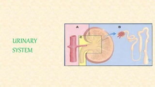
Kidneys
Partly skin
Soluble substances and water
Lungs Excretion of CO2, water vapour, ammonia, ketone bodies,
alcohol, aromatic oils
Skin Water, salts
Liver Fatty substances through bile
Colon Heavy matters
Table 1: Various excretory organs and theirs excretory products
5. Main Function of kidneys: Formation of urine
Production of waste products such as urea, creatinine, and ammonia by body cells.
They must be removed from the blood before they accumulate to toxic levels.
Several other important functions of kidneys:
1. Regulation of the volume of blood by excretion or conservation of water.
2. Regulation of the electrolyte content of the blood by the excretion or conservation of
minerals
3. Regulation of the acid–base balance of the blood by excretion or conservation of ions such
as H ions or HCO3 ions
6. 4. Regulation of all of the above in tissue fluid.
The process of urine formation helps to maintain
normal composition
volume and
pH of both blood and tissue fluid
Removal of those substances that would upset the normal constancy and balance of these
extracellular fluids.
7. Kidneys:
Shape: Bean Shaped
Location:
In the upper abdominal cavity on either side of the vertebral column, behind the
peritoneum (retroperitoneal).
The upper portions of the kidneys rest on the lower surface of the diaphragm and are
enclosed and protected by the lower rib cage.
The kidneys are embedded in adipose tissue that acts as a cushion and is in turn covered by
a fibrous connective tissue membrane called the renal fascia, which helps hold the kidneys in
place.
8. • Adrenal gland (Suprarenal) is situated above the kidneys.
• Front of right kidney bears the impression of part of duodenum.
• Left one is crossed in front by the pancreas transversely.
Internal structure of the kidney:
10. Outer layer:
• The outer tissue layer is called the renal cortex; it is made of renal corpuscles and
convoluted tubules.
Middle layer:
• The lateral and middle areas are tissue layers, and the medial area at
the hilus is a cavity.
Inner layer:
• The lateral and middle areas are tissue layers, and the medial area at the hilus is a cavity.
The renal medulla consists of wedge-shaped pieces called renal pyramids. The tip of each
pyramid is its apex or papilla.
11. •Renal pelvis:
• A cavity formed by the expansion of the ureter within the kidney at the hilus.
Calyces:
• Funnel shaped extensions of the renal pelvis.
• It enclose the papillae of the renal pyramids.
• Urine flows from the renal pyramids into the calyces, then to the renal pelvis and out into
the ureter.
12. Renal Circulation:
Kidney has rich arterial supply through respective renal arteries.
Blood returns through respective renal veins.
Renal Artery
Anterior Division Posterior division
Each division further passes between the pyramides as “Interlobar arteries.” These arteries
give “Arcuate arteries.”
These arteries run across the pyramides as “Interlobular arteries” and give off afferent
vessels of glomerulus.
Afferent vessles before going to glomerulus contain “ Juxtaglomerular cells.”
13. Juxta glomerular cells: Secrete Renin Control blood flow
Angeotensin Regulate Blood Pressure
Efferent arterioles: The vessels coming from glomerulus
Peritubular network: Efferent artriole break up into capillary network around tubules.
Interlobular veins: Venous plexuses formed from Peritubular network.
Interlobar veins
Renal Veins
14. Characteristics of renal circulation:
1. Renal Portal system: Blood has to circulate through the tuft of glomeruli and also through
peritubular capillaries. The glomeruli filters and capillaries reabsorb.
2. All circulation blood must pass through the glomeruli.
3. Pressure in renal vessel run higher than in systemic peritubular capillaries.
4. The rate and volume of blood flow is also high.
16. Structural and functional unit of kidney.
The nephron is the structural and functional unit of the kidney.
Each kidney contains approximately 1 million nephrons.
Urine formation occurs in nephron.
Each nephron has two major portions:
a renal corpuscle and
a renal tubule.
Renal corpuscle :
It consists of a glomerulus surrounded by a Bowman’s capsule.
The glomerulus is a capillary network that arises from an afferent arteriole and empties
into an efferent arteriole.
17. The diameter of the efferent arteriole is smaller than that of the afferent arteriole, which
helps maintain a fairly high blood pressure in the glomerulus.
Bowman’s capsule (or glomerular capsule) is the expanded end of a renal tubule; it
encloses the glomerulus.
The inner layer of Bowman’s capsule is made of podocytes; the name means “foot cells,”
and the “feet” of the podocytes are on the surface of the glomerular capillaries.
The arrangement of podocytes creates pores, spaces between adjacent “feet,” which make
this layer very permeable.
The outer layer of Bowman’s capsule has no pores and is not permeable.
18. The space between the inner and outer layers of Bowman’s capsule contains renal filtrate,
the fluid that is formed from the blood in the glomerulus and will eventually become urine.
Renal Tubule:
The renal tubule continues from Bowman’s capsule and consists of the following parts:
proximal convoluted tubule (in the renal cortex), loop of Henle (or loop of the nephron, in
the renal medulla), and distal convoluted tubule (in the renal cortex).
The distal convoluted tubules from several nephrons empty into a collecting tubule.
19. Several collecting tubules then unite to form a papillary duct that empties urine into a
calyx of the renal pelvis.
All parts of the renal tubule are surrounded by peritubular capillaries, which arise from
the efferent arteriole.
The peritubular capillaries will receive the materials reabsorbed by the renal tubules; this
is described in the section on urine formation.
20. • Structural and functional unit of kidney.
• About one million nephrons in one kidney.
• Each nephron consists of:
• Bowman’s capsule
• Glomerulus
• Proximal convolated tubule
• Loop of Henle’s
• Distal convolated tubule
• Collecting tubule
Nephron
21. • Bowman’s capsule:
• It is proximal blind dilated end which covers up
like a cap on glomerular tufts, so that the
glomerular blood is filtered into nephron.
• Glomerulus:
• It is tuft of capillaries with afferent capillary coming in from the circulation and
efferent capillary going out into circulation.
• Glomeruli acts as a ultra-filters.
• Deproteinised plasma is filtered through the glomerulus into nephron.
Nephron
22. • Renal tubule:
• Tubular part of nephron is about 0.3 cm in length.
• About 2 million of them in both kidneys.
• If put end to end its length would be about 60 km.
• Following parts: 1. Narrow neck
2. First or proximal convolated tubule
3. Henle’s loop or Loop of Henle’s
4. Second or proximal convolated tubule
5. Straight or collecting tubule
6. Duct of Bellini
Nephron
23. • Neck:
• It is constricted and lined by cubical cells.
• Proximal convolated tubule:
• It is lined by cubical cells.
• Cells are markedly active and secrete some
enzyme like carbonic-anhydrase etc. which help in maintenance of acid base
balance in blood.
Nephron
24. • Henle’s loop:
• U-shaped tube and has a descending limb and
an ascending limb.
• Histologically, it resembles a capillary but loop
is little wider than capillary.
• It is constricted part of nephron.
• It is physiologically important because it reabsorbs a no. of important
substances that are filtered by glomerulus, back to blood.
Nephron
25. • Distal convolated tubule:
• The absorption of Na+ is under the control of
aldosterone which takes place in this part.
• Collecting tubule:
• Collecting tubule of many nephrons combine in
one straight tubule.
• Distal convolated tubule:
• This arises from final combination of many collecting tubules and finally drains
into apex of pyramid of kidney.
Nephron
26. • It maintains (A) water equilibrium (B) pH equilibrium (C) osmotic equilibrium
(D) ionic equilibrium of blood.
• It excrete waste products in dissolved form. These are nitrogenous and sulphur
containing end products of protein metabolism.
• It aids to keep up the optimum concentrations of certain important substances.
• It excretes poisonous and foreign substances from body which includes drugs,
toxins, etc.
Functions of kidney
27. • It synthesises new substances, namely ammonia, hippuric acid and inorganic
phosphates. That manufactured ammonia aids to maintain acid base equilibrium.
• Thus, kidney excrete, synthesis, secrete and equilibrate to keep the consistency
of blood by process of filtration, secretion, reabsorption, synthesis and
excretion.
Functions of kidney
28. • There are mainly three processes involve in urine formation,
• Glomerular filtration
• Tubular reabsorption
• Tubular secretion
Physiology of urine formation
29. • It is filtration of body fluids and solutes from blood out
of glomerular capillaries into Bowman’s capsule
• All substances from blood are filtered out except the
proteins, cells and colloids.
• Colloids exert inward pull due to their osmotic pressure (30 mmHg) against
hydrostatic pressure (75 mmHg).
• This results in 45 mmHg of net filtering force.
Glomerular filtration
30. • Glomerular filtration rate depends on following factors,
Permeability of capillaries
Area of filtration
Intra-capillary pressure
Intra-capsular pressure
Osmotic pressure of blood plasma
Glomerular filtration
31. • Nearly 175 litres of deproteinised plasma , is filtered
out through glomeruli in 24 hrs but only about one and
half litre of urine is finally excreted daily.
• Hence, the rest of fluid with important constituents is
reabsorbed back in the blood to body’s advantage.
• Following substances are reabsorbed at various levels,
• Glucose:
• Reabsorbed completely from proximal tubules.
• Along with it, galactose and fructose also absorbed.
Tubular reabsorption
32. • Water:
• More than 85% of water reabsorbed from tubules.
• This takes place in proximal, distal and even in
collecting tubule.
• Of total reabsorption, seventh-eighth is reabsorbed by
proximal tubule.
• This reabsorption is controlled by antidiuretic hormone of posterior pituitary
gland.
Tubular reabsorption
33. • Antidiuretic hormone regulates extracellular water by
adjusting amount of water reabsorbed into blood by
distal and collecting tubule.
• When this fails, reabsorption does not occur and
copious urination follows. This disease is called as
“Diabetes Insipidus”.
Tubular reabsorption
34. • Salts:
• NaCl is reabsorbed in proximal and distal tubules.
• Potassium is completely reabsorbed from proximal
tubule.
• Bicarbonate is also reabsorbed by tubule.
• Phosphate is reabsorbed in proximal tubules.
• Aldosterone regulates the extracellular fluid volume by adjusting the amount of
sodium reabsorbed by blood from kidneys.
Tubular reabsorption
35. • Miscellaneous substances:
• Uric acid, sulphates, vitamin C, creatine, amino acids,
acetoacitic acid, beta-hydroxybutyric acid, etc. are also
reabsorbed from tubules, by loop of Henle’s.
Tubular reabsorption
36. • Further, the substances which are not found in filtrate
are also found in urine.
• These are directly excreted from the blood by tubules
themselves through enzyme mechanism.
• This is called as tubular secretion.
• Tubular cells also manufacture certain new substances.
• This is active process in which substances like potassium, hydrogen, creatinine,
and drugs like phenol, penicillin, p-amino hippuric acid, diodone etc. are
excreted by tubular cells from blood.
Tubular secretion
37. • Synthesis of new substances:
• New substances are all manufactured in tubular cells.
• Ammonia is synthesized by process of deamination.
• Hippuric acid is synthesized by combination of glycine
and benzoic acid.
• Phosphates are converted from organic form into
inorganic phosphates by phosphatase enzyme system.
Tubular secretion
38. • In healthy person, pH of extracellular fluid ranges from 7.35 to 7.45.
• Maintanance of this narrow range is essential for survival and depends on three
major mechanisms:
1. Buffer system
2. Respiration
3. Kidney excretion
Acid-Base Balance
39. • Most buffer systems of body are weak acids and weak bases.
• They include
• Carbonic acid-bicarbonate system
• Phosphate system
• Hemoglobin-oxyhemoglobin system
• Protein system
Buffer system
40. • It is primarily based on the carbonic acid and sodium bicarbonate.
• The following equations illustrate the mechanism,
HCl + NaHCO3 NaCl + H2CO3
NaOH + H2CO3 NaHCO3 + H2O
• Normal body processes tend to acidify the blood rather than to make it more
alkaline.
• In other words, body needs more bicarbonate salt than carbonic acid and when
the extracellular pH is normal (7.4), bicarbonate molecules out number carbonic
acid (20:1).
Carbonic acid-bicarbonate buffer system
41. • It has two components namely sodium dihydrogen phosphate and sodium
monohydrogen phosphate acting as weak acid and weak base respectively.
NaOH + NaH2PO4 H2O + Na2HPO4
HCl + Na2HPO4 NaCl + NaH2PO4
• This system is important mechanism in kidney.
• NaH2PO4 is formed when excess of H+ ion in kidney combines with Na2HPO4.
• The Na+ released from this reaction forms sodium bicarbonate and is passed in
to blood.
• The H+ ion that replaces Na+ becomes part of NaH2PO4 and is passed to urine.
• Thus reaction tends to reduce acidity of blood by increasing alkalises to urine.
Phosphate buffer system
42. • It is an effective method for buffering carbonic acid in blood.
• It is Chloride shift that occur in respiratory system.
• When CO2 enters the blood, H2CO3 is formed using carbonic anhydrase
enzyme. This reacts with KHbO2 to liberate O2 and so it reduces haemoglobin
to HHb, also K+ and HCO3- ions are formed. HCO3- diffuses in plasma where
it combines with NaCl to form NaHCO3 and Cl- is set free.
Haemoglobin-oxyhaemoglobin buffer system
43. • When O2 enters the blood, it combines with reduced haemoglobin to form
HHbO2. This reacts with KCl to form KHb+O2, while with NaHCO3 to form
NaCl and displaces HCO3.
• This bicarbonate ions diffuses in RBC to combine with H+ ions and forms
H2CO3.
• Carbonic acid (H2CO3) dissociates into H2O and CO2 which diffuse out in
plasma and from there to air.
Haemoglobin-oxyhaemoglobin buffer system
44. Protein buffer system
• It is most abundant buffer in body cells and plasma.
• The amino acids of protein contains carboxyl group (COOH) and at least one
amine group (NH2).
• The carboxyl group acts like acid and amine group as a base and thereby can
react with excess hydroxide and hydrogen ion respectively.
R R
NH2-C-COO- + H+ COOH-C-NH3 + OH-
H H
• Thus proteins act as both acidic and basic buffers.
45. Respiration
• It also plays an important role in maintaining pH of blood.
• An increase in CO2 concentration in body fluids can lower the pH as follows:
CO2 + H2O H2CO3 H+ + HCO3-
• A decrease in CO2 concentration in body fluids can raise the pH.
• Thus, pH of body fluids may be adjusted by a change in rate of breathing.
• Increase in rate will exhale out CO2 resulting in rise in pH and slowing down of
breathing will decrease the pH.
• In fact, the respiratory centres in medulla oblongata of brain are stimulated
when pH of blood falls.
46. Kidney
• Plays important role in maintaining pH.
• Mechanism involved in kidney are excretion of H+ ions and reabsorption of
HCO3-.
• Besides this, formation of ammonia is another important mechanism that helps
in acid-base balance.
47. Kidney
• The epithelial cells of all tubules besides those of the thin segment of loop of
Henle can synthesize ammonia.
• Ammonia(NH3) then reacts with H+ to form ammonium ion (NH4+).
• This is then excreted into urine in combination with chloride ion (as NH4Cl).
• The net effect is thus to increase sodium bicarbonate concentration in
extracellular fluid.
• Thus, increase in H+ ion concentration causes formation of more ammonia to
regulate acid-base balance.
48. Acid-base imbalances
• Normally, the buffer system, respiratory system and kidneys function in
coordination very efficiently to maintain the normal blood pH (7.35-7.45).
• However, under abnormal condition there may be decrease in pH (7.35 to 6.8)
causing acidosis or an increase in pH (7.45 to 8.00) called alkalosis.
Condition pH of blood H+(mmol/l) HCO3(mmol/l) pCO2(mmHg)
Respiratory acidosis <7.35 >44 29 >60
Respiratory alkalosis >7.45 <36 22 <20
Metabolic acidosis <7.35 >44 <18 <35
Metabolic alkalosis >7.45 <36 >32 >45
49. Respiratory acidosis
• Due to hypoventilation.
• In uncompensated respiratory, the bicarbonate –carbonic acid ratio is changed
from 20:1 to 10:1 or 8:1.
• There is increase in pCO2 in the blood and hence carbonic acid.
• The causes of respiratory acidosis are emphysema, pulmonary edema, injury to
respiratory center of medulla or disorders of muscles involved in breathing .
50. Respiratory alkalosis
• Due to hyperventilation.
• There is decrease in pCO2 in the blood and hence shift of bicarbonate-carbonic
acid ratio towards 20:0.5.
• The causes of respiratory alkalosis are oxygen deficiency, severe anxiety, aspirin
overdose, etc.
51. Metabolic acidosis
• Due to abnormal increase in acidic metabolic products other than CO2 or loss of
bicarbonate ions from body.
• Causes are ketosis, lactic acidosis, renal failure, salicylate overdose, cardiac
arrest, diarrhoea etc.
• In uncompensated metabolic acidosis the ratio of bicarbonate- carbonic acid
may become 12:1.
52. Metabolic alkalosis
• Due to loss of acid from body or excessive accumulation of alkalise in blood..
• Causes are excessive vomiting, hyperaldosteronism, Cushing syndrom etc.
• In uncompensated metabolic alkalosis the ratio of bicarbonate- carbonic acid
may become 31:1.
53. Counter-current multiplier system
• Descending limb of loop of Henle is relatively
permeable to Na+ and Cl-
• In ascending limb, active transport of Cl- takes place
from the filtrate of tubule to the interstitial fluid of
medulla.
• Transport of Cl- is followed passively by Na+.
• Thus, as fluid moves downwards in descending limb
loop of Henle, there is an increase in Na+ and Cl- in tubule.
• In ascending limb of loop of Henle , there is active transport of Cl- and hence
outward movement of Na+.
54. Counter-current multiplier system
• This causes fall in concentration of NaCl as fluid
moves upwards.
• In otherwords, there are two parallel streams of
liquid flowing opposite to each other (counter-
current) resulting in an increased NaCl
concentration (multiplier) in the medullary
interstitial fluid.
• This is known as counter-current multiplier system.
55. Diseases of kidney
• Descending limb of loop of Henle is relatively
permeable to Na+ and Cl-
• In ascending limb, active transport of Cl- takes place
from the filtrate of tubule to the interstitial fluid of
medulla.
• Transport of Cl- is followed passively by Na+.
• Thus, as fluid moves downwards in descending limb
loop of Henle, there is an increase in Na+ and Cl- in tubule.
• In ascending limb of loop of Henle , there is active transport of Cl- and hence
outward movement of Na+.