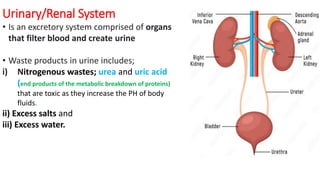
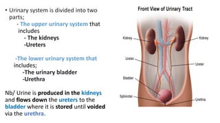
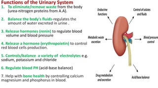
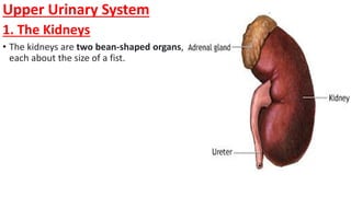
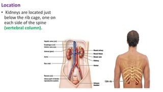
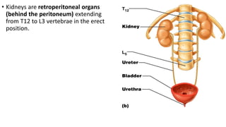
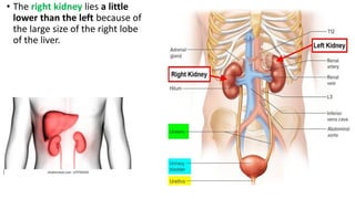
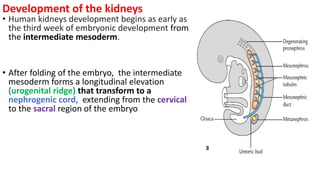
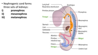
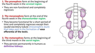
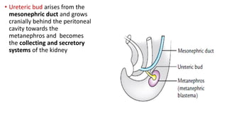
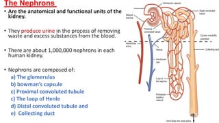
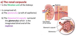
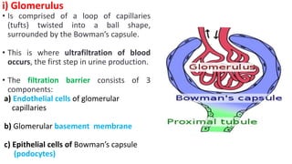
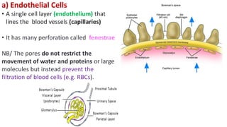
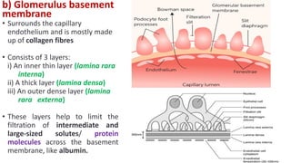
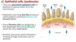
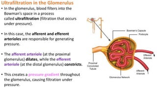
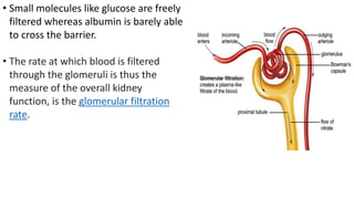
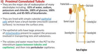
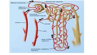
URINARY/RENAL SYSTEM
By Dr. Ann Mwangi
SPHS, USIU
Urinary/Renal System
• Is an excretory system comprised of organs
that filter blood and create urine
• Waste products in urine includes;
i) Nitrogenous wastes; urea and uric acid
(end products of the metabolic breakdown of proteins)
that are toxic as they increase the PH of body
fluids.
ii) Excess salts and
iii) Excess water.
• Urinary system is divided into two
parts;
- The upper urinary system that
includes
- The kidneys
-Ureters
-The lower urinary system that
includes;
-The urinary bladder
-Urethra
Nb/ Urine is produced in the kidneys
and flows down the ureters to the
bladder where it is stored until voided
via the urethra.
Functions of the Urinary System
1. To eliminate/remove waste from the body
(urea-nitrogen proteins from A.A).
2. Balance the body's fluids-regulates the
amount of water excreted in urine .
3. Release hormones (renin) to regulate blood
volume and blood pressure
4. Release a hormone (erythropoietin) to control
red blood cells production.
5. Controls/balance a variety of electrolytes e.g.
sodium, potassium and chloride
6. Regulate blood PH (acid-base balance)
7. Help with bone health by controlling calcium
magnesium and phosphorus in blood.
Clinical Terms
• Hematuria: blood in the urine.
• Nephrectomy: surgical removal of a kidney.
• Glomerulonephritis: inflammation of the glomeruli.
• Cystectomy: surgical removal of the urinary bladder.
• Dysuria: painful or difficult urination.
• Uremia: condition in which substances ordinarily excreted in the
urine accumulates in the blood.
• Incontinence: inability to control urination.
• Urinalysis: analysis of urine to diagnose health or disease (to detect
protein, glucose, blood or pus).
Urologist: physician who specializes in diseases of the urinary
structures in both male and female.
Upper Urinary System
1. The Kidneys
• The kidneys are two bean-shaped organs,
each about the size of a fist.
Location
• Kidneys are located just
below the rib cage, one on
each side of the spine
(vertebral column).
• Kidneys are retroperitoneal organs
(behind the peritoneum) extending
from T12 to L3 vertebrae in the erect
position.
• The right kidney lies a little
lower than the left because of
the large size of the right lobe
of the liver.
Development of the kidneys
• Human kidneys development begins as early as
the third week of embryonic development from
the intermediate mesoderm.
• After folding of the embryo, the intermediate
mesoderm forms a longitudinal elevation
(urogenital ridge) that transform to a
nephrogenic cord, extending from the cervical
to the sacral region of the embryo
.
• Nephrogenic cord forms
three sets of kidneys:
i) pronephros
ii) mesonephros
iii) metanephros
1. The pronephros form at the beginning of
the fourth week in the cervical region.
• They are non-functional and completely
regresses
2. The mesonephros form at the end of the
fourth week in the thoracolumbar region.
• They become functional for a short period of
time and completely regresses except their
caudal excretory tubule (mesonephric duct)
that takes part in the formation of vasa
efferentia of the testis.
3. The metanephros forms at the beginning of
the third month in the sacral region.
• They persists permanently in humans as
definitive kidneys.
• Ureteric bud arises from the
mesonephric duct and grows
cranially behind the peritoneal
cavity towards the
metanephros and becomes
the collecting and secretory
systems of the kidney
• The growing end of the ureteric
bud becomes dilated like a
funnel to form the renal pelvis.
• The renal pelvis undergoes
repeated divisions to form major
calyces, minor calyces, collecting
ducts, and collecting tubules
Structure of Kidney
• Kidney is formed of;
A). Four outer coverings
B). Outer part of the kidney –
Cortex
C). Inner part-Medulla
D). Renal Sinus-fatty
compartment along the
hilum of kidney that contains:
• Renal pelvis,
• Major and minor calyces (cup-like
structurers)
• Major branches of the renal
artery and vein
• Lymphatic vessels.
A. The Coverings of the kidney
• There are four kidney coverings including;
i). Pararenal fat/perinephric fat
• Is the collection of adipose tissue
located superficial to the renal fascia
(peritoneum)
ii) Renal fascia/gerota’s fascia
• Is a layer of fibrous connective tissue
encapsulating the kidneys and the
adrenal glands.
iii). Perirenal fat capsule/Adipose capsule of
kidney-located between the kidney capsule
and the renal fascia, that protects kidney from
trauma and damage.
iv). Renal capsule-Is the innermost
membranous sheath, surrounding the space
of the renal cortex.
• The capsule is composed of tough fibres,
chiefly collagen and elastin (fibrous
proteins), that help to support the kidney
mass and protect its vital tissues from injury.
B. The Renal Cortex
• Is the outer portion of the kidney between
the renal capsule and the renal medulla
where ultrafiltration occurs
• It forms a continuous smooth outer zone
with a number of projections (cortical
columns) that extend down between the
pyramids in the medulla.
• Each column consists
of fibrous materials
that allows the cortex
to be better
anchored.
• The columns also provides a
space for blood vessels;
arterioles and venules from
the renal artery and vein, as
well as the glomerular
capillaries, to perfuse the
nephrons of the kidney.
Contents of the Cortex
i) Renal corpuscle
ii) Proximal convoluted
tubules.
ii) Distal convoluted tubules
NB/ The 3 structurers above are parts
of a nephron
iv) Erythropoietin
The Nephrons
• Are the anatomical and functional units of the
kidney.
• They produce urine in the process of removing
waste and excess substances from the blood.
• There are about 1,000,000 nephrons in each
human kidney.
• Nephrons are composed of:
a) The glomerulus
b) bowman’s capsule
c) Proximal convoluted tubule
c) The loop of Henle
d) Distal convoluted tubule and
e) Collecting duct
a) The renal corpuscle
• Is the filtration unit of the kidneys
• Is comprised of;
a) The glomerulus (a tuft of capillaries)
b) The (bowman’s) capsule- surround
the glomerulus which is the
invaginated blind end of the
nephron
i) Glomerulus
• Is comprised of a loop of capillaries
(tufts) twisted into a ball shape,
surrounded by the Bowman’s capsule.
• This is where ultrafiltration of blood
occurs, the first step in urine production.
• The filtration barrier consists of 3
components:
a) Endothelial cells of glomerular
capillaries
b) Glomerular basement membrane
c) Epithelial cells of Bowman’s capsule
(podocytes)
a) Endothelial Cells
• A single cell layer (endothelium) that
lines the blood vessels (capillaries)
• It has many perforation called fenestrae
NB/ The pores do not restrict the
movement of water and proteins or large
molecules but instead prevent the
filtration of blood cells (e.g. RBCs).
b) Glomerulus basement
membrane
• Surrounds the capillary
endothelium and is mostly made
up of collagen fibres
• Consists of 3 layers:
i) An inner thin layer (lamina rara
interna)
ii) A thick layer (lamina densa)
iii) An outer dense layer (lamina
rara externa)
• These layers help to limit the
filtration of intermediate and
large-sized solutes/ protein
molecules across the basement
membrane, like albumin.
c) Epithelial cells /podocytes
• They are specialised epithelial cells of
Bowman’s capsule which form the
visceral layer.
• Podocytes have long foot-like processes
called pedicles and leave filtration slits
between them.
• These filtration slits are bridged by a
thin diaphragm (the slit diaphragm)
which has very small pores.
• Similarly, the pores restrict filtration/
crossing of large molecules such as
proteins.
Ultrafiltration in the Glomerulus
• In the glomerulus, blood filters into the
Bowman’s space in a process
called ultrafiltration (filtration that occurs
under pressure).
• In this case, the afferent and efferent
arterioles are responsible for generating
pressure.
• The afferent arteriole (at the proximal
glomerulus) dilates, while the efferent
arteriole (at the distal glomerulus) constricts.
• This creates a pressure gradient throughout
the glomerulus, causing filtration under
pressure.
• Small molecules like glucose are freely
filtered whereas albumin is barely able
to cross the barrier.
• The rate at which blood is filtered
through the glomeruli is thus the
measure of the overall kidney
function, is the glomerular filtration
rate.
ii) Proximal Convoluted Tubule
• They are the major site of reabsorption of many
electrolytes including; 65% of water, sodium,
potassium and chloride, 100% of glucose, 100%
amino acids, and 85-90% of bicarbonate.
• They are lined with simple cuboidal epithelial
cells which have a brush border (microvilli-covered
surfaces) to increase the surface area.
• The epithelial cells have large amounts
of mitochondria present to support the processes
involved in transporting ions and substances.
• The solutes and water move from the PCT to the
interstitium (space between tubules and
capillaries) and then into peritubular capillaries
iii) The Loop of Henry
• Is U shaped portion of a nephron that leads from
the proximal convoluted tubule to the distal
convoluted tubule.
• The loop of Henle has a thick descending limb, a
thin descending limb, a thick ascending limb.
• The fluid entering the descending limb contains
sodium chloride and other salts, urea, and other
chemicals that have been filtered out from the
blood.
• The cells here are permeable to water and thus the
salt and urea concentration rises within the fluid by
the time it reaches the bend.
• The ascending limb is permeable to sodium
chloride and magnesium, which passes out of the
tubule into the medullary tissue surrounding it.
iv) Distal Convoluted Tubule
• Is a portion between the loop of Henle and
the collecting tubule.
• It is partly responsible for the regulation
of potassium, sodium, calcium, and pH.
• It regulates pH by absorbing bicarbonate and
secreting protons (H+) into the filtrate
• Sodium and potassium levels are controlled
by secreting K+ and absorbing Na+. Sodium
absorption by the distal tubule is mediated by
the hormone aldosterone by increasing its
reabsorption
• It also participates in calcium regulation by
reabsorbing Ca2+ in response to the
parathyroid hormone
iv) The Renal Collecting Ducts
• A distal convoluted tubule connects to
the collecting duct system
• They connect nephrons to a minor calyx
• They participate in electrolyte and fluid
balance through reabsorption and
excretion processes regulated by
the hormones aldosterone
and vasopressin
2 Types of Nephrons
• By the location of renal corpuscles within the
cortex, two types of nephrons can be
distinguished:
1. Cortical nephrons and
2. juxtamedullary nephrons.
• Cortical nephron- contains a short loop of
Henle which only extends into the outer
region of the renal medulla (all vertebrates)
• Juxtamedullary nephron- contains a longer
loop of Henle which extends deeper into the
inner medulla (camel) making urine as
concentrated as possible and limiting the
amount of water and salt they lose. This
helps desert animals live for long periods of
time on minimal amounts of water.
4. Erythropoietin
• Is a hormone necessary for
stimulating red blood cell
production by promoting the
proliferation and
differentiation of
erythrocytes in the bone
marrow
• It is produced in the renal
cortex

4a. Urinary System explanation with all parts

  • 1.
    URINARY/RENAL SYSTEM By Dr.Ann Mwangi SPHS, USIU
  • 2.
    Urinary/Renal System • Isan excretory system comprised of organs that filter blood and create urine • Waste products in urine includes; i) Nitrogenous wastes; urea and uric acid (end products of the metabolic breakdown of proteins) that are toxic as they increase the PH of body fluids. ii) Excess salts and iii) Excess water.
  • 3.
    • Urinary systemis divided into two parts; - The upper urinary system that includes - The kidneys -Ureters -The lower urinary system that includes; -The urinary bladder -Urethra Nb/ Urine is produced in the kidneys and flows down the ureters to the bladder where it is stored until voided via the urethra.
  • 4.
    Functions of theUrinary System 1. To eliminate/remove waste from the body (urea-nitrogen proteins from A.A). 2. Balance the body's fluids-regulates the amount of water excreted in urine . 3. Release hormones (renin) to regulate blood volume and blood pressure 4. Release a hormone (erythropoietin) to control red blood cells production. 5. Controls/balance a variety of electrolytes e.g. sodium, potassium and chloride 6. Regulate blood PH (acid-base balance) 7. Help with bone health by controlling calcium magnesium and phosphorus in blood.
  • 6.
    Clinical Terms • Hematuria:blood in the urine. • Nephrectomy: surgical removal of a kidney. • Glomerulonephritis: inflammation of the glomeruli. • Cystectomy: surgical removal of the urinary bladder. • Dysuria: painful or difficult urination.
  • 7.
    • Uremia: conditionin which substances ordinarily excreted in the urine accumulates in the blood. • Incontinence: inability to control urination. • Urinalysis: analysis of urine to diagnose health or disease (to detect protein, glucose, blood or pus). Urologist: physician who specializes in diseases of the urinary structures in both male and female.
  • 8.
    Upper Urinary System 1.The Kidneys • The kidneys are two bean-shaped organs, each about the size of a fist.
  • 9.
    Location • Kidneys arelocated just below the rib cage, one on each side of the spine (vertebral column).
  • 10.
    • Kidneys areretroperitoneal organs (behind the peritoneum) extending from T12 to L3 vertebrae in the erect position.
  • 11.
    • The rightkidney lies a little lower than the left because of the large size of the right lobe of the liver.
  • 12.
    Development of thekidneys • Human kidneys development begins as early as the third week of embryonic development from the intermediate mesoderm. • After folding of the embryo, the intermediate mesoderm forms a longitudinal elevation (urogenital ridge) that transform to a nephrogenic cord, extending from the cervical to the sacral region of the embryo .
  • 13.
    • Nephrogenic cordforms three sets of kidneys: i) pronephros ii) mesonephros iii) metanephros
  • 14.
    1. The pronephrosform at the beginning of the fourth week in the cervical region. • They are non-functional and completely regresses 2. The mesonephros form at the end of the fourth week in the thoracolumbar region. • They become functional for a short period of time and completely regresses except their caudal excretory tubule (mesonephric duct) that takes part in the formation of vasa efferentia of the testis. 3. The metanephros forms at the beginning of the third month in the sacral region. • They persists permanently in humans as definitive kidneys.
  • 15.
    • Ureteric budarises from the mesonephric duct and grows cranially behind the peritoneal cavity towards the metanephros and becomes the collecting and secretory systems of the kidney
  • 16.
    • The growingend of the ureteric bud becomes dilated like a funnel to form the renal pelvis. • The renal pelvis undergoes repeated divisions to form major calyces, minor calyces, collecting ducts, and collecting tubules
  • 18.
    Structure of Kidney •Kidney is formed of; A). Four outer coverings B). Outer part of the kidney – Cortex C). Inner part-Medulla D). Renal Sinus-fatty compartment along the hilum of kidney that contains: • Renal pelvis, • Major and minor calyces (cup-like structurers) • Major branches of the renal artery and vein • Lymphatic vessels.
  • 19.
    A. The Coveringsof the kidney • There are four kidney coverings including; i). Pararenal fat/perinephric fat • Is the collection of adipose tissue located superficial to the renal fascia (peritoneum) ii) Renal fascia/gerota’s fascia • Is a layer of fibrous connective tissue encapsulating the kidneys and the adrenal glands.
  • 21.
    iii). Perirenal fatcapsule/Adipose capsule of kidney-located between the kidney capsule and the renal fascia, that protects kidney from trauma and damage. iv). Renal capsule-Is the innermost membranous sheath, surrounding the space of the renal cortex. • The capsule is composed of tough fibres, chiefly collagen and elastin (fibrous proteins), that help to support the kidney mass and protect its vital tissues from injury.
  • 22.
    B. The RenalCortex • Is the outer portion of the kidney between the renal capsule and the renal medulla where ultrafiltration occurs • It forms a continuous smooth outer zone with a number of projections (cortical columns) that extend down between the pyramids in the medulla.
  • 23.
    • Each columnconsists of fibrous materials that allows the cortex to be better anchored.
  • 24.
    • The columnsalso provides a space for blood vessels; arterioles and venules from the renal artery and vein, as well as the glomerular capillaries, to perfuse the nephrons of the kidney.
  • 25.
    Contents of theCortex i) Renal corpuscle ii) Proximal convoluted tubules. ii) Distal convoluted tubules NB/ The 3 structurers above are parts of a nephron iv) Erythropoietin
  • 27.
    The Nephrons • Arethe anatomical and functional units of the kidney. • They produce urine in the process of removing waste and excess substances from the blood. • There are about 1,000,000 nephrons in each human kidney. • Nephrons are composed of: a) The glomerulus b) bowman’s capsule c) Proximal convoluted tubule c) The loop of Henle d) Distal convoluted tubule and e) Collecting duct
  • 28.
    a) The renalcorpuscle • Is the filtration unit of the kidneys • Is comprised of; a) The glomerulus (a tuft of capillaries) b) The (bowman’s) capsule- surround the glomerulus which is the invaginated blind end of the nephron
  • 29.
    i) Glomerulus • Iscomprised of a loop of capillaries (tufts) twisted into a ball shape, surrounded by the Bowman’s capsule. • This is where ultrafiltration of blood occurs, the first step in urine production. • The filtration barrier consists of 3 components: a) Endothelial cells of glomerular capillaries b) Glomerular basement membrane c) Epithelial cells of Bowman’s capsule (podocytes)
  • 30.
    a) Endothelial Cells •A single cell layer (endothelium) that lines the blood vessels (capillaries) • It has many perforation called fenestrae NB/ The pores do not restrict the movement of water and proteins or large molecules but instead prevent the filtration of blood cells (e.g. RBCs).
  • 31.
    b) Glomerulus basement membrane •Surrounds the capillary endothelium and is mostly made up of collagen fibres • Consists of 3 layers: i) An inner thin layer (lamina rara interna) ii) A thick layer (lamina densa) iii) An outer dense layer (lamina rara externa) • These layers help to limit the filtration of intermediate and large-sized solutes/ protein molecules across the basement membrane, like albumin.
  • 32.
    c) Epithelial cells/podocytes • They are specialised epithelial cells of Bowman’s capsule which form the visceral layer. • Podocytes have long foot-like processes called pedicles and leave filtration slits between them. • These filtration slits are bridged by a thin diaphragm (the slit diaphragm) which has very small pores. • Similarly, the pores restrict filtration/ crossing of large molecules such as proteins.
  • 33.
    Ultrafiltration in theGlomerulus • In the glomerulus, blood filters into the Bowman’s space in a process called ultrafiltration (filtration that occurs under pressure). • In this case, the afferent and efferent arterioles are responsible for generating pressure. • The afferent arteriole (at the proximal glomerulus) dilates, while the efferent arteriole (at the distal glomerulus) constricts. • This creates a pressure gradient throughout the glomerulus, causing filtration under pressure.
  • 34.
    • Small moleculeslike glucose are freely filtered whereas albumin is barely able to cross the barrier. • The rate at which blood is filtered through the glomeruli is thus the measure of the overall kidney function, is the glomerular filtration rate.
  • 35.
    ii) Proximal ConvolutedTubule • They are the major site of reabsorption of many electrolytes including; 65% of water, sodium, potassium and chloride, 100% of glucose, 100% amino acids, and 85-90% of bicarbonate. • They are lined with simple cuboidal epithelial cells which have a brush border (microvilli-covered surfaces) to increase the surface area. • The epithelial cells have large amounts of mitochondria present to support the processes involved in transporting ions and substances. • The solutes and water move from the PCT to the interstitium (space between tubules and capillaries) and then into peritubular capillaries
  • 37.
    iii) The Loopof Henry • Is U shaped portion of a nephron that leads from the proximal convoluted tubule to the distal convoluted tubule. • The loop of Henle has a thick descending limb, a thin descending limb, a thick ascending limb. • The fluid entering the descending limb contains sodium chloride and other salts, urea, and other chemicals that have been filtered out from the blood. • The cells here are permeable to water and thus the salt and urea concentration rises within the fluid by the time it reaches the bend. • The ascending limb is permeable to sodium chloride and magnesium, which passes out of the tubule into the medullary tissue surrounding it.
  • 38.
    iv) Distal ConvolutedTubule • Is a portion between the loop of Henle and the collecting tubule. • It is partly responsible for the regulation of potassium, sodium, calcium, and pH. • It regulates pH by absorbing bicarbonate and secreting protons (H+) into the filtrate • Sodium and potassium levels are controlled by secreting K+ and absorbing Na+. Sodium absorption by the distal tubule is mediated by the hormone aldosterone by increasing its reabsorption • It also participates in calcium regulation by reabsorbing Ca2+ in response to the parathyroid hormone
  • 39.
    iv) The RenalCollecting Ducts • A distal convoluted tubule connects to the collecting duct system • They connect nephrons to a minor calyx • They participate in electrolyte and fluid balance through reabsorption and excretion processes regulated by the hormones aldosterone and vasopressin
  • 40.
    2 Types ofNephrons • By the location of renal corpuscles within the cortex, two types of nephrons can be distinguished: 1. Cortical nephrons and 2. juxtamedullary nephrons. • Cortical nephron- contains a short loop of Henle which only extends into the outer region of the renal medulla (all vertebrates) • Juxtamedullary nephron- contains a longer loop of Henle which extends deeper into the inner medulla (camel) making urine as concentrated as possible and limiting the amount of water and salt they lose. This helps desert animals live for long periods of time on minimal amounts of water.
  • 41.
    4. Erythropoietin • Isa hormone necessary for stimulating red blood cell production by promoting the proliferation and differentiation of erythrocytes in the bone marrow • It is produced in the renal cortex