ENDOCRINE SYSTEM
•Download as PPTX, PDF•
0 likes•268 views

it is useful for the pharmacy students

Recommended
More Related Content
What's hot
What's hot (20)
Similar to ENDOCRINE SYSTEM
Similar to ENDOCRINE SYSTEM (20)
More from SURESH BABU EMANDI DEPARTMENT OF PHARMACOGNOSY Vikas Institute of Pharmaceutical scienes
More from SURESH BABU EMANDI DEPARTMENT OF PHARMACOGNOSY Vikas Institute of Pharmaceutical scienes (20)
Recently uploaded
Recently uploaded (20)
ENDOCRINE SYSTEM
- 1. SURESH BABU EMANDI M.Pharm Vikas Institute of Pharmaceutical Sciences Near Air Port, Rajahmundry-533103. ENDOCRINE SYSTEM
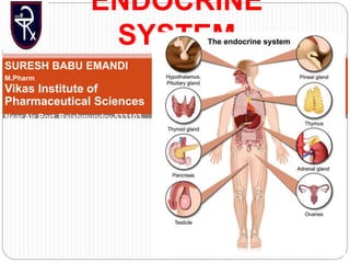
- 2. The endocrine system is a complex network of glands and organs. It uses hormones to control and coordinate your body's metabolism, energy level, reproduction, growth and development, and response to injury, stress, and mood. The following
- 3. INTRODUCTION All the physiological activities of the body are regulated by two major systems: 1. Nervous system 2. Endocrine system. These two systems interact with one another and regulate the body functions. This section deals with endocrine system and Section 10 deals with nervous system. Endocrine system functions by secreting some chemical substances called hormones
- 4. „CELL-TO-CELL SIGNALING Cell to cell signaling refers to the transfer of information from one cell to another. It is also called cell signaling or intercellular communication. The cells of the body communicate with each other through some chemical substances called chemical messengers. „ CHEMICAL MESSENGERS Chemical messengers are the substances involved in cell signaling. These messengers are mainly secreted from endocrine glands. Some chemical messengers are secreted by nerve endings and the cells of several other tissues also. All these chemical messengers carry the message (signal) from the signaling cells (controlling cells) to the target cells. The messenger substances may be the hormones or hormone like substances.
- 5. Classification of Chemical Messengers Generally the chemical messengers are classified into two types: 1. Classical hormones secreted by endocrine glands 2. Local hormones secreted from other tissues. chemical messengers are classified into four types: 1. Endocrine messengers 2. Paracrine messengers 3. Autocrine messengers 4. Neurocrine messengers.
- 6. Endocrine glands play an important role in homeostasis and control of various other activities in the body through their hormones. Hormones are transported by blood to target organs or tissues in different parts of the body, where the actions are executed.
- 7. HORMONES SECRETED FROM GLANDS
- 9. HORMONES SECRETED BY THE OTHER ORGANS
- 10. LOCAL HORMONES
- 11. CHEMISTRY OF HORMONES Hormones are chemical messengers, synthesized by endocrine glands. Based on chemical nature, hormones are classified into three types 1. Steroid hormones 2. Protein hormones 3. Derivatives of the amino acid called tyrosine
- 13. PITUTARY GLAND
- 14. Pituitary gland or hypophysis is a small endocrine gland with a diameter of 1 cm and weight of 0.5 to 1 g. It is situated in a depression called ‘sella turcica’, present in the sphenoid bone at the base of skull. It is connected with the hypothalamus by the pituitary stalk or hypophyseal stalk DIVISIONS OF PITUITARY GLAND Pituitary gland is divided into two divisions: 1. Anterior pituitary or adenohypophysis 2. Posterior pituitary or neurohypophysis.
- 15. Both the divisions are situated close to each other. Still both are entirely different in their development, structure and function. Between the two divisions, there is a small and relatively avascular structure called pars intermedia. Actually, it forms a part of anterior pituitary
- 16. DEVELOPMENT OF PITUITARY GLAND Both the divisions of pituitary glands develop from different sources. Anterior pituitary is ectodermal in origin and arises from the pharyngeal epithelium as an upward growth known as Rathke pouch. Posterior pituitary is neuroectodermal in origin and arises from hypothalamus as a downward diverticulum. Rathke pouch and the downward diverticulum from hypothalamus grow towards each other and meet in the midway between the roof of the buccal cavity and base of brain. There, the two structures lie close together.
- 17. REGULATION OF SECRETION „Hypothalamo-hypophyseal Relationship The relationship between hypothalamus and pituitary gland is called hypothalamo-hypophyseal relationship. Hormones secreted by hypothalamus are transported to anterior pituitary and posterior pituitary. But the mode of transport of these hormones is different. Hormones from hypothalamus are transported to anterior pituitary through hypothalamo-hypophysial portal blood vessels. But, the hormones from hypothalamus to posterior pituitary are transported by nerve fibers of hypothalamo-hypophyseal tract
- 18. ANTERIOR PITUITARY OR ADENOHYPOPHYSIS Anterior pituitary is also known as the master gland because it regulates many other endocrine glands through its hormones. „PARTS Anterior pituitary consists of three parts 1. Pars distalis 2. Pars tuberalis 3. Pars intermedia.
- 19. HISTOLOGY Anterior pituitary has two types of cells, which have different staining properties: 1. Chromophobe cells 2. Chromophil cells. Chromophobe Cells Chromophobe cells do not possess granules and stain poorly. These cells form 50% of total cells in anterior pituitary. Chromophobe cells are not secretory in nature, but are the precursors of chromophil cells. Chromophil Cells Chromophil cells contain large number of granules and are darkly stained.
- 20. REGULATION OF ANTERIOR PITUITARY SECRETION Hypothalamus controls anterior pituitary by secreting the releasing and inhibitory hormones (factors), which are called neurohormones. These hormones from hypothalamus are transported anterior pituitary through hypothalamo-hypophyseal portal vessels. Some special nerve cells present in various parts hypothalamus send their nerve fibers (axons) to median eminence and tuber cinereum. These nerve cells synthesize the hormones and release them into median
- 21. Releasing and Inhibitory Hormones Secreted by Hypothalamus 1. Growth hormone-releasing hormone (GHRH): Stimulates the release of growth hormone 2. Growth hormone-releasing polypeptide (GHRP): Stimulates the release of GHRH and growth hormone 3. Growth hormone-inhibitory hormone (GHIH) or somatostatin: Inhibits the growth hormone release 4. Thyrotropic-releasing hormone (TRH): Stimulates the release of thyroid stimulating hormone 5. Corticotropin-releasing hormone (CRH): Stimulates the release of adrenocorticotropin 6. Gonadotropin-releasing hormone (GnRH): Stimulates the release of gonadotropins, FSH and LH 7. Prolactin-inhibitory hormone (PIH): Inhibits prolactin secretion. It is believed that PIH is dopamine.
- 22. HORMONES SECRETED BY ANTERIOR PITUITARY Six hormones are secreted by the anterior pituitary: 1. Growth hormone (GH) or somatotropic hormone (STH) 2. Thyroid-stimulating hormone (TSH) or thyrotropic hormone 3. Adrenocorticotropic hormone (ACTH) 4. Follicle-stimulating hormone (FSH) 5. Luteinizing hormone (LH) in females or interstitial cell- stimulating hormone (ICSH) in males 6. Prolactin. Recently, the hormone β-lipotropin is found to be secreted by anterior pituitary.
- 23. Tropic Hormones First five hormones of anterior pituitary stimulate the other endocrine glands. Growth hormone also stimulates the secretory activity of liver and other tissues. Therefore, these five hormones are called tropic hormones. Prolactin is concerned with milk secretion. Gonadotropic Hormones Follicle-stimulating hormone and the luteinizing hormone are together called gonadotropic hormones or gonadotropins because of their action on gonads.
- 24. GROWTH HORMONE „ Source of Secretion Growth hormone is secreted by somatotropes which are the acidophilic cells of anterior pituitary. Chemistry, Blood Level and Daily Output GH is protein in nature, having a single-chain polypeptide with 191 amino acids. Its molecular weight is 21,500. Basal level of GH concentration in blood of normal adult is up to 300 g/dL and in children, it is up to 500 ng/ dL. Its daily output in adults is 0.5 to1.0 mg.
- 25. Transport Growth hormone is transported in blood by GH-binding proteins (GHBPs). Half-life and Metabolism Half-life of circulating growth hormone is about 20 minutes. It is degraded in liver and kidney.
- 26. Actions of Growth Hormone GH is responsible for the general growth of the body. Hypersecretion of GH causes enormous growth of the body, leading to gigantism. Deficiency of GH in children causes stunted growth, leading to dwarfism. GH is responsible for the growth of almost all tissues of the body, which are capable of growing. It increases the size and number of cells by mitotic division. GH also causes specific differentiation of certain types of cells like bone cells and muscle cells. GH also acts on the metabolism of all the three major types of foodstuffs in the body, viz. proteins, lipids and carbohydrates.
- 27. GH mobilizes fats from adipose tissue. So, the concentration of fatty acids increases in the body fluids. These fatty acids are used for the production of energy by the cells. In embryonic stage, GH is responsible for the differentiation and development of bone cells. In later stages, GH increases the growth of the skeleton. It increases both the length as well as the thickness of the bones.
- 28. HORMONES OF THYROID GLAND Thyroid gland secretes three hormones: 1. Tetraiodothyronine or T4(thyroxine) 2. Tri-iodothyronine or T3 3. Calcitonin. T4 is other wise known as thyroxine and it forms about 90% of the total secretion, whereas T3 is only 9% to 10%.
- 29. Thyroid Gland Thyroid is an endocrine gland situated at the root of the neck on either side of the trachea. It has two lobes, which are connected in the middle by an isthmus. It weighs about 20 to 40 g in adults. Thyroid is larger in females than in males. The structure and the function of the thyroid gland change in different stages of the sexual cycle in females. Its function increases slightly during pregnancy and lactation and decreases during menopause.
- 30. HISTOLOGY Thyroid gland is composed of large number of closed follicles. These follicles are lined with cuboidal epithelial cells, which are called the follicular cells. Follicular cavity is filled with a colloidal substance known as thyroglobulin, which is secreted by the follicular cells. Follicular cells also secrete tetraiodothyronine (T4 or thyroxine) and tri-iodothyronine (T3). In between the follicles, the parafollicular cells are present.
- 31. HORMONES OF THYROID GLAND Thyroid gland secretes three hormones: 1. Tetraiodothyronine or T4(thyroxine) 2. Tri-iodothyronine or T3 3. Calcitonin. T4 is otherwise known as thyroxine and it forms about 90% of the total secretion, whereas T3 is only 9% to 10%. Details of calcitonin are given in next chapter.
- 32. SYNTHESIS OF THYROID HORMONES Synthesis of thyroid hormones takes place in thyroglobulin, present in follicular cavity. Iodine and tyrosine are essential for the formation of thyroid hormones. Iodine is consumed through diet. It is converted into iodide and absorbed from GI tract. Tyrosine is also consumed through diet and is absorbed from the GI tract. For the synthesis of normal quantities of thyroid hormones, approximately 1 mg of iodine is required per week or about 50 mg per year. To prevent iodine deficiency, common table salt is iodized with one part of sodium iodide to every 100,000 parts of sodium chloride
- 33. STAGES OF SYNTHESIS OF THYROID HORMONES Synthesis of thyroid hormones occurs in five stages: 1. Thyroglobulin synthesis 2. Iodide trapping 3. Oxidation of iodide 4. Transport of iodine into follicular cavity 5. Iodination of tyrosine 6. Coupling reactions
- 34. Thyroglobulin Synthesis 1. Endoplasmic reticulum and golgi apparatus in the follicular cells of thyroid gland synthesize and secrete thyroglobulin continuously. Thyroglobulin molecule is a large glycoprotein containing 140 molecules of amino acid tyrosine. After synthesis, thyroglobulin is stored in the follicle
- 35. 2. Iodide Trapping Iodide is actively transported from blood into follicular cell, against electrochemical gradient. This process is called iodide trapping. Iodide is transported into the follicular cell along with sodium by sodium-iodide symportpump, which is also called iodide pump. Normally, iodide is 30 times more concentrated in the thyroid gland than in the blood. During hyperactivity of the thyroid gland, the concentration of iodide increases 200 times more.
- 36. 3. Oxidation of Iodide Iodide must be oxidized to elementary iodine, because only iodine is capable of combining with tyrosine to form thyroid hormones. The oxidation of iodide into iodine occurs inside the follicular cells in the presence of thyroid peroxidase. Absence or inactivity of this enzyme stops the synthesis of thyroid hormones.
- 37. 4. Transport of Iodine into Follicular Cavity From the follicular cells, iodine is transported into the follicular cavity by an iodide-chloride pump called pendrin.
- 38. 5. Iodination of Tyrosine Combination of iodine with tyrosine is known as iodination. It takes place in thyroglobulin. First, iodine is transported from follicular cells into the follicular cavity, where it binds with thyroglobulin. This process is called organification of thyroglobulin. Then, iodine (I) combines with tyrosine, which is already present in thyroglobulin. Iodination process is accelerated by the enzyme iodinase, which is secreted by follicular cells. Iodination of tyrosine occurs in several stages. Tyrosine is iodized first into monoiodotyrosine (MIT) and later into di-iodotyrosine (DIT). MIT and DIT are called the iodotyrosine residues
- 39. 6. Coupling Reactions Iodotyrosine residues get coupled with one another. The coupling occurs in different configurations, to give rise to different thyroid hormones. Coupling reactions are: i. One molecule of DIT and one molecule of MIT combine to form tri-iodothyronine (T3)
- 40. ii. Sometimes one molecule of MIT and one molecule of DIT combine to produce another form of T3 called reverse T3 or rT3 . Reverse T3 is only 1% of thyroid output iii. Two molecules of DIT combine to form tetraiodothyronine (T4 ), which is thyroxine. Tyrosine + I = Monoiodotyrosine (MIT) MIT + I = Di-iodotyrosine (DIT) DIT + MIT = Tri-iodothyronine (T3 ) MIT + DIT = Reverse T3
- 42. REGULATION OF SECRETION OF THYROID HORMONES Secretion of thyroid hormones is controlled by anterior pituitary and hypothalamus through feedback mechanism. Many factors are involved in the regulation of thyroid secretion. ROLE OF PITUITARY GLAND Thyroid-stimulating Hormone Thyroid-stimulating hormone (TSH) secreted by anterior pituitary is the major factor regulating the synthesis and release of thyroid hormones. It is also necessary for the growth and the secretory activity of the thyroid gland. Thus, TSH influences every stage of formation and release of thyroid hormones. Chemistry Thyroid-stimulating hormone is a peptide hormone with one α-chain and one β-chain. Half-life and Plasma Level Half-life of TSH is about 60 minutes. The normal plasma level of
- 43. Actions of Thyroid-stimulating Hormone Thyroid-stimulating hormone increases: 1. The number of follicular cells of thyroid 2. The conversion of cuboidal cells in thyroid gland into columnar cells and thereby it causes the development of thyroid follicles 3. Size and secretory activity of follicular cells 4. Iodide pump and iodide trapping in follicular cells 5. Thyroglobulin secretion into follicles 6. Iodination of tyrosine and coupling to form the hormones
- 45. HYPERTHYROIDISM Increased secretion of thyroid hormones is called hyperthyroidism. Causes of Hyperthyroidism Hyperthyroidism is caused by: 1. Graves’ disease 2. Thyroid adenoma.
- 46. Graves’ disease Graves’ disease is an autoimmune disease and it is the most common cause of hyperthyroidism. Normally, TSH combines with surface receptors of thyroid cells and causes the synthesis and secretion of thyroid hormones. In Graves’ disease, the B lymphocytes (plasma cells) produce autoimmune antibodies called thyroid-stimulating autoantibodies (TSAbs).
- 47. Signs and Symptoms of Hyperthyroidism 1. Intolerance to heat as the body produces lot of heat due to increased basal metabolic rate caused by excess of thyroxine. 2. Increased sweating due to vasodilatation 3. Decreased body weight due to fat mobilization 4. Diarrhea due to increased motility of GI tract 5. Muscular weakness because of excess protein catabolism
- 48. 6.Nervousness, extreme fatigue, inability to sleep, mild tremor in the hands and psychoneurotic symptoms such as hyperexcitability, extreme anxiety or worry. All these symptoms are due to the excess stimulation of neurons in the central nervous system 7. Toxic goiter (see below) 8. Oligomenorrhea or amenorrhea. 9. Exophthalmos. 10. Polycythemia 11. Tachycardia and atrial fibrillation
- 49. HYPOTHYROIDISM Decreased secretion of thyroid hormones is called hypothyroidism. Hypothyroidism leads to myxedema in adults. and cretinism in children.
- 50. Myxedema Myxedema is the hypothyroidism in adults, characterized by generalized edematous appearance. Causes for myxedema Myxedema occurs due to diseases of thyroid gland, genetic disorder or iodine deficiency. It is also caused by deficiency of thyroid-stimulating hormone or thyrotropin-releasing hormone. Common cause of myxedema is the autoimmune disease called Hashimoto’s thyroiditis
- 51. Signs and symptoms of myxedema Typical feature of this disorder is an edematous appearance throughout the body. It is associated with the following symptoms: 1. Swelling of the face 2. Bagginess under the eyes 3. Non-pitting type of edema 4.Atherosclerosis: It is the hardening of the walls of arteries because of accumulation of fat deposits and other substances.
- 52. General features of hypothyroidism In adults are: 1. Anemia 2. Fatigue and muscular sluggishness 3. Extreme somnolence with sleeping up to 14 to 16 hours per day 4. Menorrhagia and polymenorrhea 5. Decreased cardiovascular functions such as reduction in rate and force of contraction of the heart, cardiac output and blood volume 6. Increase in body weight 7. Constipation 8. Mental sluggishness 9. Depressed hair growth 10. Scaliness of the skin 11. Frog-like husky voice
- 53. Cretinism Cretinism is the hypothyroidism in children, characterized by stunted growth. Causes for cretinism Cretinism occurs due to congenital absence of thyroid gland, genetic disorder or lack of iodine in the diet.
- 54. Features of cretinism 1. A newborn baby with thyroid deficiency may appear normal at the time of birth because thyroxine might have been supplied from mother. But a few weeks after birth, the baby starts developing the signs like sluggish movements and croaking sound while crying. Unless treated immediately, the baby will be mentally retarded permanently. 2. Skeletal growth is more affected than the soft tissues. So, there is stunted growth with bloated body.
- 55. The tongue becomes so big that it hangs down with dripping of saliva. The big tongue obstructs swallowing and breathing. The tongue produces characteristic guttural breathing that may sometimes choke the baby
- 56. GOITER Goiter means enlargement of the thyroid gland. It occurs both in hypothyroidism and hyperthyroidism. Goiter in Hyperthyroidism – Toxic Goiter Toxic goiter is the enlargement of thyroid gland with increased secretion of thyroid hormones, caused by thyroid tumor. Goiter in Hypothyroidism – Non-toxic Goiter Non-toxic goiter is the enlargement of thyroid gland without increase in hormone secretion. It is also called
- 57. Based on the cause, the non-toxic hypothyroid goiter is classified into two types. 1. Endemic colloid goiter 2. Idiopathic non-toxic goiter
- 58. Parathyroid glands Human beings have four parathyroid glands, which are situated on the posterior surface of upper and lower poles of thyroid gland .
- 59. Parathyroid glands are very small in size, measuring about 6 mm long, 3 mm wide and 2 mm thick, with dark brown color.
- 60. Histology Each parathyroid gland is made up of chief cells and oxyphil cells. Chief cells secrete parathormone. Oxyphil cells are the degenerated chief cells and their function is known. However, these cells may secrete parathormone during pathological condition called parathyroid adenoma. The number of oxyphil cells increases after puberty.
- 61. PARATHORMONE Parathormone secreted by parathyroid gland is essential for the maintenance of blood calcium level within a very narrow critical level. Maintenance of blood calcium level is necessary because calcium is an important inorganic ion for many physiological functions
- 62. Source of Secretion Parathormone (PTH) is secreted by the chief cells of the parathyroid glands. Chemistry Parathormone is protein in nature, having 84 amino acids. Its molecular weight is 9,500
- 63. ACTIONS OF PARATHORMONE ON BLOOD CALCIUM LEVEL Primary action of PTH is to maintain the blood calcium level within the critical range of 9 to 11 mg/dL. The blood calcium level has to be maintained critically because, it is very important for many of the activities in the body. PTH maintains blood calcium level by acting on: 1. Bones 2. Kidney 3. Gastrointestinal tract
- 64. On Bone Parathormone enhances the resorption of calcium from the bones (osteoclastic activity) by acting on osteoblasts and osteoclasts of the bone. Resorption of calcium from bones occurs in two phases: i. Rapid phase ii. Slow phase.
- 65. 2. On Kidney PTH increases the reabsorption of calcium from the renal tubules along with magnesium ions and hydrogen ions. It increases calcium reabsorption mainly from distal convoluted tubule and proximal part of collecting duct. PTH also increases the formation of 1,25- dihydroxycholecalciferol (activated form of vitamin D) from 25-hydroxycholecalciferol in kidneys
- 66. 3. On Gastrointestinal Tract PTH increases the absorption of calcium ions from the GI tract indirectly. It increases the formation of 1,25- dihydroxycholecalciferol in the kidneys. This vitamin, in turn increases the absorption of calcium from GI tract. Thus, the activated vitamin D is very essential for the absorption of calcium from the GI tract. And PTH is essential for the formation of activated vitamin D.
- 67. Schematic diagram showing activation of vitamin D