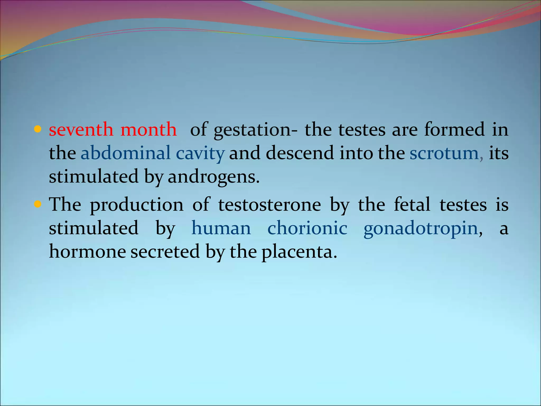
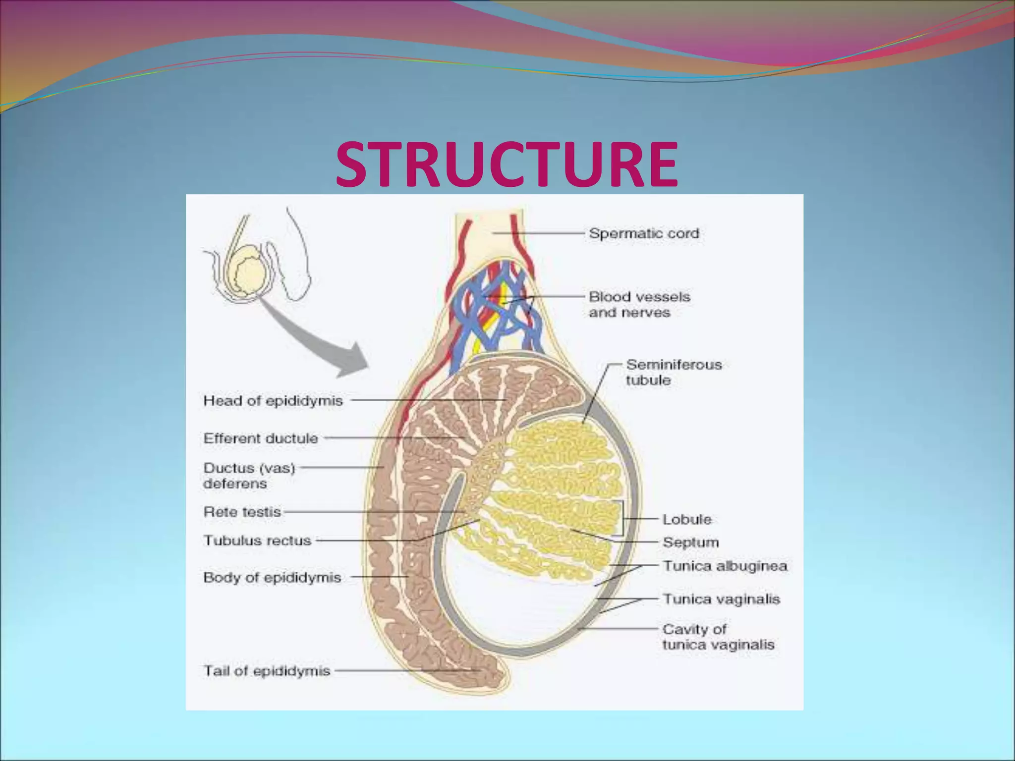
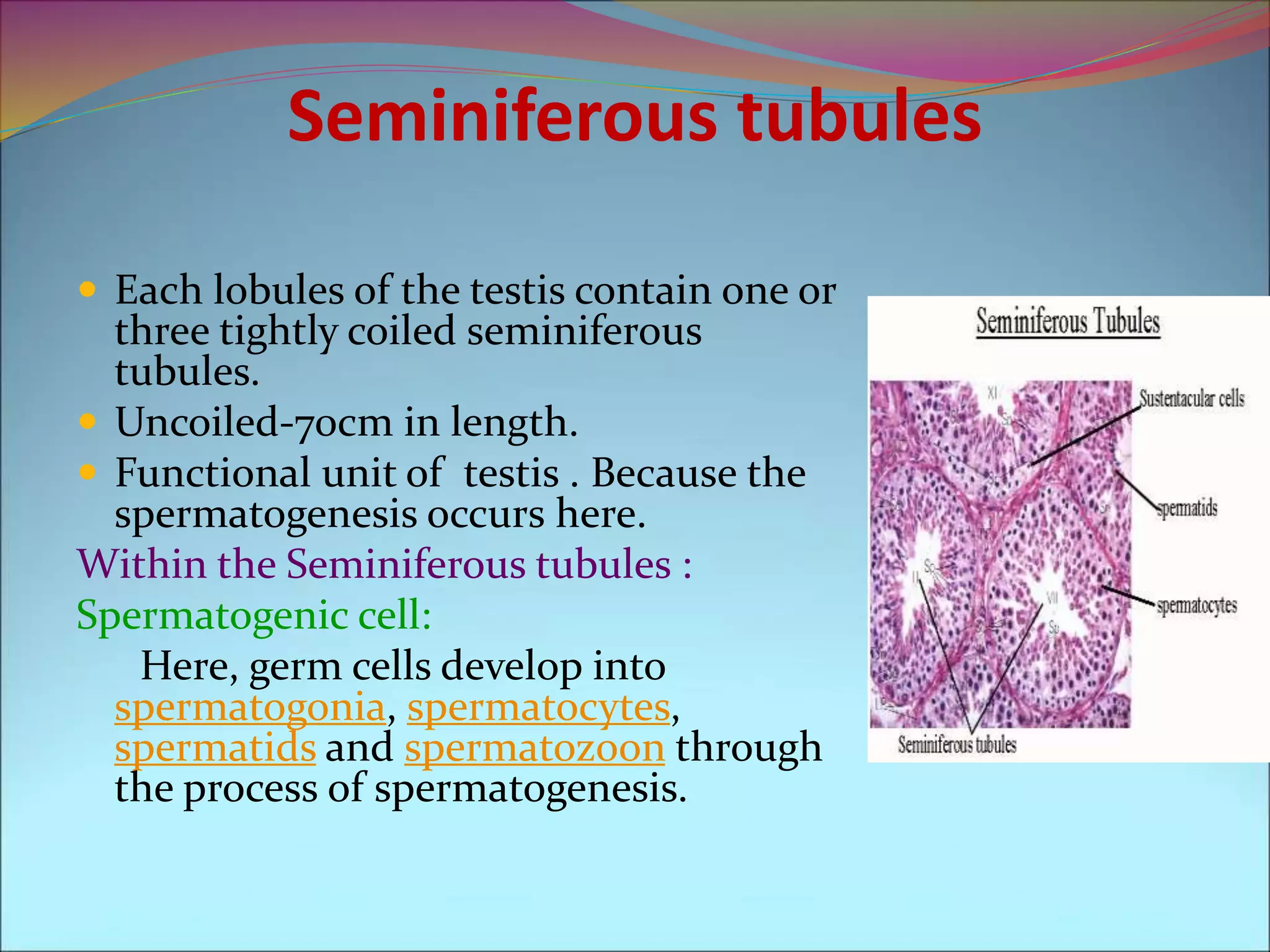
SYNOPSIS
 Introduction to testis
 Embryonic development of testis
 Anatomy
 Scrotum layers
Tunica vaginalis
Tunica albuginea
Tunica vasculosa
 Seminiferous tubules
Spermatogenic cell
Sertoli cell
Leydig cells
 Rete testis
 Efferent ductules
 Testicular arteris
 Summary
 References.
TESTIS
 Male gonad.
 Mixed exocrine and
endocrine gland because
they produce sperm and
Secreting male sex
hormones.
 They are contained within
the scrotal sac, which is
located directly behind the
penis and in front of the
anus.
EMBRYONIC DEVELOPMENT OF TESTES
 During mammalian development, the gonads are at
first capable of becoming either ovaries or testes.
 week 4, the gonadal rudiments are present within the
intermediate mesoderm adjacent to the developing
kidneys.
 week 6, sex cords develop within the forming testes.
 In males, the sex-specific gene SRY that is found on
the Y-chromosome initiates sex determination by
downstream regulation of sex-determining factors,
(such as GATA4, SOX9 and AMH), which leads to
development of the male phenotype.
 first trimester gestation -Testosterone and its potent
derivative, dihydrotestosterone, play key roles in the
formation of male genitalia in the fetus but do not play
a role in the actual formation of the testes.
 seventh month of gestation- the testes are formed in
the abdominal cavity and descend into the scrotum, its
stimulated by androgens.
 The production of testosterone by the fetal testes is
stimulated by human chorionic gonadotropin, a
hormone secreted by the placenta.
Anatomy:
 Paired, oval gland in the scrotum.
Length = 4-5cm
Diameter = 2.5cm
Mass = 10-15gm(single)
Temperature = 35°c
STRUCTURE
SCROTUM LAYERS
The scrotum consists of
loose skin and
subcutaneous layers and
supports the testis.
Tunica vaginalis
Tunica albuginea
Tunica vasculosa
Tunica vaginalis
 Outer thin ,serous sac .
 Derived from peritoneum.
 Forms during the descent of the testis.
 Partially covers the testes.
Tunica albuginea
 Internal to the tunica vaginalis.
 Tough, White fibrous capsule composed of dense irregular
connective tissue called tunica albuginea.
It extents inward
Form septa
Divide testis into serious of internal compartments called
lobules.
Each 200-300 lobules contain 1-3 tightly coiled tubules
called seminiferous tubules(semin-seed,fer-carry).
Tunica vasculosa
Testicular artery descends to the inferior pole just deep
to tunica albuginea.
Seminiferous tubules
 Each lobules of the testis contain one or
three tightly coiled seminiferous
tubules.
 Uncoiled-70cm in length.
 Functional unit of testis . Because the
spermatogenesis occurs here.
Within the Seminiferous tubules :
Spermatogenic cell:
Here, germ cells develop into
spermatogonia, spermatocytes,
spermatids and spermatozoon through
the process of spermatogenesis.
Sertoli cell
 Sertoli cell (a kind of sustentacular cell)
is a 'nurse' cell of the testes that is part of a
seminiferous tubule.
 It is activated by follicle-stimulating
hormone and has FSH-receptor on its
membranes.
 Extend from the basement membrane to
the lumen of the tubule.
 Internal to the basement membrane and
spermatogonia, tight junction joins
neighboring sertoli cells to one another.
 These junction form obstruction known as
blood- testes barrier.
EXOCRINE FUNCTIONS:
 Mechanical support.
 Nourishment.
 Phagocytosis of cytoplasmic fragments.
 Release of spermatozoa.
 Blood testis barrier.
ENDOCRINE FUNCTIONS:
 anti-Müllerian hormone (AMH)
inhibin and activin.
androgen binding protein.
Estradiol.
Transferrin.
Between tubules (interstitial cells):
Leydig cells :
 cells localized between
seminiferous tubules .
 produce and secrete
testosterone and other
androgens
 sexual development and
puberty,
 secondary sexual
characteristics.
 supporting spermatogenesis.
 Testosterone also controls
testicular volume.
Rete testis:
 Once the sperm produced,they move through the
Seminiferous tubules and enter the rete testes for
further maturation.
 Cilia are located on some cells of the rete testis
persumably for moving the sperm.
Efferent ductules:
 sperm Transported out of the testis into epididymis
through the series of efferent ductules.
Testicular arteris:
 Arises from the abdominal aorta below the origin of
renal arteris.
 Testis receive blood through the testicular arteris.
Summary
It’s a male gonad, paired oval gland in the scrotum.
seventh month of gestation- the testes are formed in
the abdominal cavity and descend into the scrotum, its
stimulated by androgens.
Scrotum layer
Seminiferous tubules.
 Rete testis
 Efferent ductules
 Testicular arteris
Function-spermatogenesis and steroidogenesis.
Testis by Dr Pandian M

Testis by Dr Pandian M

  • 2.
    SYNOPSIS  Introduction totestis  Embryonic development of testis  Anatomy  Scrotum layers Tunica vaginalis Tunica albuginea Tunica vasculosa  Seminiferous tubules Spermatogenic cell Sertoli cell Leydig cells  Rete testis  Efferent ductules  Testicular arteris  Summary  References.
  • 3.
    TESTIS  Male gonad. Mixed exocrine and endocrine gland because they produce sperm and Secreting male sex hormones.  They are contained within the scrotal sac, which is located directly behind the penis and in front of the anus.
  • 4.
    EMBRYONIC DEVELOPMENT OFTESTES  During mammalian development, the gonads are at first capable of becoming either ovaries or testes.  week 4, the gonadal rudiments are present within the intermediate mesoderm adjacent to the developing kidneys.  week 6, sex cords develop within the forming testes.
  • 5.
     In males,the sex-specific gene SRY that is found on the Y-chromosome initiates sex determination by downstream regulation of sex-determining factors, (such as GATA4, SOX9 and AMH), which leads to development of the male phenotype.  first trimester gestation -Testosterone and its potent derivative, dihydrotestosterone, play key roles in the formation of male genitalia in the fetus but do not play a role in the actual formation of the testes.
  • 6.
     seventh monthof gestation- the testes are formed in the abdominal cavity and descend into the scrotum, its stimulated by androgens.  The production of testosterone by the fetal testes is stimulated by human chorionic gonadotropin, a hormone secreted by the placenta.
  • 7.
    Anatomy:  Paired, ovalgland in the scrotum. Length = 4-5cm Diameter = 2.5cm Mass = 10-15gm(single) Temperature = 35°c
  • 8.
  • 9.
    SCROTUM LAYERS The scrotumconsists of loose skin and subcutaneous layers and supports the testis. Tunica vaginalis Tunica albuginea Tunica vasculosa
  • 10.
    Tunica vaginalis  Outerthin ,serous sac .  Derived from peritoneum.  Forms during the descent of the testis.  Partially covers the testes.
  • 11.
    Tunica albuginea  Internalto the tunica vaginalis.  Tough, White fibrous capsule composed of dense irregular connective tissue called tunica albuginea. It extents inward Form septa Divide testis into serious of internal compartments called lobules. Each 200-300 lobules contain 1-3 tightly coiled tubules called seminiferous tubules(semin-seed,fer-carry).
  • 12.
    Tunica vasculosa Testicular arterydescends to the inferior pole just deep to tunica albuginea.
  • 13.
    Seminiferous tubules  Eachlobules of the testis contain one or three tightly coiled seminiferous tubules.  Uncoiled-70cm in length.  Functional unit of testis . Because the spermatogenesis occurs here. Within the Seminiferous tubules : Spermatogenic cell: Here, germ cells develop into spermatogonia, spermatocytes, spermatids and spermatozoon through the process of spermatogenesis.
  • 14.
    Sertoli cell  Sertolicell (a kind of sustentacular cell) is a 'nurse' cell of the testes that is part of a seminiferous tubule.  It is activated by follicle-stimulating hormone and has FSH-receptor on its membranes.  Extend from the basement membrane to the lumen of the tubule.  Internal to the basement membrane and spermatogonia, tight junction joins neighboring sertoli cells to one another.  These junction form obstruction known as blood- testes barrier.
  • 15.
    EXOCRINE FUNCTIONS:  Mechanicalsupport.  Nourishment.  Phagocytosis of cytoplasmic fragments.  Release of spermatozoa.  Blood testis barrier. ENDOCRINE FUNCTIONS:  anti-Müllerian hormone (AMH) inhibin and activin. androgen binding protein. Estradiol. Transferrin.
  • 16.
    Between tubules (interstitialcells): Leydig cells :  cells localized between seminiferous tubules .  produce and secrete testosterone and other androgens  sexual development and puberty,  secondary sexual characteristics.  supporting spermatogenesis.  Testosterone also controls testicular volume.
  • 17.
    Rete testis:  Oncethe sperm produced,they move through the Seminiferous tubules and enter the rete testes for further maturation.  Cilia are located on some cells of the rete testis persumably for moving the sperm.
  • 18.
    Efferent ductules:  spermTransported out of the testis into epididymis through the series of efferent ductules. Testicular arteris:  Arises from the abdominal aorta below the origin of renal arteris.  Testis receive blood through the testicular arteris.
  • 19.
    Summary It’s a malegonad, paired oval gland in the scrotum. seventh month of gestation- the testes are formed in the abdominal cavity and descend into the scrotum, its stimulated by androgens. Scrotum layer Seminiferous tubules.  Rete testis  Efferent ductules  Testicular arteris Function-spermatogenesis and steroidogenesis.