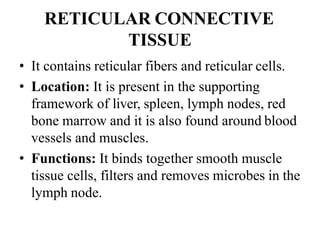
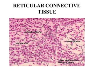
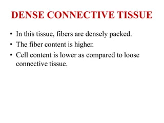
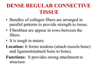
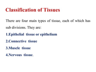
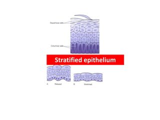
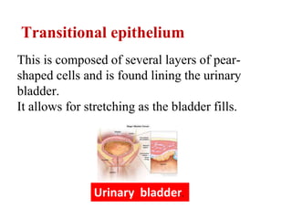
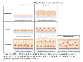
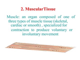
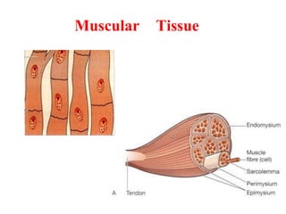
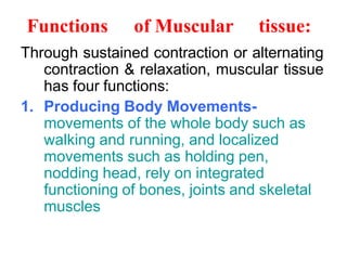
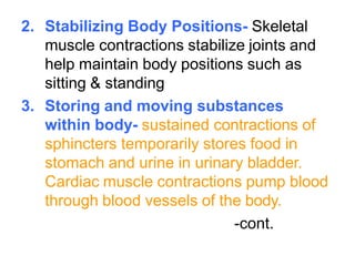
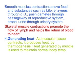
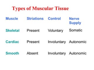
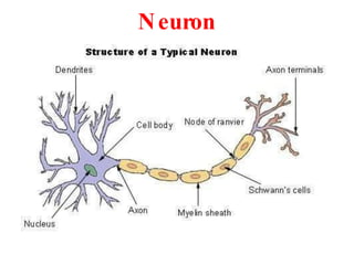
Tissue level of organization
Tissues
Tissue
The tissues of the body consist of large numbers of cells
and they are classified according to the size, shape and
functions of these cells.
Classification of Tissues
There are four main types of tissue, each of which has
sub divisions. They are:
1.Epithelial tissue or epithelium
2.Connective tissue
3.Muscle tissue
4.Nervous tissue.
Epithelial tissue provides a covering
(skin, the linings of the various passages inside the body).
Connective tissue supports other tissues and binds them
together (bone, blood, and lymph tissues).
Muscle tissue includes striated (also called voluntary)
muscles that move the skeleton, and smooth muscle,
such as the muscles that surround the stomach.
Nerve tissue is made up of nerve cells (neurons) and is
used to carry "messages" to and from various parts of
the body.
1. Epithelial Tissue
Epithelial tissue, commonly referred to as epithelium
is made up of one or more layers of cells that covers or
lines the external and internal surfaces of various body
parts.
It consists of variously shaped cells closely arranged
in one or more layers.
Epithelial tissue
This group of tissues is found covering the body and
lining cavities and tubes.
It is also present in glands.
Functions of Epithelium tissues
• Protection of underlying structures from, dehydration,
chemical and mechanical damage
• Secretion
• Absorption
The cells are very closely packed and the intercellular
substance, called the matrix.
The cells usually lie on a basement membrane.
Epithelial tissue may be:
• simple: a single layer of cells
• stratified: several layers of cells.
Simple Epithelium
 Simple epithelium consists of a single layer of identical
cells and is divided into four types.
 It is usually found on absorptive or secretory
surfaces, the single layer enhances these processes.
 The types are named according to the shape of the cells,
which differs according to their functions.
 The more active the tissue, the taller are the cells.
Classification of epithelium tissues
Epithelium tissues are classified in to Two types
1.Simple Epithelium
 Squamous
 Cuboidal
 Columnar
 Ciliated
 Pseudostratified
 Glandular
2.Compound epithelium
1.Squamous (pavement) epithelium
This is composed of a single layer of flattened cells.
The cells fit closely together like flat stones, forming a thin
and very smooth membrane.
Squamous (pavement) epithelium
This epithelium occurs in the walls of blood
vessels, air sacs of the lungs where oxygen
and carbon dioxide are exchanged,lymph
vessels, wall of Bowman’s capsule, loops of the
nephrons of Kidneys.
Functions of squamous epithelium
Exchange of gases.
Filtration in Bowman’s Capsule.
Exchange of materials in blood capillaries and
tissue fluid
Diffusion takes place freely through this thin,
smooth, inactive lining of the following
structures:
Heart
Blood vessels
Lymph vessels where it is also known as
Endothelium
alveoli of the lungs
2.Cuboidal (cubical) epithelium
Cuboidal cells epithelium are single-layered cells that are
shaped like a cuboid.
Cuboidal (cubical) epithelium
These are found in small collecting ducts of
kidneys, pancreas, and salivary glands
Cuboidal epithelium is actively involved in
secretion,absorption and excretion
Functions of cuboidal epithelium
The main function of the cuboidal cell
epithelium is to help in absorption, excretion,
and secretion.
3.Columnar Epithelium
This is formed by a single layer of cells,
rectangular in shape, on a basement membrane.
It is found lining the organs of the alimentary tract
and consists of a mixture of cells.
Some absorb the products of digestion and others
secrete mucus.
Mucus is a thick sticky substance secreted by
modified columnar cells called goblet cells.
4.Ciliated epithelium
This is formed by columnar cells each of which
has many fine, hair-like processes, called cilia.
The cilia consist of microtubules inside the
plasma membrane that extends from the free
border (luminal border) of the columnar
cells.
.
Ciliated epithelium
The wave-like movement of many cilia propels
the contents of the tubes, which they line in one
direction only
Ciliated epithelium is found lining the uterine
tubes and most of the respiratory passages.
In the uterine tubes the cilia propel ova towards
the uterus and in the respiratory passages they
propel mucus towards the throat.
Functions ofCiliated Epithelium
It is responsiblefor passingof ovumthroughfallopian
tube.
In respiratory tract, it helpsin expellingthe mucus and
particles trappedin it, towardsthe pharynx.
Stratified Epithelia
Stratified epithelia consist of several layers of cells of
various shapes.
The superficial layers grow up from below.
Basement membranes are usually absent.
The main function of stratified epithelium is to protect
underlying structures from mechanical wear and ear.
There are two main types: stratified squamous and
transitional
Stratified squamous epithelium
This is composed of a number of layers of cells
of different shapes representing newly formed
and mature cells.
In the deepest layers the cells are mainly
columnar.as they grow towards the surface, they
become flattened and are then shed.
Stratified epithelium
Non-keratinised stratified epithelium.
This is found on wet surfaces that may be
subjected to wear and tear but are protected
from drying.
e.g. the conjunctiva of the eyes, the lining of the
mouth, the pharynx, the esophagus and vagina.
Keratinized stratified epithelium
This is found on dry surfaces that are subjected
to wear and tear, i.e. skin, hair and nails. The
surface layer consists of dead epithelial cells to
which the protein keratin has been added.
This forms a tough, relatively waterproof
protective layer that prevents drying of the
underlying live cells.
The surface layer of skin is rubbed off and is
replaced from below
Transitional epithelium
This is composed of several layers of pear-
shaped cells and is found lining the urinary
bladder.
It allows for stretching as the bladder fills.
Urinary bladder.
Connective tissue
Connective tissue is the most abundant tissue in the
body.
The cells forming the connective tissues are more
widely separated from each other than those
forming the epithelium, and intercellular substance
(matrix) is present in considerably larger amounts.
There may or may not be fibres present in the
matrix, which may be of a semisolid jelly-like
consistency or dense and rigid, depending upon
the position and function of the tissue.
2. MuscularTissue
Muscle: an organ composed of one of
three types of muscle tissue (skeletal,
cardiac or smooth) , specialized for
contraction to produce voluntary or
involuntary movement
Muscular Tissue
Muscular Tissue
A group of cells (fibers) specialized to
produce motion in response to muscle
action potentials by its qualities of
contractility, extensibility, elasticity and
excitability
Properties of Muscular Tissue
1. Excitability: an ability of muscle to
generate impulse.
2. Contractility: it is either shortening or
development of tension or both.
A. Isotonic Contraction-contraction in which
tension remains same whereas changes
occurs in the length of muscle fiber. E.g.
flexion of arm
B. Isometric Contraction-contraction in
which length of muscle fibers remain
same and tension is increased. E.g.
holding book by hand, pulling any heavy
object
3. Muscle Tone- the muscle fibers always
maintain a state of slight contraction with
certain degree of vigor and tension. This
is a state of partial contraction of
muscles. It is achieved by the contraction
of a few muscle fibres at a time.
4. Extensibility- an ability of muscle fibers
to stretch without being damaged
5. Elasticity- an ability of muscle fibers to
return to its original length and shape
after contraction or extension
Functions of Muscular tissue:
Through sustained contraction or alternating
contraction & relaxation, muscular tissue
has four functions:
1. Producing Body Movements-
movements of the whole body such as
walking and running, and localized
movements such as holding pen,
nodding head, rely on integrated
functioning of bones, joints and skeletal
muscles
2. Stabilizing Body Positions- Skeletal
muscle contractions stabilize joints and
help maintain body positions such as
sitting & standing
3. Storing and moving substances
within body- sustained contractions of
sphincters temporarily stores food in
stomach and urine in urinary bladder.
Cardiac muscle contractions pump blood
through blood vessels of the body.
-cont.
Smooth muscles contractions move food
and substances such as bile, enzymes
through g.i.t., push gametes through
passageway of reproductive system,
propel urine through urinary system.
Skeletal muscle contractions promote the
flow of lymph and helps the return of blood
to heart.
4.Generating heat- As muscular tissue
contracts, it produces heat, by
thermogenesis. Heat generated by muscle
is used to maintain normal body temp.
Types of Muscular Tissue
Muscle Striations Control Nerve
Supply
Skeletal Present Voluntary Somatic
Cardiac Present Involuntary Autonomic
Smooth Absent Involuntary Autonomic
Cells of connective tissue
Connective tissue, excluding blood (Ch. 4), is
found in all organs supporting the specialized
tissue.
The different types of cell involved include:
 fibroblasts
 fat cells
 macrophages
 leukocytes
COMPOSITION OF
CONNECTIVE TISSUE
• Collagen Fibers: Large fibers made of the
protein collagen and are typically the most
abundant fibers. Promote tissue flexibility.
• Elastic Fibers: Intermediate fibers made of the
protein elastin. Branching fibers that allow for
stretch and recoil
• Reticular Fibers: Small delicate, branched fibers
that have same chemical composition of
collagen. Forms structural framework for organs
such as spleen and lymph nodes.
CLASSIFICATION OF CONNECTIVE
TISSUE
1) Loose connective tissue :
a) Areolar connective tissue
b) Adipose connective tissue
c) Reticular connective tissue
2) Dense connective tissue:
a) Dense regular connective tissue
b) Dense irregular connective tissue
c) Elastic connective tissue
CLASSIFICATION OF
CONNECTIVE TISSUE
3) Cartilage:
a) Hyaline cartilage
b) Fibro cartilage
c) Elastic cartilage
4) Bone tissue
Liquid connective tissue
a) Blood tissue
b) Lymph
Loose connective tissue
Areolar Connective tissue:
1)They forms a loose network in intracellular
material .
2)It consists of collagen, elastic fibers, reticular
fibers and several kinds of cells.
Location: Below the skin, fill space between
muscles, supports blood vessels and nerves in
alimentary canal.
Functions: It gives strength, elasticity and
support to tissue.
Areolar Connective tissue:
ADIPOSE CONNECTIVE TISSUE
• It consists of adipocytes which stores fat.
Location: It is present in subcutaneous layer deep in
the skin, around the heart and kidneys
Functions :
• Prevents heat loose from body.
• Act as reservoir of energy.
• It give shape to the limbs and body.
• It protects underlying organ from injury.
ADIPOSE CONNECTIVE TISSUE
RETICULAR CONNECTIVE
TISSUE
• It contains reticular fibers and reticular cells.
• Location: It is present in the supporting
framework of liver, spleen, lymph nodes, red
bone marrow and it is also found around blood
vessels and muscles.
• Functions: It binds together smooth muscle
tissue cells, filters and removes microbes in the
lymph node.
RETICULAR CONNECTIVE
TISSUE
DENSE CONNECTIVE TISSUE
• In this tissue, fibers are densely packed.
• The fiber content is higher.
• Cell content is lower as compared to loose
connective tissue.
DENSE REGULAR CONNECTIVE
TISSUE
• Bundles of collagen fibers are arranged in
parallel patterns to provide strength to tissue.
• Fibroblast are appear in rows between the
fibers.
• It is tough in nature.
Location: It forms tendons (attach muscle bone)
and ligaments(attach bone to bone).
Functions: It provides strong attachment to
structure.
DENSE REGULAR CONNECTIVE
TISSUE
DENSE IRREGULAR
CONNECTIVE TISSUE
• It contains collagen fibers which are irregular
arranged and a few fibroblasts are appear in
rows between the fibers.
Location: It present in dermis layer of skin,
membrane capsules around kidneys, liver,
testes and lymph node, heart valves.
Functions: It provides strength to different
organs.
DENSE IRREGULAR
CONNECTIVE TISSUE
ELASTIC CONNECTIVE TISSUE
• It consists of freely branching elastic fibers.
• Fibroblast are present in space between fibers.
• It is yellowish in colour.
Location: It is present in tissues , walls of elastic
arteries, trachea, bronchial tubes and vocal
cords.
Functions: It allows stretching of various
organs.
ELASTIC CONNECTIVE TISSUE
CARTILAGE
• It consist of network of closely packed
collagen fibers and elastic fibers.
• The cells of mature cartilage called as
chondrocytes.
HYALINE CARTILAGE
• It is bluish white in color.
• It consists of fine collagen fibers and many
chondrocytes.
Location: It is present at the end of long bones,
anterior ends of ribs, nose and parts of larynx,
trachea, bronchi, bronchial tubes.
Function: It provides small surface for
movement at joints, flexibility and support.
HYALINE CARTILAGE
FIBRO CARTILAGE
• It is strongest form of cartilage.
• The chondrocytes are scattered among the
bundle collagen fibers within the extracellular
matrix.
Location: It is present in inter-verteblar disc.
Functions: It covers and protects bony structures
of body.
FIBRO CARTILAGE
ELASTIC CARTILAGE
• The chondrocytes are located within a
threadlike network of elastic fibers within
extracellular matrix.
Location: It is present in pinna of ear and top of
larynx.
Functions: It provides strength and elasticity
and maintain the shape of certain organs such
as the external ear.
ELASTIC CARTILAGE
4. Nervous Tissue
The human body is composed of approximately
200 distinctly different types of cells. These cells
are organized into four basic tissues that in turn,
are assembled to form organs.
Nervous tissue consists of two kinds of
nerve cells:
Neurons are the basic structural unit of the
nervous system.
Each cell consists of the following parts; the cell
body contains the nucleus and other cellular
organelles.
The dendrites are typically short,
slender extensions of the cell body that
receive stimuli.
The axon is typically a long, slender
extension of the cell body that sends
stimuli.
The axon branches are, typically, smaller
extensions of the axon.
Neuroglia, or glial cells, provides support
functions for the neurons, such as insulation or
anchoring neurons to blood vessels.
Neuron
References:
1. Presentation on Introduction To Human Anatomy
& Physiology, By Mr. Abhay Shripad Joshi.
2. Human Anatomy and Physiology-I,
By Dr. Mahesh Prasad, Dr. Antesh Kumar
Jha, Mr. Ritesh Kumar Srivastav, Nirali
Prakashan, As per PCI Syllabus.
Page No. 1.23 to 1.28.
3. www.google.com.
THANK YOU

TISSUES

  • 1.
    Tissue level oforganization Tissues
  • 2.
    Tissue The tissues ofthe body consist of large numbers of cells and they are classified according to the size, shape and functions of these cells.
  • 3.
    Classification of Tissues Thereare four main types of tissue, each of which has sub divisions. They are: 1.Epithelial tissue or epithelium 2.Connective tissue 3.Muscle tissue 4.Nervous tissue.
  • 4.
    Epithelial tissue providesa covering (skin, the linings of the various passages inside the body). Connective tissue supports other tissues and binds them together (bone, blood, and lymph tissues).
  • 5.
    Muscle tissue includesstriated (also called voluntary) muscles that move the skeleton, and smooth muscle, such as the muscles that surround the stomach. Nerve tissue is made up of nerve cells (neurons) and is used to carry "messages" to and from various parts of the body.
  • 6.
    1. Epithelial Tissue Epithelialtissue, commonly referred to as epithelium is made up of one or more layers of cells that covers or lines the external and internal surfaces of various body parts. It consists of variously shaped cells closely arranged in one or more layers.
  • 7.
    Epithelial tissue This groupof tissues is found covering the body and lining cavities and tubes. It is also present in glands. Functions of Epithelium tissues • Protection of underlying structures from, dehydration, chemical and mechanical damage • Secretion • Absorption
  • 8.
    The cells arevery closely packed and the intercellular substance, called the matrix. The cells usually lie on a basement membrane. Epithelial tissue may be: • simple: a single layer of cells • stratified: several layers of cells.
  • 9.
    Simple Epithelium  Simpleepithelium consists of a single layer of identical cells and is divided into four types.  It is usually found on absorptive or secretory surfaces, the single layer enhances these processes.  The types are named according to the shape of the cells, which differs according to their functions.  The more active the tissue, the taller are the cells.
  • 10.
    Classification of epitheliumtissues Epithelium tissues are classified in to Two types 1.Simple Epithelium  Squamous  Cuboidal  Columnar  Ciliated  Pseudostratified  Glandular 2.Compound epithelium
  • 11.
    1.Squamous (pavement) epithelium Thisis composed of a single layer of flattened cells. The cells fit closely together like flat stones, forming a thin and very smooth membrane.
  • 12.
    Squamous (pavement) epithelium Thisepithelium occurs in the walls of blood vessels, air sacs of the lungs where oxygen and carbon dioxide are exchanged,lymph vessels, wall of Bowman’s capsule, loops of the nephrons of Kidneys.
  • 13.
    Functions of squamousepithelium Exchange of gases. Filtration in Bowman’s Capsule. Exchange of materials in blood capillaries and tissue fluid
  • 14.
    Diffusion takes placefreely through this thin, smooth, inactive lining of the following structures: Heart Blood vessels Lymph vessels where it is also known as Endothelium alveoli of the lungs
  • 15.
    2.Cuboidal (cubical) epithelium Cuboidalcells epithelium are single-layered cells that are shaped like a cuboid.
  • 16.
    Cuboidal (cubical) epithelium Theseare found in small collecting ducts of kidneys, pancreas, and salivary glands Cuboidal epithelium is actively involved in secretion,absorption and excretion
  • 17.
    Functions of cuboidalepithelium The main function of the cuboidal cell epithelium is to help in absorption, excretion, and secretion.
  • 18.
    3.Columnar Epithelium This isformed by a single layer of cells, rectangular in shape, on a basement membrane.
  • 19.
    It is foundlining the organs of the alimentary tract and consists of a mixture of cells. Some absorb the products of digestion and others secrete mucus. Mucus is a thick sticky substance secreted by modified columnar cells called goblet cells.
  • 20.
    4.Ciliated epithelium This isformed by columnar cells each of which has many fine, hair-like processes, called cilia. The cilia consist of microtubules inside the plasma membrane that extends from the free border (luminal border) of the columnar cells. .
  • 21.
    Ciliated epithelium The wave-likemovement of many cilia propels the contents of the tubes, which they line in one direction only
  • 22.
    Ciliated epithelium isfound lining the uterine tubes and most of the respiratory passages. In the uterine tubes the cilia propel ova towards the uterus and in the respiratory passages they propel mucus towards the throat.
  • 23.
    Functions ofCiliated Epithelium Itis responsiblefor passingof ovumthroughfallopian tube. In respiratory tract, it helpsin expellingthe mucus and particles trappedin it, towardsthe pharynx.
  • 24.
    Stratified Epithelia Stratified epitheliaconsist of several layers of cells of various shapes. The superficial layers grow up from below. Basement membranes are usually absent. The main function of stratified epithelium is to protect underlying structures from mechanical wear and ear. There are two main types: stratified squamous and transitional
  • 25.
    Stratified squamous epithelium Thisis composed of a number of layers of cells of different shapes representing newly formed and mature cells. In the deepest layers the cells are mainly columnar.as they grow towards the surface, they become flattened and are then shed.
  • 26.
  • 27.
    Non-keratinised stratified epithelium. Thisis found on wet surfaces that may be subjected to wear and tear but are protected from drying. e.g. the conjunctiva of the eyes, the lining of the mouth, the pharynx, the esophagus and vagina.
  • 28.
    Keratinized stratified epithelium Thisis found on dry surfaces that are subjected to wear and tear, i.e. skin, hair and nails. The surface layer consists of dead epithelial cells to which the protein keratin has been added.
  • 29.
    This forms atough, relatively waterproof protective layer that prevents drying of the underlying live cells. The surface layer of skin is rubbed off and is replaced from below
  • 30.
    Transitional epithelium This iscomposed of several layers of pear- shaped cells and is found lining the urinary bladder. It allows for stretching as the bladder fills. Urinary bladder.
  • 31.
    Connective tissue Connective tissueis the most abundant tissue in the body. The cells forming the connective tissues are more widely separated from each other than those forming the epithelium, and intercellular substance (matrix) is present in considerably larger amounts.
  • 32.
    There may ormay not be fibres present in the matrix, which may be of a semisolid jelly-like consistency or dense and rigid, depending upon the position and function of the tissue.
  • 34.
    2. MuscularTissue Muscle: anorgan composed of one of three types of muscle tissue (skeletal, cardiac or smooth) , specialized for contraction to produce voluntary or involuntary movement
  • 35.
  • 36.
    Muscular Tissue A groupof cells (fibers) specialized to produce motion in response to muscle action potentials by its qualities of contractility, extensibility, elasticity and excitability
  • 37.
    Properties of MuscularTissue 1. Excitability: an ability of muscle to generate impulse.
  • 38.
    2. Contractility: itis either shortening or development of tension or both. A. Isotonic Contraction-contraction in which tension remains same whereas changes occurs in the length of muscle fiber. E.g. flexion of arm B. Isometric Contraction-contraction in which length of muscle fibers remain same and tension is increased. E.g. holding book by hand, pulling any heavy object
  • 39.
    3. Muscle Tone-the muscle fibers always maintain a state of slight contraction with certain degree of vigor and tension. This is a state of partial contraction of muscles. It is achieved by the contraction of a few muscle fibres at a time. 4. Extensibility- an ability of muscle fibers to stretch without being damaged 5. Elasticity- an ability of muscle fibers to return to its original length and shape after contraction or extension
  • 40.
    Functions of Musculartissue: Through sustained contraction or alternating contraction & relaxation, muscular tissue has four functions: 1. Producing Body Movements- movements of the whole body such as walking and running, and localized movements such as holding pen, nodding head, rely on integrated functioning of bones, joints and skeletal muscles
  • 41.
    2. Stabilizing BodyPositions- Skeletal muscle contractions stabilize joints and help maintain body positions such as sitting & standing 3. Storing and moving substances within body- sustained contractions of sphincters temporarily stores food in stomach and urine in urinary bladder. Cardiac muscle contractions pump blood through blood vessels of the body. -cont.
  • 42.
    Smooth muscles contractionsmove food and substances such as bile, enzymes through g.i.t., push gametes through passageway of reproductive system, propel urine through urinary system. Skeletal muscle contractions promote the flow of lymph and helps the return of blood to heart. 4.Generating heat- As muscular tissue contracts, it produces heat, by thermogenesis. Heat generated by muscle is used to maintain normal body temp.
  • 43.
    Types of MuscularTissue Muscle Striations Control Nerve Supply Skeletal Present Voluntary Somatic Cardiac Present Involuntary Autonomic Smooth Absent Involuntary Autonomic
  • 44.
    Cells of connectivetissue Connective tissue, excluding blood (Ch. 4), is found in all organs supporting the specialized tissue. The different types of cell involved include:  fibroblasts  fat cells  macrophages  leukocytes
  • 45.
    COMPOSITION OF CONNECTIVE TISSUE •Collagen Fibers: Large fibers made of the protein collagen and are typically the most abundant fibers. Promote tissue flexibility. • Elastic Fibers: Intermediate fibers made of the protein elastin. Branching fibers that allow for stretch and recoil • Reticular Fibers: Small delicate, branched fibers that have same chemical composition of collagen. Forms structural framework for organs such as spleen and lymph nodes.
  • 46.
    CLASSIFICATION OF CONNECTIVE TISSUE 1)Loose connective tissue : a) Areolar connective tissue b) Adipose connective tissue c) Reticular connective tissue 2) Dense connective tissue: a) Dense regular connective tissue b) Dense irregular connective tissue c) Elastic connective tissue
  • 47.
    CLASSIFICATION OF CONNECTIVE TISSUE 3)Cartilage: a) Hyaline cartilage b) Fibro cartilage c) Elastic cartilage 4) Bone tissue Liquid connective tissue a) Blood tissue b) Lymph
  • 48.
    Loose connective tissue AreolarConnective tissue: 1)They forms a loose network in intracellular material . 2)It consists of collagen, elastic fibers, reticular fibers and several kinds of cells. Location: Below the skin, fill space between muscles, supports blood vessels and nerves in alimentary canal. Functions: It gives strength, elasticity and support to tissue.
  • 49.
  • 50.
    ADIPOSE CONNECTIVE TISSUE •It consists of adipocytes which stores fat. Location: It is present in subcutaneous layer deep in the skin, around the heart and kidneys Functions : • Prevents heat loose from body. • Act as reservoir of energy. • It give shape to the limbs and body. • It protects underlying organ from injury.
  • 51.
  • 52.
    RETICULAR CONNECTIVE TISSUE • Itcontains reticular fibers and reticular cells. • Location: It is present in the supporting framework of liver, spleen, lymph nodes, red bone marrow and it is also found around blood vessels and muscles. • Functions: It binds together smooth muscle tissue cells, filters and removes microbes in the lymph node.
  • 53.
  • 54.
    DENSE CONNECTIVE TISSUE •In this tissue, fibers are densely packed. • The fiber content is higher. • Cell content is lower as compared to loose connective tissue.
  • 55.
    DENSE REGULAR CONNECTIVE TISSUE •Bundles of collagen fibers are arranged in parallel patterns to provide strength to tissue. • Fibroblast are appear in rows between the fibers. • It is tough in nature. Location: It forms tendons (attach muscle bone) and ligaments(attach bone to bone). Functions: It provides strong attachment to structure.
  • 56.
  • 57.
    DENSE IRREGULAR CONNECTIVE TISSUE •It contains collagen fibers which are irregular arranged and a few fibroblasts are appear in rows between the fibers. Location: It present in dermis layer of skin, membrane capsules around kidneys, liver, testes and lymph node, heart valves. Functions: It provides strength to different organs.
  • 58.
  • 59.
    ELASTIC CONNECTIVE TISSUE •It consists of freely branching elastic fibers. • Fibroblast are present in space between fibers. • It is yellowish in colour. Location: It is present in tissues , walls of elastic arteries, trachea, bronchial tubes and vocal cords. Functions: It allows stretching of various organs.
  • 60.
  • 61.
    CARTILAGE • It consistof network of closely packed collagen fibers and elastic fibers. • The cells of mature cartilage called as chondrocytes.
  • 62.
    HYALINE CARTILAGE • Itis bluish white in color. • It consists of fine collagen fibers and many chondrocytes. Location: It is present at the end of long bones, anterior ends of ribs, nose and parts of larynx, trachea, bronchi, bronchial tubes. Function: It provides small surface for movement at joints, flexibility and support.
  • 63.
  • 64.
    FIBRO CARTILAGE • Itis strongest form of cartilage. • The chondrocytes are scattered among the bundle collagen fibers within the extracellular matrix. Location: It is present in inter-verteblar disc. Functions: It covers and protects bony structures of body.
  • 65.
  • 66.
    ELASTIC CARTILAGE • Thechondrocytes are located within a threadlike network of elastic fibers within extracellular matrix. Location: It is present in pinna of ear and top of larynx. Functions: It provides strength and elasticity and maintain the shape of certain organs such as the external ear.
  • 67.
  • 68.
    4. Nervous Tissue Thehuman body is composed of approximately 200 distinctly different types of cells. These cells are organized into four basic tissues that in turn, are assembled to form organs.
  • 69.
    Nervous tissue consistsof two kinds of nerve cells: Neurons are the basic structural unit of the nervous system. Each cell consists of the following parts; the cell body contains the nucleus and other cellular organelles.
  • 70.
    The dendrites aretypically short, slender extensions of the cell body that receive stimuli. The axon is typically a long, slender extension of the cell body that sends stimuli.
  • 71.
    The axon branchesare, typically, smaller extensions of the axon. Neuroglia, or glial cells, provides support functions for the neurons, such as insulation or anchoring neurons to blood vessels.
  • 72.
  • 73.
    References: 1. Presentation onIntroduction To Human Anatomy & Physiology, By Mr. Abhay Shripad Joshi. 2. Human Anatomy and Physiology-I, By Dr. Mahesh Prasad, Dr. Antesh Kumar Jha, Mr. Ritesh Kumar Srivastav, Nirali Prakashan, As per PCI Syllabus. Page No. 1.23 to 1.28. 3. www.google.com.
  • 74.